


A Versatile In Vitro Quantitative Assay for Macrophage Efferocytosis in Diverse Research Applications
适用于多种研究场景的巨噬细胞胞葬作用体外定量检测方法

(*contributed equally to this work) 发布: 2026年05月20日第16卷第10期 DOI: 10.21769/BioProtoc.5690 浏览次数: 1002
评审: Philipp WörsdörferScott McCombAnonymous reviewer(s)
 PDF
PDF  Q&A
Q&A
参见作者原研究论文
The authors used this protocol in:

Jan 2026
Advertisement








Abstract
Macrophage efferocytosis is a previously unrecognized key pathogenic event, engulfing apoptotic targets, preventing inflammation and necrosis, and maintaining immune homeostasis. The phagocytic function can be disrupted by harmful factors and toxic substances. This protocol describes a versatile visualized in vitro method that can be used for the detection of general efferocytosis. This method is applicable to a wide range of research scenarios. As a representative application, it can be used to evaluate macrophage efferocytosis dysfunction in diseases linked to harmful exposures, including atherosclerosis, chronic inflammation, and malignant tumors. Among them, the detection of the effects of oxidized low-density lipoprotein (ox-LDL) and arsenite on macrophage efferocytosis capacity is an exemplary application of this protocol. Primary macrophages collected from mice were labeled with a cell-tracking dye and exposed to ox-LDL or arsenite, then co-cultured with apoptotic thymocytes or hepatocytes (labeled with another cell-tracking dye) for 2 h at a ratio of 5:1. Macrophage efferocytosis was visualized using a laser confocal microscope. The results indicate that arsenite impaired macrophage efferocytosis, leading to insufficient clearance of apoptotic thymocytes or hepatocytes. This method can be extended to subsequent studies, including those involving different types of phagocytes, apoptotic cell models, and research related to exposure to various factors.
Key features
• This protocol harvests primary mouse macrophages, treats them with different factors (ox-LDL, sodium arsenite), and assesses efferocytosis of apoptotic/necrotic thymocytes or hepatocytes.
• Dye-swap red–green staining eliminates fluorescence interference and staining bias and validates dye effects on efferocytosis.
Keywords: Efferocytosis (胞葬作用) Macrophages (巨噬细胞)
Thymocytes (胸腺细胞)
Hepatocytes (肝细胞)
Apoptotic targets (凋亡靶细胞)
Macrophages (巨噬细胞)
Thymocytes (胸腺细胞)
Hepatocytes (肝细胞)
Apoptotic targets (凋亡靶细胞)
Graphical overview
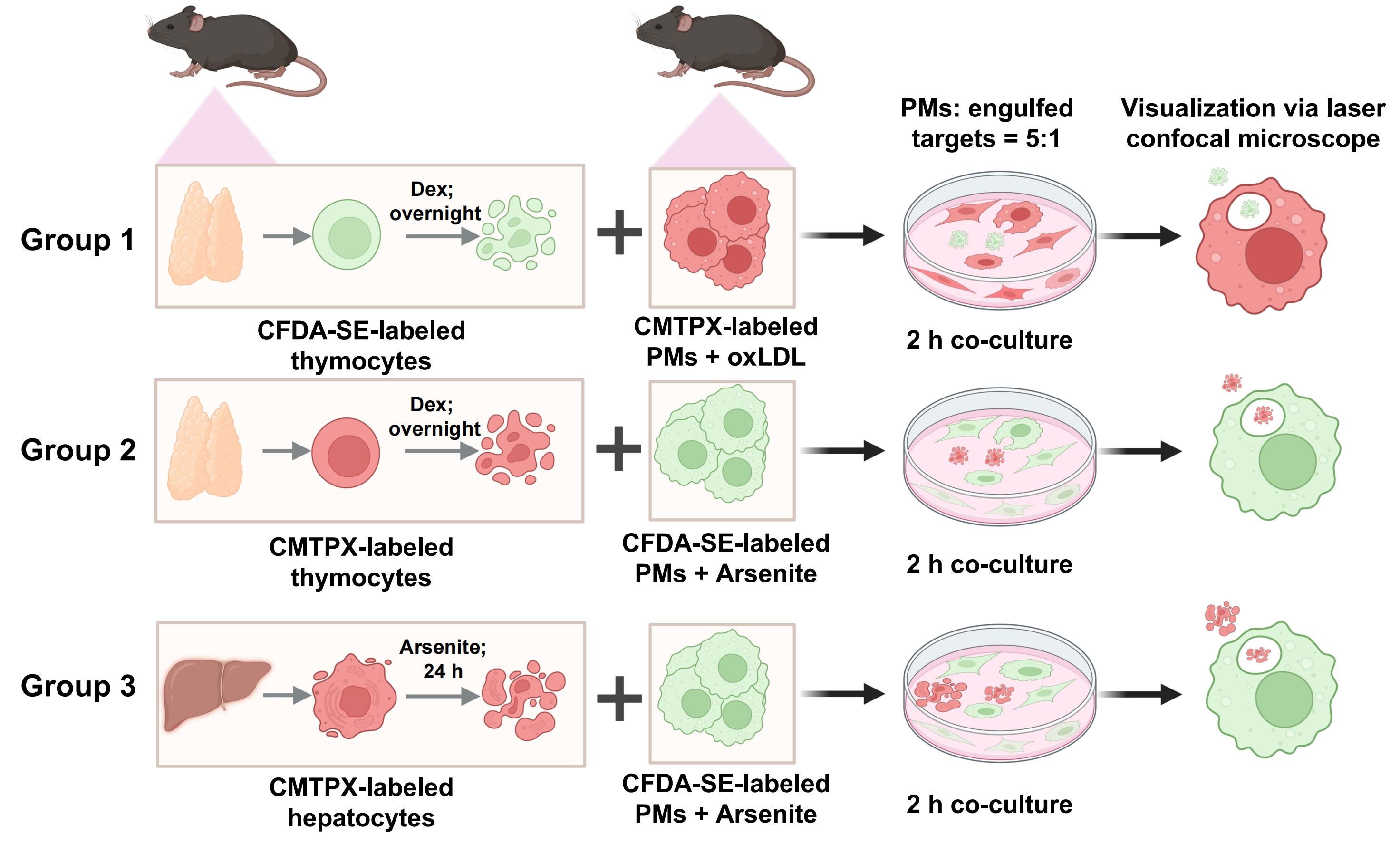
Confocal laser scanning microscopy is employed to evaluate the efferocytosis of various apoptotic cells by primary peritoneal macrophages (PMs) in response to different factors [oxidized low-density lipoprotein (ox-LDL) and sodium arsenite]. This schematic illustrates the step-by-step experimental procedure, including efferocytosis of dexamethasone-induced apoptotic/necrotic thymocytes by macrophages under ox-LDL exposure, efferocytosis of dexamethasone-induced apoptotic/necrotic thymocytes by macrophages under sodium arsenite exposure, and efferocytosis of sodium arsenite-induced apoptotic/necrotic hepatocytes by macrophages under sodium arsenite exposure.
Background
Efferocytosis, a specialized type of phagocytosis, is defined as the phagocytosis and clearance of apoptotic, aged, or injured cells [1,2]. Impaired efferocytosis causes an inflammatory response and leads to disease states [2,3]. Macrophages are the major component involved in efferocytosis [4]. Impaired macrophage efferocytosis exacerbates inflammation and tissue damage, thereby advancing the progression of various inflammatory disorders, such as atherosclerosis, autoimmune conditions, and cancer [2,5–8]. Under physiological conditions, macrophages residing in almost every organ and tissue are the major phagocytes to perform efferocytosis [9–11]. Some tissue-resident macrophages have specific names, such as alveolar macrophages in the lungs, microglia in the brain, and Kupffer cells in the liver [1]. All of these cells are involved in removing apoptotic cells through efferocytosis in different tissues [1].
Various factors (e.g., alkaloids, advanced glycation end products, modified low-density lipoproteins, heavy metals, pesticides, and pollutant residues) have a pathogenetic role in the development and progression of impaired efferocytosis-related diseases, including diabetes [12], cardiovascular diseases [13], and neurological disorders [14]. Previous studies have demonstrated that 7-ketocholesterol, 12(S)-hydroxyeicosatetraenoic acid, 20-OH-RvE4, SiO2, ethanol, and acetaldehyde impair the efferocytosis capacities of mouse or human macrophages [15–19]. Arsenic, classified as a metalloid, is a naturally occurring element that is widely distributed in the Earth's crust [20]. Environmental arsenic exposure correlates with multi-system disorders, including skin [21], nervous [22], respiratory [23], cardiovascular [24,25], and immune systems [26]. Arsenic is one of WHO’s 10 chemicals of major public health concern [27]. In this study, sodium arsenite was used as a representative factor to elucidate its effect on macrophage efferocytosis. Low-density lipoproteins (LDLs), especially oxidized LDL (ox-LDL), play a key role in promoting atherosclerosis [28,29]. High levels of ox-LDL are considered a risk factor for cardiovascular events due to their central role in atherosclerotic plaque formation [30]. In this study, ox-LDL was selected as another distinct representative factor to investigate its regulatory effects on macrophage efferocytosis.
In the present study, a visualized in vitro protocol is applied to evaluate the effects of different factors on efferocytosis. This approach involves isolating primary cells, co-culturing cell-tracking dye-labeled macrophages with apoptotic cells, and recording the results using laser confocal microscopy. This approach offers several advantages, including convenience and visualization, particularly of the intricate details of cellular processes [31,32]. The superior resolution and depth of field provided by confocal microscopy allow for the observation of intracellular structures, facilitating the analysis of morphological changes during efferocytosis, such as the formation of pseudopodia and the engulfment of apoptotic targets. These details enable researchers to directly observe the process of macrophages engulfing apoptotic targets and the nuances of their interactions.
This protocol presents an updated efferocytosis assay protocol, based on existing approaches [33,34]. The protocol details two distinct approaches—the first using two distinct representative factors (ox-LDL and arsenite) and the second using two kinds of apoptotic cells (thymocytes and hepatocytes). These approaches clarify the efferocytotic capacity of macrophages toward apoptotic cells generated under different conditions. Furthermore, they provide a reliable method for evaluating the effects of different factors on macrophage efferocytosis.
Materials and reagents
Reagents
1. Isoflurane (RWD, catalog number: R510-22-10)
2. Annexin V-FITC/PI Apoptosis Detection kit (Elabscience, catalog number: E-CK-A211)
3. CellTraceTM CMTPX Red cell tracer (Yeasen Biotechnology, catalog number: 40717ES)
4. CFDA SE Cell Proliferation and Cell Tracking kit (Yeasen Biotechnology, catalog number: 40714ES), comprising CFDA-SE fluorescent probe (40714-A), CFDA-SE solvent (40714-B), and 5× cell staining buffer (40714-C)
5. Collagenase Type IV (Sigma, catalog number: C5138, specific activity ≥125 CDU/mg)
6. Dexamethasone (GlpBio, catalog number: GC40775)
7. EDTA (Invitrogen, catalog number: 91222920)
8. M199 medium (GlpBio, catalog number: C11150500BT)
9. Oxidized LDL (ox-LDL) (Yiyuan Biotech, catalog number: YB-002)
10. Penicillin-streptomycin solution (pen/strep) (Procell Life Science & Technology, catalog number: PB180120)
11. RPMI 1640 culture medium (Gibco, catalog number: C11875500BT)
12. Sodium arsenite (Sigma-Aldrich, catalog number: S9663)
13. Thioglycollate medium (Sigma-Aldrich, catalog number: T9032)
14. Phosphate buffer saline (PBS), pH 7.4 (Biopico Life Sciences, catalog number: 090501)
15. 1 M HEPES solution (Procell Life Science & Technology, catalog number: PB180325)
16. 10× Hanks’ balanced salt solution (HBSS), Ca2+/Mg2+-free (Sigma-Aldrich, catalog number: H4641)
17. Calcium chloride, anhydrous (CaCl2) (Sangon Biotech, catalog number: A501330)
18. Red blood cell lysis buffer (Elabscience, catalog number: E-CK-A105)
19. Fetal bovine serum (FBS) (Procell Life Science & Technology, catalog number: 164210)
20. Sodium pyruvate solution (Procell Life Science & Technology, catalog number: PB180422)
21. GlutaMAX (Gibco, catalog number: 35050061)
22. Puralube vet ointment (Dechra, catalog number: 1703321138)
Solutions
1. 3% (w/v) thioglycollate medium (see Recipes)
2. 1,000× CFDA-SE stock solution (see Recipes)
3. CFDA-SE working solution (see Recipes)
4. Wash buffer (see Recipes)
5. Collagen buffer (see Recipes)
Recipes
1. 3% (w/v) thioglycollate medium
Prepare fresh 3% (w/v) thioglycollate medium aseptically in a biosafety cabinet by dissolving 0.03 g of thioglycollate medium powder (4 °C storage) in 1 mL of sterile PBS. Immediately apply the prepared solution to maintain optimal induction efficiency.
2. 1,000× CFDA-SE stock solution
To prepare the 1,000× CFDA-SE stock solution, add 500 μL of CFDA-SE solvent to one tube of CFDA-SE fluorescent probe (1.4 mg) and mix thoroughly to yield a clear stock solution with a concentration of 2.8 mg/mL. Aliquot the stock solution immediately after preparation and store it at ≤ -20 °C (or -70 °C for an extended shelf life) under dry and light-protected conditions; repeated freeze-thaw cycles must be strictly avoided. The stock solution is optimal for use within 1 month and has a maximum shelf life of 2 months when stored as specified above.
3. CFDA-SE working solution
Prepare 1× cell staining buffer (the diluent for CFDA-SE) by taking the supplied 5× cell staining buffer and diluting it 1:5 with sterile deionized water (e.g., 2 mL of 5× buffer mixed with 8 mL of sterile deionized water) with thorough agitation. Store the diluted 1× buffer at 4 °C, protected from light, and use it within 1 week for optimal performance. For the 2× CFDA-SE working solution, take the 1,000× CFDA-SE stock solution thawed to 20–25 °C, dilute it 1:500 with freshly prepared 1× cell staining buffer (e.g., 2 μL of 1,000× stock added to 1 mL of 1× buffer), and mix gently to form a homogeneous 2× working solution.
Critical: The working solution must be freshly prepared immediately before use, as CFDA-SE is susceptible to hydrolysis in aqueous solutions; all preparation steps should be performed in the dark to prevent fluorophore quenching. All kit components are pre-sterilized, and no additional sterilization is required.
4. Wash buffer
| Reagent | Final concentration | Volume |
|---|---|---|
| 1 M HEPES | 50 mM | 5 mL |
| 0.5 M EDTA (pH 7.98) | 5 mM | 1 mL |
| 10× HBSS | 1× | 10 mL |
| ddH2O | 84 mL | |
| Total | 100 mL |
Prepare the solution according to the table above and autoclave before use.
5. Collagen buffer
| Reagent | Final concentration | Volume |
|---|---|---|
| 1 M HEPES | 50 mM | 1.5 mL |
| CaCl2 (50 mM) | 5 mM | 3 mL |
| 10× HBSS | 1× | 3 mL |
| ddH2O | 22.5 mL | |
| Total | 30 mL |
Prepare the solution according to the table above and autoclave prior to use.
Note: The 50 mM CaCl2 working solution is prepared by diluting 2 mL of 1 M CaCl2 stock solution (11.1 g of CaCl2 dissolved in 100 mL of ddH2O) into 38 mL of ddH2O. Due to the poor aqueous stability of collagenase, add 30 mg of collagenase powder to 30 mL of the pre-autoclaved solution immediately before primary hepatocyte isolation, yielding a working concentration of 1 mg/mL (≥125 CDU/mL) to ensure on-site preparation and immediate application.
Laboratory supplies
1. Scissors (Beyotime, catalog number: FS500)
2. Tweezers (Beyotime, catalog number: FS500)
3. 1 and 5 mL syringes (Heshou, Changzhou Yuekang Medical Devices Co., Ltd.)
4. 70 μm cell strainer (BIOFIL, catalog number: 01.TC.010.0053)
5. 15 mL centrifuge tube (BIOFIL, catalog number: CFT011150)
6. P1250 pipette tips (A-gen Biotechnology, catalog number: T-1250-B)
7. P200 pipette tips (Biosharp, catalog number: BS-200-T)
8. P10 pipette tips (Axygen, catalog number: T-300)
9. 24 G catheter (Yikang group)
10. 35 mm non-treated glass-bottom cell culture dish for laser confocal microscopy (BIOFIL, catalog number: BDD012035)
11. 60 mm cell culture dish (BIOFIL, catalog number: TCD000060)
Equipment
1. Automatic cell counter (Thermo Fisher Scientific, model: Countess 3)
2. Confocal laser scanning microscope (Nikon, model: A1R)
3. Flow cytometer (BD, model: FACSCelesta)
Software and datasets
1. FlowJo software (Version X; TreeStar, Ashland, OR, USA)
2. GraphPad Prism 5 software (GraphPad Software Inc., San Diego, CA, USA)
Procedure
登录/注册后免费查看全文
文章信息
稿件历史记录
提交日期: Feb 14, 2026
接收日期: Apr 9, 2026
在线发布日期: Apr 24, 2026
出版日期: May 20, 2026
版权信息
© 2026 The Author(s); This is an open access article under the CC BY-NC license (https://creativecommons.org/licenses/by-nc/4.0/).
如何引用
Xu, X., Su, T., Sun, Q., Wang, X., Liu, Y. and Wang, H. (2026). A Versatile In Vitro Quantitative Assay for Macrophage Efferocytosis in Diverse Research Applications. Bio-protocol 16(10): e5690. DOI: 10.21769/BioProtoc.5690.

 Download Citation in RIS Format
分类
免疫学 > 免疫细胞功能 > 巨噬细胞
细胞生物学 > 基于细胞的分析方法 > 细胞吞排作用
环境生物学
您对这篇实验方案有问题吗?
在此处发布您的问题,我们将邀请本文作者来回答。同时,我们会将您的问题发布到Bio-protocol Exchange,以便寻求社区成员的帮助。
 发布问题
发布问题


