


Detecting Touch-Induced Calcium Dynamics With Live-Cell Imaging in Torenia Stigma
利用活细胞成像检测蓝猪耳柱头中的触碰诱导钙动态变化

发布: 2026年05月05日第16卷第9期 DOI: 10.21769/BioProtoc.5683 浏览次数: 410
评审: Shweta PanchalPooja VermaSamik Bhattacharya
 PDF
PDF  Q&A
Q&A
参见作者原研究论文
The authors used this protocol in:

Jul 2025
Advertisement








Abstract
Calcium ions serve as a universal secondary messenger, integrating diverse external signals, such as light, herbivory, and mechanical stimuli, within plant cells. However, the visualization and mechanistic dissection of calcium signaling specifically in response to mechanical stimulation remain technically challenging and underexplored in most plants. Previous studies have been largely confined to a few model systems, including Arabidopsis; here, we introduce a live-cell imaging approach using the stigmas of Torenia fournieri. This in vitro system enables multiscale observation of calcium signal patterns following controlled mechanical stimulation. This versatile platform not only simplifies the design of calcium imaging assays but also provides a tractable system for functionally validating other key molecular components in this signaling pathway.
Key features
• Live-cell imaging is employed to monitor calcium signals in response to mechanical stimulation, enabling examination at both the whole-organism and cellular levels.
• Stigma vitality is maintained under controlled in vitro conditions throughout the imaging.
Keywords: Calcium (钙) Touch (触碰)
Live-cell imaging (活细胞成像)
Stigma (柱头)
Torenia (蓝猪耳)
Touch (触碰)
Live-cell imaging (活细胞成像)
Stigma (柱头)
Torenia (蓝猪耳)
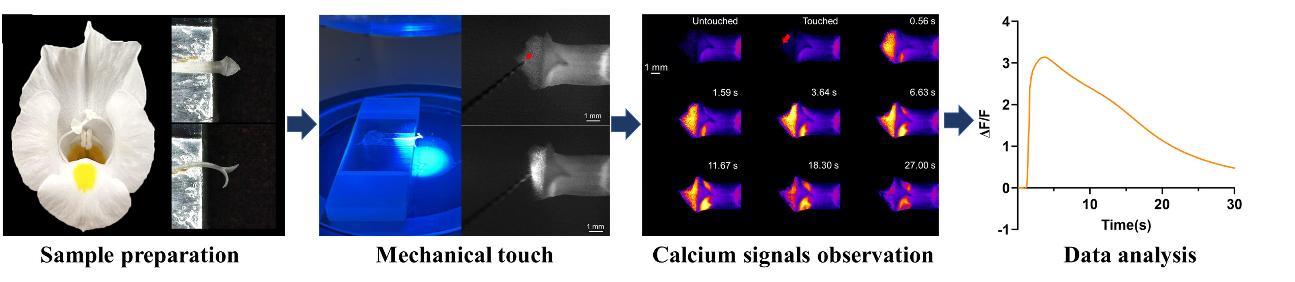
Graphical overview
Background
Plants constantly perceive mechanical stimuli throughout their life cycle, from wind-blown leaves and probing roots to pollen hydration and seed germination. Indeed, a plant's existence can be viewed as a continuous history of responding to such mechanical cues. Upon stimulation, plants upregulate touch-related TOUCH/TCH genes, which mediate contact-induced morphogenesis by integrating hormonal pathways involving auxin, cytokinin, ethylene, and jasmonic acid [1,2]. Beyond these slower morphological changes, some plants exhibit rapid movements in response to touch. Leaves of species like the Venus flytrap and Mimosa pudica execute rapid movements for insect capture or herbivory avoidance, respectively [3,4]. Thus, external mechanical forces fundamentally shape a plant's adaptive strategies.
Calcium ions (Ca2+) serve as one of the most important second messengers within plant cells. Advances in genetically encoded fluorescent indicators now allow real-time observation of Ca2+ dynamics in living tissues. Studies utilizing these tools have demonstrated that diverse stimuli trigger pronounced fluctuations in cytosolic Ca2+ ([Ca2+]cyt) concentrations [5–7]. Mechanical stimuli are particularly effective inducers of such Ca2+ signals. In Arabidopsis, touching the leaf tip generates a calcium signal at the site of contact, which subsequently spreads through the vascular bundles to the petiole [5]. When a single trichome is touched, the resulting calcium signal radiates outward from the trichome as a center [6]. In the touch-sensitive plant Mimosa pudica, touching causes the leaflets to close within seconds. During this process, [Ca2+]cyt levels rise from the veins to the pulvinus, significantly amplifying in that location [4]. Shortly thereafter, the signal transmits hierarchically to the next pulvini, ultimately causing the entire compound leaf to fold [4]. Notably, the speed of calcium signal transmission in Mimosa is much faster than in Arabidopsis leaves. Similarly, in the Venus flytrap, after the first touch, [Ca2+]cyt concentration rises starting at the touched trigger hair and spreads in the leaf [3]. Following a second touch, the [Ca2+]cyt concentration reaches the threshold to trigger closure [3]. Interestingly, this signal remains confined to the stimulated trap and does not propagate via the vasculature or to adjacent leaves [3]. These findings establish that mechanical touch directly initiates Ca2+ fluctuation and propagation, which is essential for activating downstream stress-response genes or driving rapid cellular deformations.
Although different plants employ Ca2+ as a messenger to transduce external mechanical cues into internal responses, the specific pathways and transduction mechanisms exhibit notable variation. Deciphering the Ca2+ signatures elicited by touch across different species is therefore key to understanding the conservation and specificity within this critical signaling network. Here, we have identified the touch-responsive behavior of stigmas in Torenia fournieri, a classical model in the study of plant reproduction [9,10]. The bilobed stigma of T. fournieri closes within seconds after mechanical stimulation, and we have demonstrated that the generation and propagation of touch-induced Ca2+ signals are essential for this stigma movement [11]. In this study, we present an integrated experimental protocol that details how to trigger stigma movement through controlled mechanical stimulation and how to visualize the associated [Ca2+]cyt signal on living stigmas during this rapid response. Given that this protocol allows stimulation of a single papilla cell on the inner epidermal surface of the Torenia fournieri stigma, it may offer a valuable reference for mechanical stimulation of single-cell structures in plants, including leaf trichomes of Arabidopsis.
Materials and reagents
Biological materials
1. Transgenic T. fournieri plants expressing the GCaMP6f-NES probe (UBIQUITIN 10 promoter) [12] produce flowers with open stigmas capable of normal movement; this probe can be used by other groups
Reagents
1. Distilled water (Sangon Biotech, catalog number: E607017-0500)
2. Absolute ethanol (Sangon Biotech, catalog number: A500737-0005)
Laboratory supplies
1. PCR tubes (200 μL) (Sangon Biotech, catalog number: F611542-0001)
2. Scissors (Sangon Biotech, catalog number: F519231-0001)
3. Microsurgical tissue forceps (Vetus, catalog number: SS-JP)
4. Sharp-point tungsten needles (ETRA, model: 0.1 mm × 25 mm)
5. Double-sided tape (3M, catalog number: 55236)
6. Single-sided tape (Scotch, catalog number: 810-CQ33)
7. Microscope slide (25 mm × 75 mm, 1–1.2 mm) (Citotest, catalog number: 10127105P-G)
Equipment
1. Fluorescence stereomicroscope (Olympus, model: SZX16) equipped with a 1× objective lens (Olympus, model: SDFPLAPO1XPF) and a sCMOS camera (Olympus, model: DP74)
Software and datasets
1. CellSens Dimension (Olympus, V4.4.1)
2. GraphPad Prism (GraphPad Software, V10.0)
Procedure
登录/注册后免费查看全文
文章信息
稿件历史记录
提交日期: Jan 3, 2026
接收日期: Apr 3, 2026
在线发布日期: Apr 17, 2026
出版日期: May 5, 2026
版权信息
© 2026 The Author(s); This is an open access article under the CC BY license (https://creativecommons.org/licenses/by/4.0/).
如何引用
Zhou, X., Ma, X., Yang, S. and Su, S. (2026). Detecting Touch-Induced Calcium Dynamics With Live-Cell Imaging in Torenia Stigma. Bio-protocol 16(9): e5683. DOI: 10.21769/BioProtoc.5683.

 Download Citation in RIS Format
分类
植物科学 > 植物细胞生物学 > 细胞成像
细胞生物学 > 细胞成像 > 活细胞成像
您对这篇实验方案有问题吗?
在此处发布您的问题,我们将邀请本文作者来回答。同时,我们会将您的问题发布到Bio-protocol Exchange,以便寻求社区成员的帮助。
 发布问题
发布问题


