


A Cell-Based Protocol to Assess Manganese Content and Relative Transport Activity of Manganese Transporters
基于细胞体系评估锰含量及锰转运蛋白相对转运活性的实验方案

(*contributed equally to this work, § Technical contact) 发布: 2026年05月05日第16卷第9期 DOI: 10.21769/BioProtoc.5680 浏览次数: 902
评审: Joyce ChiuAlexander DupuySubash Chandra Godar
 PDF
PDF  Q&A
Q&A
参见作者原研究论文
The authors used this protocol in:

Sep 2025
Advertisement









Abstract
Manganese (Mn) is an essential trace element whose intracellular homeostasis is tightly controlled by specialized membrane transporters. Dysregulation of Mn transport leads to pathological Mn accumulation and severe human disease; however, efficient and quantitative cell-based methods for assessing Mn2+ transporter activity remain limited. Here, we present an optimized cellular Fura-2 manganese extraction assay (CFMEA) that enables robust quantification of cellular Mn content and provides a normalized framework for assessing relative Mn2+ transport activity in a high-throughput format. This protocol integrates Fura-2-based fluorescence detection of Mn2+ at the Ca2+ isosbestic excitation wavelength with dsDNA quantification to normalize dsDNA levels in cell extracts and immunoblotting to account for transporter protein expression levels. Cells expressing Mn2+ transporters are exposed to MnCl2 in 96-well plates, washed to remove extracellular Mn2+, and lysed in a Fura-2-containing extraction buffer. Fluorescence quenched by Mn2+ is quantified and converted to cellular Mn content using a cell-free Mn-Fura-2 standard curve and then normalized to dsDNA content and protein abundance to determine relative transporter activity. This workflow provides a relatively sensitive, reproducible, and low-cost approach for comparative analysis of Mn2+ transporters and their variants across multiple cell types. The protocol is demonstrated using the Mn2+ efflux transporter SLC30A10 in HEK293T cells and is readily adaptable for studying other Mn2+ transport pathways.
Key features
• High-throughput, cell-based assay for quantifying cellular manganese content and assessing relative Mn2+ transporter function.
• Enhanced accuracy and reproducibility by integrating double-stranded DNA quantification and protein normalization into the cellular Fura-2 manganese extraction assay (CFMEA) workflow.
• Workflow compatible with diverse cell types and Mn2+ transporters, including systems overexpressing SLC30A10 in HEK293Tcells.
Keywords: Mn2+ transporters (Mn2+ 转运蛋白) Mn content (锰含量)
Relative Mn2+ transport activity (Mn2+ 相对转运活性)
CFMEA (CFMEA)
dsDNA quantitative assay (dsDNA 定量检测)
Mn content (锰含量)
Relative Mn2+ transport activity (Mn2+ 相对转运活性)
CFMEA (CFMEA)
dsDNA quantitative assay (dsDNA 定量检测)
Graphical overview
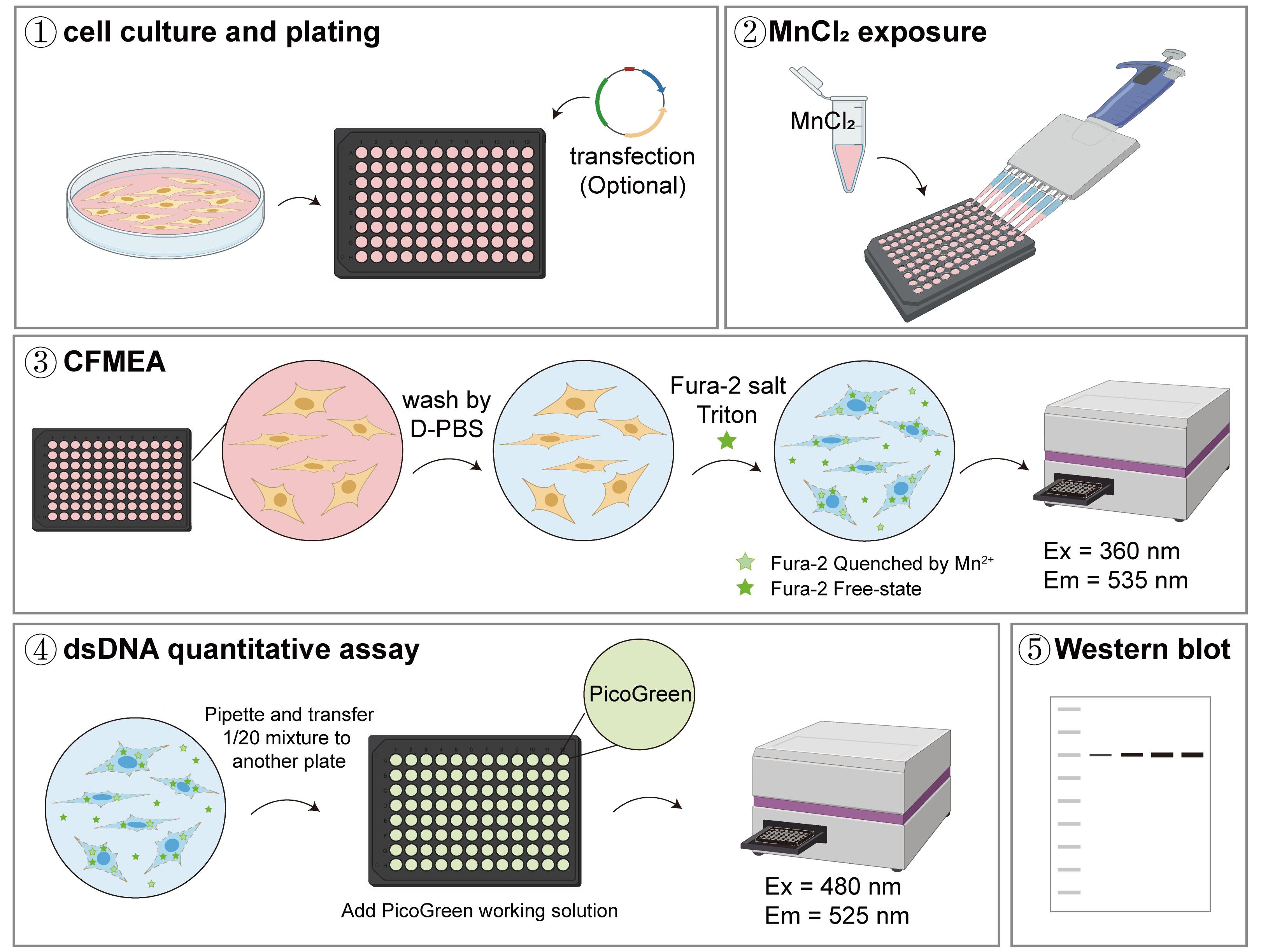
Assessing manganese content and relative transport activity of manganese transporters. (1) Cells were plated in a poly-D-lysine (PDL)-coated black-walled/clear-bottom 96-well plate. (2) When cultures reached 60%–80% confluency, they were exposed to MnCl2 for the specified duration. (3–5) Following exposure, wells were washed with D-PBS to remove extracellular Mn2+, and cells were lysed in a buffer containing Fura-2 dye. Fura-2 fluorescence was measured to quantify Mn2+-induced changes in the signal from cell lysates. The resulting values were normalized to double-stranded DNA content and transporter protein expression to determine relative manganese transport activity.
Background
Manganese (Mn) is an essential trace element that is mainly concentrated in bone, liver, brain, kidneys, and pancreas [1,2]. As a key cofactor for several metalloenzymes, including arginase 1/2 (ARG1/2), glutamine synthetase, and manganese superoxide dismutase (MnSOD), Mn plays an important role in preventing abnormal protein aggregation and limiting reactive oxygen species (ROS)-mediated cellular stress [3–5]. Dysregulation of Mn homeostasis, whether through deficiency or excess, can have severe health consequences. Mn deficiency is associated with osteoporosis and dyslipidemia, while excessive Mn accumulation in the nervous system causes a debilitating neurological disorder known as manganism [6–9].
Given the profound toxicity associated with dysregulated Mn levels, accurately assessing cellular Mn content is of paramount importance. Kwakye et al. (2011) developed the cellular Fura-2 manganese extraction assay (CFMEA) for quantifying the cellular Mn levels in cell-based systems [10–12]. CFMEA employs the fluorescent probe Fura-2, which is conventionally used as a Ca2+ indicator but exhibits more than 99% quenching of its fluorescence upon Mn2+ binding compared to the Ca2+-bound form. Importantly, the excitation wavelength at which Mn2+ quenches (i.e., decreases) the fluorescence of Fura-2 coincides with the Ca2+ isosbestic point (360 nm). At this specific wavelength, the fluorescence intensity of calcium-bound Fura-2 is indistinguishable from that of calcium-free Fura-2, rendering the signal independent of Ca2+ concentration. This unique property enables CFMEA to reliably measure cellular Mn content without interference from Ca2+ dynamics.
Compared with conventional techniques such as atomic absorption spectrometry (AAS), inductively coupled plasma mass spectrometry (ICP-MS), and inductively coupled plasma optical emission spectrometry (ICP-OES), CFMEA offers distinct practical advantages. While these instrumental methods provide high sensitivity and precision for metal quantification, even achieving detection limits in the nanogram range, they require specialized equipment and complex sample preparation, involving cell digestion or acid extraction, and are not readily amenable to rapid or real-time assessment of Mn2+ transport kinetics in cultured cells. In contrast, CFMEA is straightforward, rapid, and can be performed using standard laboratory fluorescence detection systems without the need for sample purification or extraction. These features make CFMEA particularly well-suited for efficiently determining changes in the Mn2+ content within cells [10–12,14]. However, unlike ICP-MS, which provides highly accurate absolute quantification of manganese at trace levels, CFMEA is primarily designed for relative measurements. It is most suitable for comparing manganese transport activity (e.g., uptake or efflux rates) between experimental conditions rather than determining absolute manganese concentrations at nanogram sensitivity.
Mn homeostasis is dynamically regulated by multiple membrane transporters. In mammals, Mn uptake from the extracellular space is primarily facilitated by transporters such as Zrt- and Irt-like protein 8 (ZIP8, also known as SLC39A8), Zrt- and Irt-like protein 14 (ZIP14, also known as SLC39A14), divalent metal transporter 1 (DMT1, also known as SLC11A2), and transferrin receptor (TfR). Conversely, the efflux of Mn, which is essential for reducing intracellular Mn levels, is predominantly mediated by SLC30A10 (ZnT10) [14,15]. Loss-of-function mutations in SLC30A10, ZIP8, or ZIP14 alter intracellular Mn levels, leading to severe clinical manifestations, such as hypermanganesemia with dystonia 1 (HMNDYT1) or hypermanganesemia with dystonia-2 (HMNDYT2) [16–19]. Therefore, investigating the activity of Mn2+ transporters is critical for understanding the mechanisms underlying disease pathogenesis.
A key limitation in the field is the lack of convenient, efficient methods to measure the relative transport activity of Mn2+ transporters. However, besides providing detailed methods to measure the cellular Mn content using CFMEA, this protocol also details how to normalize changes in Mn levels to dsDNA levels in cell extracts using PicoGreen and ultimately determine the relative transport activity of Mn2+ transporters by normalizing protein expression levels using western blotting. CFMEA detects Mn2+ concentrations from 100 to 500 μM, corresponding to 10%–95% quenching of maximal fluorescence, covering physiologically relevant intracellular Mn2+ levels. The PicoGreen dsDNA normalization provides a broad linear detection range of 1–1,000 ng/mL, enabling precise correction for well-to-well variations in cell number. By normalizing Mn content to both dsDNA and transporter protein expression, the method captures changes in intracellular Mn2+ following transporter overexpression in a relatively sensitive manner and allows relative quantification of transport activity per unit of protein. Here, we use the Mn efflux transporter SLC30A10 and its mutants expressed in HEK293T cells as examples to demonstrate the feasibility of the method.
Materials and reagents
Biological materials
1. Human embryonic kidney 293T cells (HEK293T) (Cell Bank of the Chinese Academy of Sciences, catalog number: SCSP-502)
Reagents
Cell culture and transfection
1. Plasmid constructs: the gene encoding the full-length human SLC30A10 (Uniprot: Q6XR72) was cloned into a modified pEG vector that incorporates a 3×Flag tag, a Twin-Strep tag, and an HRV 3C protease cleavage site at the N-terminus. Mutants were generated by site-directed mutagenesis using the wild-type plasmid as the template.
2. Poly-D-lysine (PDL) (Beyotime Biotechnology, catalog number: ST508)
3. Dulbecco's modified Eagle medium (DMEM) (Thermo Fisher Scientific, Gibco, catalog number: C11995500BT)
4. Fetal bovine serum (FBS) (Moregate Biotech, catalog number: FBSF)
5. Penicillin/streptomycin, sterile (100×) (MeilunBio, catalog number: MA0110)
6. Dulbecco’s phosphate-buffered saline (D-PBS) (Sangon Biotech, catalog number: E607009)
7. 0.25% (w/v) trypsin-EDTA, phenol red (modified) (MeilunBio, catalog number: MA0233-1)
8. 0.4% (w/v) Trypan Blue solution (YEASEN, catalog number: 40207ES20)
9. LipofectamineTM 3000 (Thermo Fisher Scientific, Gibco, catalog number: L3000015)
10. Opti-MEMTM (Thermo Fisher Scientific, Gibco, catalog number: L3000015)
11. Manganese (II) chloride tetrahydrate (MnCl2) (Sigma-Aldrich, catalog number: 221279, CAS number: 13446-34-9)
Manganese content and dsDNA quantification
12. Fura-2, pentapotassium salt, cell-impermeant (Sigma-Aldrich, catalog number: 17195, CAS number: 113694-64-7)
13. 20% (v/v) Triton X-100 (prepared from Triton X) (Sangon Biotech, catalog number: A417820)
14. 1 M Tris-HCl (pH 7.5) (prepared from Tris base) (MeilunBio, catalog number: MB3739, CAS number: 77-86-1)
15. 1 M EDTA (prepared from EDTA) (Sangon Biotech, catalog number: A60007-0500, CAS number: 60-00-4)
16. Picogreen dsDNA quantitation reagent (YEASEN, catalog number: 12641ES04)
Western blotting
17. RIPA lysis buffer (Beyotime Biotechnology, catalog number: P0013B)
18. Phenylmethylsulfonyl fluoride (PMSF) (Sangon Biotech, catalog number: A430281)
19. BCA Protein Assay kit (Beyotime Biotechnology, catalog number: P0012)
20. 5× SDS-PAGE sample loading buffer (Beyotime Biotechnology, catalog number: P0015)
21. SDS-PAGE Gel Preparation kit (YEASEN, catalog number: 20328ES72)
22. Methanol anhydrous (HUSHI, catalog number: 8008041900, CAS number: 67-56-1)
23. Glycine (MeilunBio, catalog number: MB4166, CAS number: 56-40-6)
24. SDS (MeilunBio, catalog number: MB2479-1, CAS number: 151-21-3)
25. Skim milk (YEASEN, catalog number: 36120ES76)
26. EZ-buffers H 10× TBST buffer (Sangon Biotech, catalog number: C520009)
27. Bovine serum albumin (BSA) (Sigma, catalog number: A9418)
28. DYKDDDDK tag monoclonal antibody (Proteintech, catalog number: 66008-4-Ig)
29. GAPDH monoclonal antibody (Proteintech, catalog number: 60004-1-Ig)
30. Peroxidase AffiniPure goat anti-mouse IgG (H+L) (YEASEN, catalog number: 33201ES60)
31. Super ECL detection reagent ECL (YEASEN, catalog number: 36208ES60)
Solutions
1. Complete culture media (see Recipes)
2. PDL coating solution (see Recipes)
3. MnCl2 stock (see Recipes)
4. Fura-2 salt dilution buffer (see Recipes)
5. Fura-2 salt working solution (see Recipes)
6. 1× TE buffer (see Recipes)
7. Picogreen working solution (see Recipes)
8. Cell lysis buffer (see Recipes)
9. SDS-PAGE running buffer (10×) (see Recipes)
10. Western blotting transfer buffer (10×) (see Recipes)
Recipes
1. Complete culture media
| Reagent | Final concentration | Quantity or volume |
|---|---|---|
| DMEM | 89% v/v | 445 mL |
| FBS | 10% v/v | 50 mL |
| Penicillin/Streptomycin stock (100×) | 1% v/v | 5 mL |
| Total | - | 500 mL |
Store at 4 °C.
2. PDL coating solution
| Reagent | Final concentration | Quantity or volume |
|---|---|---|
| PDL stock (10 mg/mL, D-PBS) | 20 μg/mL | 40 μL |
| D-PBS | - | 19.96 mL |
| Total | - | 20 mL |
PDL (10 mg) is a sterile powder packaged in a 2 mL microcentrifuge tube. For the first use, prepare sterile PDL stock:
a. Add 1 mL of sterile D-PBS to the microcentrifuge tube.
b. Pipette gently to dissolve completely.
The PDL stock can be stored at 4 °C for up to a year. When coating, dilute the PDL stock with D-PBS to 20 μg/mL.
3. MnCl2 stock
| Reagent | Quantity or volume |
|---|---|
| Manganese (II) chloride tetrahydrate | 1.979 g |
| Double-distilled water | to 10 mL |
| Total | 10 mL |
a. Weigh 1.979 g of MnCl2 using an analytical balance. Add 8 mL of double-distilled water to the tube and dissolve completely by ultrasonic treatment in a water bath.
b. Once fully dissolved, adjust the final volume to 10 mL with double-distilled water.
c. Filter the solution through a 0.22 μm membrane filter in a biosafety cabinet.
d. Aliquot and store at 4 °C for up to 3 months or at -20 °C for up to 1 year.
e. Sonicate for at least 3 min before use.
4. Fura-2 salt dilution buffer
| Reagent | Final concentration | Quantity or volume |
|---|---|---|
| Triton X-100 (20%) | 0.1% v/v | 250 μL |
| D-PBS | 99.9% v/v | 49.75 mL |
| Total | - | 50 mL |
The Fura-2 salt dilution buffer can be stored at 4 °C for a week.
5. Fura-2 salt working solution
| Reagent | Final concentration | Quantity or volume |
|---|---|---|
| Fura-2 salt stock solution (1 mM) | 0.5 μM | 6 μL |
| Fura-2 salt dilution buffer | - | 12 mL |
| Total | - | 12.006 mL |
a. Add 1.202 mL of double-distilled water to dissolve 1 mg of Fura-2 salt powder to prepare a 1 mM stock solution. The Fura-2 salt stock solution can be stored at 4 °C for up to 6 months. It is recommended to aliquot the stock solution and protect it from light.
b. Note that the Fura-2 salt working solution should be used immediately after preparation and protected from light during use.
6. 1× TE buffer
| Reagent | Final concentration | Quantity or volume |
|---|---|---|
| Tris-HCl, pH 7.5, 1 M | 10 mM | 1 mL |
| EDTA, pH 7.5, 1 M | 1 mM | 100 μL |
| Double-distilled water | - | to 100 mL |
| Total | - | mL |
7. Picogreen working solution
| Reagent | Final concentration | Quantity or volume |
|---|---|---|
| Picogreen reagent (200×) | 0.50% v/v | 60 μL |
| 1× TE buffer | 99.50% v/v | 11.94 mL |
| Total | - | 12.00 mL |
The Picogreen working solution should be used immediately after preparation and protected from light during use.
8. Cell lysis buffer
| Reagent | Final concentration | Quantity or volume |
|---|---|---|
| RIPA | 99% v/v | 3.96 mL |
| PMSF (100 mM, 100×) | 1% v/v | 40 μL |
| Total | - | 4.00 mL |
9. SDS-PAGE running buffer (10×)
| Reagent | Final concentration | Quantity or volume |
|---|---|---|
| Tris | 0.25 M | 30.285 g |
| Glycine | 1.923 M | 144.4 g |
| SDS | 1% w/v | 10 g |
| Double-distilled water | - | to 1 L |
10. Western blotting transfer buffer (10×)
| Reagent | Final concentration | Quantity or volume |
|---|---|---|
| Tris | 0.25 M | 30.285 g |
| Glycine | 1.923 M | 144.4 g |
| Double-distilled water | - | to 1 L |
The 1× transfer buffer is prepared by mixing 10× transfer buffer, methanol, and double-distilled water in a ratio of 1:2:7 (v/v/v).
Laboratory supplies
1. 100 mm dish, tissue culture treated (Corning, catalog number: 430167)
2. 6-well plate, tissue culture treated (Yueyibio, catalog number: YB-6)
3. 96-well assay plate, black plate, clear bottom with lid, tissue culture treated, polystyrene, individually packaged (black-walled/clear-bottom 96-well plate) (Xinyou Biotechnology, catalog number: 060096)
4. 10 μL micropipette tip (Yueyibio, catalog number: T-10X)
5. 200 μL micropipette tip (Yueyibio, catalog number: T-200X)
6. 1,250 μL micropipette tip (Yueyibio, catalog number: T-1250X)
7. 1,250 μL Clip TipTM (Thermo Scientific, catalog number: 94410813)
8. 1.5 mL centrifuge tube (YEASEN, catalog number: 83503ES10)
9. 15 mL centrifuge tube (Yueyibio, catalog number: YB0019-15)
10. 50 mL centrifuge tube (Yueyibio, catalog number: YB0010-50)
11. Medical gauze block (Cofoe, model: 5CM×7CM-8P)
12. Aluminum foil (Cleanwrap, catalog number: CF-2)
13. AmershamTM HybondTM P 0.45 PVDF (Cytiva, catalog number:10600023)
Equipment
1. Biological safety cabinet (Thermo Fisher Scientific, catalog number: 1500 Series A2)
2. CO2 incubator (Haier Biomedical, catalog number: HCP 168)
3. Centrifuge (Thermo Fisher Scientific, catalog number: Multifuge X1R)
4. Inverted laboratory microscope (Leica, catalog number: DM IL LED)
5. Mini centrifuge (Haier, catalog number: LX-120T2Z)
6. Sonicator bath: Ultrasonic Cleaning System 40 KHz (SCIENTZ, catalog number: SB-5200D)
7. Vacuum pump (TIPS, catalog number: TIPS200)
8. Automatic cell counter (RWD, catalog number: C100-SE)
9. Spark multimode microplate reader (TECAN, model: Spark®)
10. LED digital dry bath (Dlab, catalog number: HB120-S)
11. Gel electrophoresis system (Bio-Rad, model: Mini-PROTEAN® Tetra System)
12. Protein transfer system (Bio-Rad, model: Trans-Blot® TurboTM)
13. Imaging system (Bio-Rad, model: Molecular Imager® ChemiDocTM XRS+)
14. E1-Clip TipTM BluetoothTM (Thermo Scientific, catalog number: 4671100BT)
Software and datasets
1. Excel 2016 (Microsoft)
2. Prism 8 (GraphPad)
3. Image LabTM Software (Bio-Rad)
4. FIJI (ImageJ2, version 1.54p) with its default distribution (National Institutes of Health); no additional plugins or add-ons beyond the standard FIJI installation are required for the analyses presented in this study
Procedure
登录/注册后免费查看全文
文章信息
稿件历史记录
提交日期: Jan 16, 2026
接收日期: Mar 22, 2026
在线发布日期: Apr 10, 2026
出版日期: May 5, 2026
版权信息
© 2026 The Author(s); This is an open access article under the CC BY-NC license (https://creativecommons.org/licenses/by-nc/4.0/).
如何引用
Zhong, H., Shen, X. and Yang, H. (2026). A Cell-Based Protocol to Assess Manganese Content and Relative Transport Activity of Manganese Transporters. Bio-protocol 16(9): e5680. DOI: 10.21769/BioProtoc.5680.

 Download Citation in RIS Format
分类
细胞生物学 > 基于细胞的分析方法 > 离子分析
细胞生物学 > 基于细胞的分析方法 > 转运
生物化学 > 蛋白质 > 活性
您对这篇实验方案有问题吗?
在此处发布您的问题,我们将邀请本文作者来回答。同时,我们会将您的问题发布到Bio-protocol Exchange,以便寻求社区成员的帮助。
 发布问题
发布问题


