


Assessment of Epithelial Barrier Integrity by TEER and FITC-Dextran Permeability Assays
基于 TEER 与 FITC-葡聚糖通透性检测的上皮屏障完整性评估方案

发布: 2026年04月20日第16卷第8期 DOI: 10.21769/BioProtoc.5673 浏览次数: 1158
评审: Joyce ChiuHsih-Yin TanAnonymous reviewer(s)
 PDF
PDF  Q&A
Q&A
参见作者原研究论文
The authors used this protocol in:

Oct 2025
Advertisement








Abstract
The integrity of epithelial barriers is essential for maintaining tissue homeostasis, particularly in the intestinal tract, where it separates the host from the complex luminal environment. Two complementary, standard methods for assessing this barrier are transepithelial electrical resistance (TEER), which provides a rapid, non-destructive measure of ionic conductance across tight junctions, and the fluorescein isothiocyanate (FITC)-dextran assay, which directly quantifies paracellular macromolecule flux. This protocol details a robust and reproducible method for performing both assays using intestinal epithelial cell monolayers (e.g., Caco-2, T84) cultured on permeable Transwell supports. We outline the procedure from cell culture and monolayer differentiation to TEER measurement with an Epithelial Volt/Ohm Meter 3 (EVOM3) and the subsequent FITC-dextran permeability assay. By combining these techniques, this protocol provides a comprehensive assessment of barrier function, making it ideal for studying tight junction dynamics and regulation under various experimental conditions, such as cytokine stimulation, drug screening, or microbial challenges.
Key features
• Combines electrical resistance (TEER) and macromolecular flux (FITC-dextran) assays for a comprehensive assessment of intestinal epithelial barrier integrity.
• Applicable to various intestinal models, including Caco-2 and T84 cell lines; can be applied to other immortalized or primary epithelial and endothelial cells.
• Utilizes the Epithelial Volt/Ohm Meter 3 (EVOM3) for accurate, non-destructive, and rapid TEER measurements in intestinal epithelial cell monolayers.
• Provides clear guidelines for EVOM3 electrode handling and measurement to ensure reproducible results.
Keywords: Intestinal barrier (肠道屏障) TEER (TEER)
Transepithelial Electrical Resistance (跨上皮电阻)
FITC-dextran (FITC-葡聚糖)
Permeability (通透性)
Caco-2 (Caco-2细胞)
T84 (T84细胞)
EVOM3 (EVOM3)
TEER (TEER)
Transepithelial Electrical Resistance (跨上皮电阻)
FITC-dextran (FITC-葡聚糖)
Permeability (通透性)
Caco-2 (Caco-2细胞)
T84 (T84细胞)
EVOM3 (EVOM3)
Graphical overview
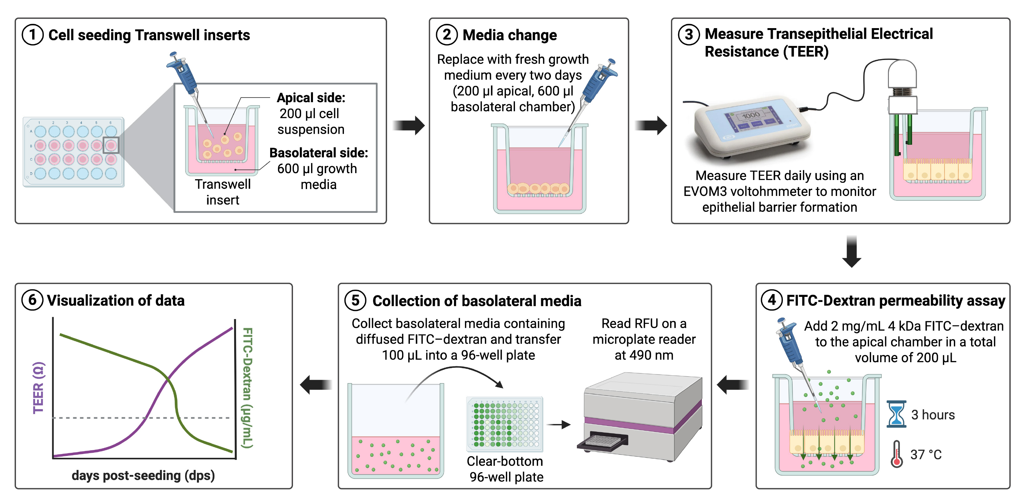
Graphical overview of transepithelial electrical resistance (TEER) measurement and fluorescein isothiocyanate (FITC)-dextran permeability assay. Schematic workflow illustrating the experimental setup and readouts used to monitor epithelial barrier integrity and function. (1) Cells are seeded onto Transwell inserts, with the cell suspension added to the apical chamber (200 μL) and growth medium to the basolateral chamber (600 μL). (2) Growth medium is replaced with fresh medium every two days in both compartments, starting one day post-seeding. (3) TEER is measured daily using an EVOM3 voltmeter to monitor the establishment and maturation of the epithelial barrier. (4) Once the monolayer is established, paracellular permeability is assessed by adding 4 kDa FITC-dextran (2 mg/mL) to the apical chamber in a total volume of 200 μL, followed by incubation for 3 h at 37 °C. (5) Basolateral media containing diffused FITC-dextran are collected and transferred (100 μL) into a clear-bottom 96-well plate, and fluorescence is measured at 490 nm using a microplate reader. (6) TEER values and FITC-dextran flux are visualized over time to assess epithelial barrier formation and permeability.
Background
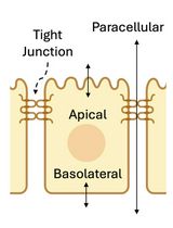
A fundamental characteristic of intestinal epithelial cells is their highly polarized structure, which establishes two distinct membrane domains: an apical surface facing the intestinal lumen and a basolateral surface facing the underlying tissue [1–3]. This structural and functional asymmetry is essential for the epithelium's dual roles: nutrient absorption and formation of a highly selective barrier against harmful luminal contents like toxins and pathogens [1–3]. This critical barrier function is maintained by the coordinated assembly of intercellular protein complexes, including tight junctions (TJs), which regulate the paracellular pathway, the space between adjacent cells [4–6]. Disruption of the intestinal barrier is a hallmark of various gastrointestinal diseases, including inflammatory bowel disease (IBD) [5,6]. Therefore, accurately quantifying barrier integrity is crucial for both basic research and preclinical studies.
In vitro models using intestinal epithelial cells cultured on porous membrane inserts (e.g., Transwell) are invaluable for these studies, as they recapitulate key features of epithelial polarity and allow for functional assessment [7]. To quantify barrier integrity, two complementary methods are widely employed. The first, transepithelial electrical resistance (TEER), measures the resistance to ion flow across the cell monolayer and serves as a highly sensitive indicator of TJ integrity [7–10]. However, TEER primarily reflects ionic conductance and does not directly measure permeability to larger, non-charged macromolecules [10]. The paracellular flux assay addresses this limitation [11]. By quantifying the passage of inert, fluorescently labeled molecules of a defined size, such as fluorescein isothiocyanate (FITC)-dextran, from the apical to the basolateral compartment, the assay provides direct information on the size-selective properties of the paracellular pathway [11]. This protocol combines both methods, using the EVOM3 Voltohmmeter for sensitive TEER readings and a standard FITC-dextran flux assay, to provide a comprehensive and reliable evaluation of intestinal epithelial barrier function and permeability.
Materials and reagents
Biological materials
1. T84 human colorectal carcinoma cells [American Type Culture Collection (ATCC), catalog number: CCL-248]
2. Caco-2 human colorectal adenocarcinoma cells [American Type Culture Collection (ATCC), catalog number: HTB-37]
Reagents
1. Dulbecco's modified Eagle medium/F12 (DMEM/F12) (Corning, catalog number: MT10092CV)
2. DMEM, high glucose, GlutaMAX supplement, pyruvate (Gibco, catalog number: 10569044)
3. Fetal bovine serum (FBS), heat-inactivated (Sigma-Aldrich, catalog number: 12306C)
4. Penicillin-streptomycin solution (10,000 U/mL) (Thermo Fisher Scientific, catalog number: 15140122)
5. Rat tail collagen type I (Thermo Fisher Scientific, catalog number: CB354249)
6. Phosphate-buffered saline (PBS), 10× powder pH 7.4 (Thermo Fisher Scientific, catalog number: BP665-1)
7. Trypsin-EDTA (0.25%) (Thermo Fisher Scientific, catalog number: 25200056)
8. Trypan Blue solution (0.4%) (Gibco, catalog number: 15250061)
9. Tergazyme, enzyme-active powdered detergent (Thermo Fisher Scientific, catalog number: 16000115)
10. Ethanol, 99.5% (Thermo Fisher Scientific, catalog number: 615090040)
11. FITC-dextran, 4 kDa (Sigma-Aldrich, catalog number: 46944)
12. Sterile distilled water (dH2O) (Thermo Fisher Scientific, catalog number: 10977015)
Solutions
1. Complete growth medium (for T84 cells) (see Recipes)
2. Complete growth medium (for Caco-2 cells) (see Recipes)
3. 1× PBS (see Recipes)
4. 60% EtOH (see Recipes)
5. 70% EtOH (see Recipes)
6. Rat tail collagen coating solution (see Recipes)
7. FITC-dextran working stock (see Recipes)
8. Tergazyme solution (1%) (see Recipes)
Recipes
1. Complete growth medium (for T84 cells)
| Reagent | Final concentration | Quantity (for 500 mL) |
|---|---|---|
| DMEM/F12 | n/a | 445 mL |
| FBS | 10% (v/v) | 50 mL |
| Penicillin-Streptomycin | 100 U/mL | 5 mL |
The prepared media can be stored at 4 °C and used for a month. Warm to 37 °C in a water bath before use.
2. Complete growth medium (for Caco-2 cells)
| Reagent | Final concentration | Quantity (for 500 mL) |
|---|---|---|
| DMEM-GlutaMAX | n/a | 445 mL |
| FBS | 10% (v/v) | 50 mL |
| Penicillin-Streptomycin | 100 U/mL | 5 mL |
The prepared media can be stored at 4 °C and used for a month. Warm to 37 °C in a water bath before use.
3. 1× PBS
| Reagent | Final concentration | Quantity (for 500 mL) |
|---|---|---|
| 10× PBS | 1/10 | 10 mL |
| Sterile dH2O | n/a | 90 mL |
| Total | n/a | 100 mL |
10× PBS powder is used to prepare a 10× PBS stock solution, which can be stored at room temperature for several months.
4. 60% EtOH
| Reagent | Final concentration | Quantity (for 500 mL) |
|---|---|---|
| EtOH, 99.5% | 60% (w/v) | 60 mL |
| Sterile dH2O | n/a | 40 mL |
| Total | n/a | 100 mL |
5. 70% EtOH
| Reagent | Final concentration | Quantity (for 500 mL) |
|---|---|---|
| EtOH, 99.5% | 70% (w/v) | 70 mL |
| Sterile dH2O | n/a | 30 mL |
| Total | n/a | 100 mL |
6. Rat tail collagen coating solution
| Reagent | Final concentration | Quantity (for 100 mL) |
|---|---|---|
| 100× rat tail collagen stock (1 mg/mL) | 10 μg/mL | 1 mL |
| 60% ethanol | n/a | 99 mL |
| Total | n/a | 100 mL |
This is the final working solution used for coating tissue culture flasks and Transwell inserts. The prepared solution can be stored at 4 °C and used for a month.
7. FITC-dextran working stock
| Reagent | Final concentration | Quantity (for 100 mL) |
|---|---|---|
| FITC-dextran | 10 mg/mL | 500 mg |
| Sterile dH2O | n/a | 50 mL |
This is the working stock at 10 mg/mL. Aliquot in microcentrifuge tubes and store at 4 °C in the dark.
8. Tergazyme solution (1%)
| Reagent | Final concentration | Quantity (for 50 mL) |
|---|---|---|
| Tergazyme powder | 1% (w/v) | 0.5 g |
| Sterile dH2O | n/a | to 50 mL |
Laboratory supplies
1. T-25 tissue culture flasks (CellTreat, catalog number: 229331)
2. 24-well cell culture plates (Corning, catalog number: 3527)
3. 6.5 mm Transwell® with 3.0 μm pore polycarbonate membrane insert, sterile (Thermo Fisher Scientific, catalog number: 3415)
4. Clear-bottom 96-well plates (Corning, catalog number: 3595)
5. 15 mL conical tubes (Thermo Fisher Scientific, catalog number: 14955237)
6. Microcentrifuge tubes, 1.5 mL (Thermo Fisher Scientific, catalog number: 05408129)
7. Serological pipettes (5, 10, 25 mL) (MedSupplyPartner, catalog numbers: CT-229005B, CT-229010B, CT-1367811)
8. Pipette tips (p20, p200, p1000), sterile (Thermo Fisher Scientific, catalog number: 12111136)
9. Aspirating pipette (Thermo Fisher Scientific, catalog number: 14955135)
10. Cell counting slides (Bio-Rad, catalog number: 1450015)
11. Aluminum foil
Equipment
1. EVOM3 Voltohmmeter with STX2 electrode (World Precision Instruments)
2. Class II Biological Safety Cabinet (Nuaire, model: NU-425-500)
3. Humidified cell culture incubator (37 °C, 5% CO2) (ThermoFisher, model: HERAcell vios 160i)
4. Automated cell counter (Bio-Rad, model: TC20)
5. 800TS microplate reader (BioSPX, model: BioTek 800TS)
6. Water bath (37 °C) (BioLab)
Procedure
登录/注册后免费查看全文
文章信息
稿件历史记录
提交日期: Jan 19, 2026
接收日期: Mar 17, 2026
在线发布日期: Apr 2, 2026
出版日期: Apr 20, 2026
版权信息
© 2026 The Author(s); This is an open access article under the CC BY-NC license (https://creativecommons.org/licenses/by-nc/4.0/).
如何引用
Keser, Y., Boulant, S. and Stanifer, M. L. (2026). Assessment of Epithelial Barrier Integrity by TEER and FITC-Dextran Permeability Assays. Bio-protocol 16(8): e5673. DOI: 10.21769/BioProtoc.5673.

 Download Citation in RIS Format
分类
细胞生物学 > 基于细胞的分析方法 > 电生理技术
细胞生物学 > 细胞结构
细胞生物学 > 基于细胞的分析方法 > 转运
您对这篇实验方案有问题吗?
在此处发布您的问题,我们将邀请本文作者来回答。同时,我们会将您的问题发布到Bio-protocol Exchange,以便寻求社区成员的帮助。
 发布问题
发布问题


