


Accessible STORM Imaging: An Optimized Workflow for Conventional Widefield Epifluorescence/TIRF Setups
适用于常规宽场表面荧光/全内反射荧光系统的优化 STORM 成像流程

(*contributed equally to this work) 发布: 2026年04月20日第16卷第8期 DOI: 10.21769/BioProtoc.5672 浏览次数: 708
评审: Elena A. OstrakhovitchKM Rifat FaysalAnonymous reviewer(s)
 PDF
PDF  Q&A
Q&A
Advertisement








Abstract
Stochastic optical reconstruction microscopy (STORM) is a single-molecule localization microscopy technique that enables visualization of cellular structures beyond the diffraction limit. This approach has revealed previously inaccessible ultrastructural details in a wide range of cellular components, including the actin cytoskeleton, clathrin-coated pits, mitochondria, and bacterial nucleoid-associated proteins. STORM relies on the sequential emission of single photons from photosensitive fluorophores, which are precisely localized before entering a dark state or undergoing photobleaching. By activating fluorophores individually and fitting their point spread functions (PSFs), the center of mass can be calculated with a localization precision of up to ~20 nm. The parallel detection of thousands of single-molecule events, each assigned to distinct spatial coordinates, enables the reconstruction of a high-resolution image. Here, we describe a simple and efficient STORM workflow—including sample preparation, image acquisition, and quality control measurements—that we used to visualize various subcellular structures, such as mitochondria, microtubules, and lysosomes labeled with the commonly employed cyanine dye Alexa Fluor 647, as well as the actin cytoskeleton stained with Alexa Fluor 488–conjugated phalloidin. Image acquisition was performed using a conventional epifluorescence/total internal reflection (TIRF) microscope adapted for STORM imaging. Key adaptations included the use of a 160×/1.43 NA oil-immersion objective and a high-power mode, which concentrates the laser beam onto a small region of the sample, ensuring sufficient light intensity to drive fluorophores into the dark state. In addition, implementing a 1.6× magnification lens and a 4×4 binning camera mode allowed us to achieve a 100-nm pixel size optimal for reliable molecule detection. We believe that this protocol will be highly valuable to the microscopy community, as it lowers technical barriers to performing STORM on widely available microscopy platforms, thereby facilitating broader implementation of this powerful super-resolution technique.
Key features
• Optimized immunofluorescence, including multiple washing steps using 0.1% Tween-20 and a secondary antibody fixation step for single and dual-color STORM.
• Imaging acquisition performed with a conventional epifluorescence/TIRF microscope adapted for STORM imaging.
• Use of a 160×/1.43 NA oil-immersion objective and a high-power mode that concentrates the laser beam onto a small region of the sample.
• Implementation of a 1.6× magnification lens and 4 × 4 camera binning to achieve 100 nm pixel size.
Keywords: STORM (STORM) SMLM (单分子定位显微成像)
Lysosomes (溶酶体)
Microtubules (微管)
Mitochondria (线粒体)
Actin (肌动蛋白)
TIRF (全内反射荧光)
Uncertainty (不确定性)
FWHM (半峰全宽)
SMLM (单分子定位显微成像)
Lysosomes (溶酶体)
Microtubules (微管)
Mitochondria (线粒体)
Actin (肌动蛋白)
TIRF (全内反射荧光)
Uncertainty (不确定性)
FWHM (半峰全宽)
Graphical overview
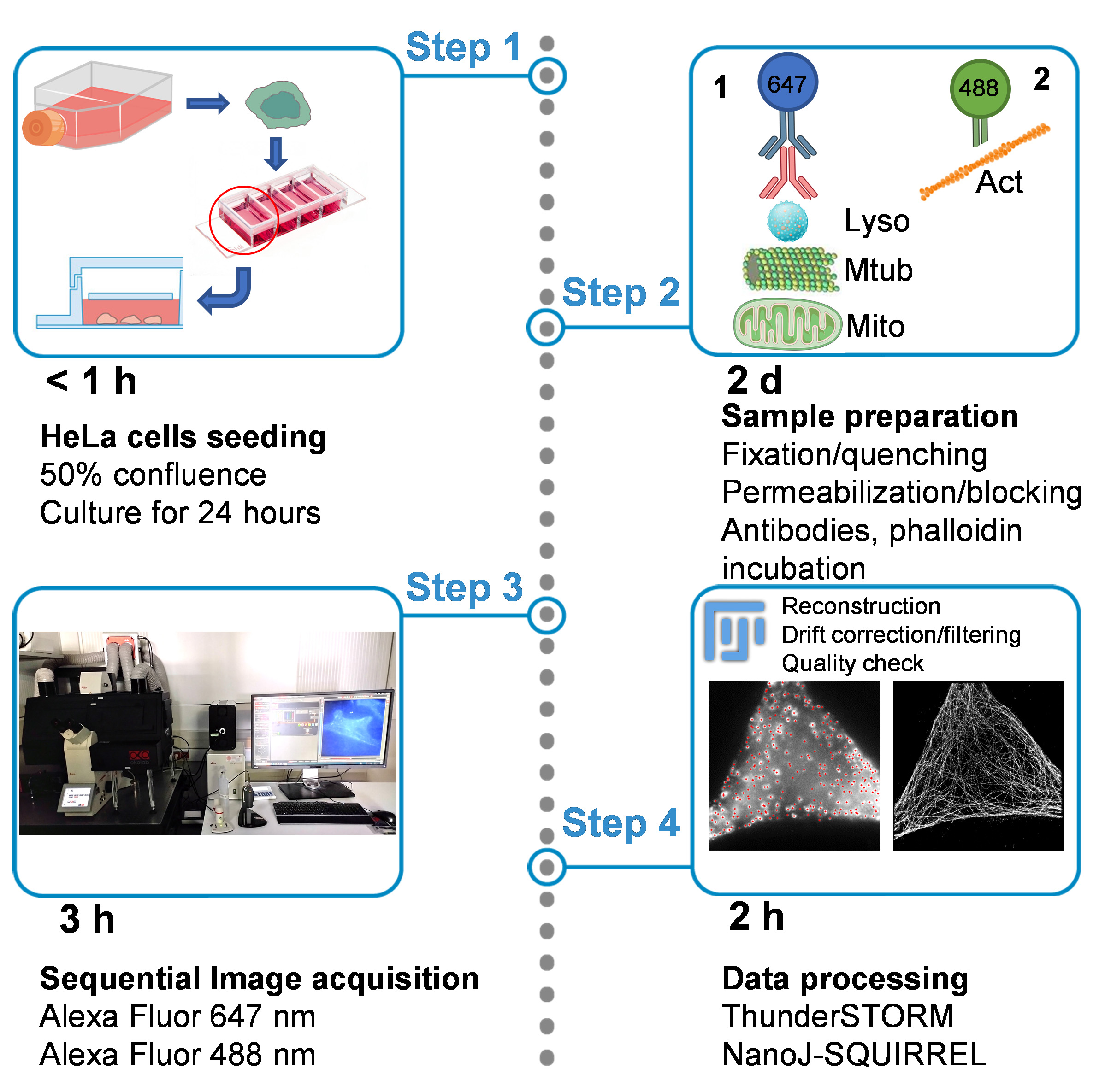
Step-by-step workflow for stochastic optical reconstruction microscopy (STORM) imaging. The graphical summary outlines all the critical steps of the workflow and highlights the time required for each stage. (Step 1) HeLa cells are seeded in dedicated four-well imaging chambers and cultured for 24 h to allow cell adhesion. (Step 2, left) Cells are then fixed and quenched, an essential step to decrease auto-fluorescence, followed by permeabilization and blocking, a crucial step to avoid unspecific antibody binding. Different samples are prepared using distinct primary antibodies to visualize different cellular structures, including lysosomes (Lyso) and microtubules (Mtub) for single-color STORM, as well as mitochondria (Mito) for dual-color STORM. We used the corresponding secondary antibody, conjugated with Alexa Fluor 647, matched to the host species of the primary antibody. (Step 2, right) For mitochondria-labeled dual-color STORM samples, filamentous actin was additionally stained using phalloidin-Alexa Fluor 488. (Step 3) Imaging acquisition is performed in STORM imaging buffer using a widefield/total internal reflection (TIRF) system. Sequential acquisition is required for dual-color samples, with Alexa Fluor 647 imaged first, followed by Alexa Fluor 488. (Step 4) The final step involves data processing with the ThunderSTORM plugin, which consists of single-molecule localization, drift correction, and filtering to generate the final STORM reconstructed image. The quality check of the resulting images is performed with the NanoJ-SQUIRREL plugin.
Background
The discovery of super-resolution imaging methods has enabled optical microscopy to overcome the conventional light diffraction limit, defined by Ernst Karl Abbe in 1873. These methods have completely revolutionized the field of optical fluorescence microscopy, tremendously enhancing its resolution power [1]. Single-molecule localization microscopy (SMLM) techniques rely on the precise determination of fluorophore positions, with subpixel accuracy, based on the emission from a small molecule subset, which can be distinguished from nearby fluorophores in the dark (non-emitting) state, thus allowing the reconstruction of a high-resolution image [2,3]. The strategy applied to achieve the fluorophore on/off state switching is specific to each SMLM technique, which mainly includes photo-activated localization microscopy (PALM) and stochastic optical reconstruction microscopy (STORM). Briefly, PALM can also be applied to live-cell imaging and relies on the use of photoactivatable/switchable proteins, which can be activated with near-UV/violet light (typically ~405 nm) or photo-converted from a shorter to a longer wavelength form. The detection of emission by single molecules is achieved using low-intensity activation or switching laser cycles, which turn on or convert only a subset of fluorophores at a time, until they are photobleached [4]. In contrast, STORM exploits organic fluorophores such as Alexa Fluor–conjugated antibodies (e.g., Alexa Fluor 647), which undergo reversible photo switching in suitable imaging buffers; therefore, it is suitable for fixed sample imaging [5].
STORM is a single-molecule high-resolution microscopy technique that allows the visualization of unresolved details of many cellular structures, including clathrin-coated pits, mitochondria, synaptic structures, and neuronal spectrin, which forms, with actin and associated proteins, a periodic structure in neurons [6–9]. In addition, the technique relies on fluorophores blinking, defined as the stochastic switching of a photosensitive molecule between fluorescent (on) and non-fluorescent (off) states, enabling the precise localization of individual emitters, before they enter a long-lived dark state or become photobleached. The localization coordinates of many individuals emitting fluorophores, each associated with respective spatial coordinates, allow the reconstruction of a high-resolution image [10]. The implementation of advanced imaging methods for broader laboratory use highlights the need for rigorous validation strategies and quantitative performance metrics supporting protocol optimization and cross-platform reproducibility in quantitative imaging workflows [11].
The current protocol describes a complete workflow, including sample preparation, image acquisition, data processing, and quality assessment to obtain high-quality single and dual-color STORM images of different cellular structures such as tubulin, lysosomes, mitochondria, and the actin cytoskeleton. Here, we employed commercially available primary antibodies and secondary antibodies conjugated to Alexa Fluor 647 switchable fluorophores, which show excellent blinking properties and represent the most widely used dyes for STORM imaging. In addition, for dual-color experiments, the actin cytoskeleton was labeled using a phalloidin-Alexa Fluor 488, which allows high-quality reconstruction of the cytoskeleton, although its blinking properties are lower than those of the Alexa Fluor 647 counterpart [12]. For imaging, an oxygen-scavenging imaging buffer, which maintains a reducing chemical environment ideal for the dye photo conversion, is used. The choice to perform dual-color STORM using an Alexa Fluor 647/Alexa Fluor 488 combination was dictated by the excitation–emission filter of our system, as well as the compatibility of the two dyes with the imaging buffer, which needs to be adjusted and optimized according to the chemical properties of the fluorophores.
Despite the growing interest in super-resolution microscopy, the implementation of STORM often requires specialized instrumentation and technical expertise that may not be readily available in many laboratories. This protocol addresses this gap by providing a workflow that enables STORM imaging using widely available microscopy platforms with minimal technical modifications. By lowering the technical barriers to SMLM implementation, this approach aims to facilitate broader adoption of super-resolution imaging across research laboratories. Such accessibility could benefit diverse applications, including studies of cellular architecture, mechanotransduction, and tissue organization, where nanoscale imaging can provide insights into biological processes such as osteocyte responses to mechanical stress [13]. STORM imaging requires customized microscopes or commercially available systems, which are specifically designed for SMLM experiments, including the ONI imager (www.oni.bio.com) or the Abbelight platform (www.abbelight.com). This represents a limitation to the spreading of this powerful microscopy technique. In order to overcome this limitation, we designed this protocol where image acquisition is performed on a conventional epifluorescence/total internal reflection (TIRF) microscope, commonly available in many laboratories or imaging facilities, adapted for STORM imaging. Crucial adaptations include the use of a 160×/1.43 NA oil-immersion objective and a high-power mode that concentrates the laser beam onto a small region of the sample, ensuring sufficient intensity to drive fluorophores into the dark state. This allows us to overcome the limitation of the low laser power compared to the system designed for STORM, which shows high-power laser beams. Indeed, power ranges (at samples) for 638 and 488 nm laser lines are on the order of 250 mW for ONI or 40–500 mW for Abbelight microscopes, compared to 25–30 mW for our system used here. In addition, implementing a 1.6× magnification lens and a 4×4 binning camera mode allowed us to achieve a 100-nm effective pixel size optimal for reliable molecule detection. Moreover, the use of TIRF microscopy, which allows regulation of laser penetration depth within the sample [14], helps with background removal, optimizing the molecule localization during image analysis. Image reconstruction and post-processing drift correction, as well as the evaluation of the quality of molecule localization, were performed using the ThunderSTORM open-source Fiji plugin tool [15]. The quality of STORM reconstructed images was assessed using the NanoJ-SQUIRREL open-source Fiji plugin [16], comparing the corresponding TIRF image.
These developments aim to improve the accessibility of STORM imaging and reduce technical barriers to its implementation, facilitating broader adoption of super-resolution microscopy in standard laboratory environments. Such efforts align with recent methodological advances emphasizing translational robustness and standardized experimental workflows to ensure reproducibility across research settings [17]. We believe that this protocol will be particularly valuable to the microscopy community, especially for beginner STORM users, as it can be implemented on widely available microscopy platforms already available in many laboratories or imaging facilities. With minor technical adjustments, these platforms can be adapted for SMLM imaging, avoiding substantial financial investments.
Materials and reagents
Biological materials
1. HeLa cell line (American Type Culture Collection, catalog number: CCL2)
Reagents
1. Dulbecco’s phosphate buffered saline (PBS) (Merck Life Science, catalog number: D8537)
2. Dulbecco’s modified Eagle medium (DMEM) high glucose (Biowest, catalog number: L0101-500)
3. Fetal bovine serum (FBS) (Sigma, catalog number: F-7524)
4. L-Glutamine 100 mM (Biowest, catalog number: X0550)
5. Sodium pyruvate 100 mM (Biowest, catalog number: L0642)
6. Ethylenediaminetetraacetic acid (EDTA) Tritiplex III (Merck, catalog number: 1.08418)
7. Sodium hydroxide (NaOH) (Merck, catalog number: 1370311002)
8. Paraformaldehyde (PFA) 16% w/v aqueous solution, methanol-free (Thermo Fisher Scientific, catalog number: 043368.9M)
9. Glutaraldehyde solution, 50 wt% in H2O (Sigma, catalog number: 340855)
10. Triton X-100 (Sigma, catalog number: T9284)
11. Sodium borohydride (Merck, catalog number: 452882)
12. Tween 20 (Sigma, catalog number: P6585)
13. D-(+)-glucose (Merck, catalog number: 1.08337.0250)
14. Cysteamine (Sigma, catalog number: 30070); store at 4 °C
15. Glucose oxidase from Aspergillus niger (Merck, catalog number: G2133); store at -20 °C
16. Catalase from bovine liver (Merck, catalog number: C40); store at -20 °C
17. Mouse anti-LAMP2 (H4B4) primary antibody (Santa Cruz, catalog number: SC-18822)
18. Goat anti-mouse Alexa Fluor 647 (Thermo Fisher Scientific, catalog number: A21236); store at -20 °C
19. Mouse anti-tubulin primary antibody (Sigma, catalog number: T5168)
20. Rabbit anti-TOMM22 primary antibody (Sigma, catalog number: HPA003037)
21. Goat anti-rabbit Alexa Fluor 647 (Thermo Fisher Scientific, catalog number: A21245)
22. Phalloidin-Alexa Fluor 488 (Thermo Fisher Scientific, catalog number: A12379); store at -20 °C
Solutions
1. HeLa cell culture medium (see Recipes)
2. EDTA 0.5 M stock (see Recipes)
3. PBS-EDTA (see Recipes)
4. Fixation solution (see Recipes)
5. Quenching solution (see Recipes)
6. Permeabilization buffer (see Recipes)
7. Blocking/antibody buffer (see Recipes)
8. Washing buffer (see Recipes)
9. Antibody fixation solution (see Recipes)
10. Cysteamine (see Recipes)
11. D-glucose 25% (see Recipes)
12. Glucose oxidase (see Recipes)
13. Catalase (see Recipes)
14. Imaging buffer (see Recipes)
Recipes
1. HeLa cell culture medium
| Reagent | Final concentration | Quantity or volume |
|---|---|---|
| DMEM (4.5 g/L D-glucose) | n/a | 440 mL |
| L-Glutamine | 1% (v/v) | 5 mL |
| Sodium pyruvate | 1% (v/v) | 5 mL |
| FBS | 10% (v/v) | 50 mL |
| Total | n/a | 500 mL |
2. EDTA 0.5 M stock
| Reagent | Final concentration | Quantity or volume |
|---|---|---|
| EDTA | 0.5 M | 9.3 g |
| MilliQ water | n/a | Up to 50 mL |
| Total | n/a | 50 mL |
Adjust pH to 7.8 using sodium hydroxide (NaOH). Filter solution with 0.22 μm filter.
3. PBS-EDTA
| Reagent | Final concentration | Quantity or volume |
|---|---|---|
| EDTA (0.5 M stock) | 10 mM | 1 mL |
| 1× PBS | n/a | 49 mL |
| Total | n/a | 50 mL |
4. Fixation solution
| Reagent | Final concentration | Quantity or volume |
|---|---|---|
| PFA 16% | 3% (v/v) | 0.544 mL |
| Glutaraldehyde (50 wt%) | 0.1% (v/v) | 0.0058 mL |
| 1× PBS | n/a | 2.35 mL |
| Total | n/a | 2.9 mL |
Critical: Prepare the fixation solution fresh before use. Store reagents at room temperature until their expiration. Opened paraformaldehyde stock ampoules can be stored at room temperature for up to 1 month.
Caution: Paraformaldehyde and glutaraldehyde are toxic and hazardous; wear a lab coat, gloves, and safety goggles. Perform all steps in a chemical safety hood and dispose of waste according to local regulations.
5. Quenching solution
| Reagent | Final concentration | Quantity or volume |
|---|---|---|
| Sodium borohydride | 0.1% (w/v) | 10 mg |
| 1× PBS | n/a | 10 mL |
| Total | n/a | 10 mL |
Critical: Prepare fresh before use. Store sodium borohydride at room temperature until expiring date.
Caution: Sodium borohydride is toxic and hazardous, and releases flammable hydrogen gas upon dissolution. Handle in a chemical safety hood and dispose of waste according to local regulations.
6. Permeabilization solution
| Reagent | Final concentration | Quantity or volume |
|---|---|---|
| Triton X-100 | 0.1% (v/v) | 0.003 mL |
| 1× PBS | n/a | 2.997 mL |
| Total | n/a | 3 mL |
7. Blocking/antibody solution
| Reagent | Final concentration | Quantity or volume |
|---|---|---|
| FBS | 10% (v/v) | 0.3 mL |
| 1× PBS | n/a | 2.7 mL |
| Total | n/a | 3 mL |
8. Washing buffer
| Reagent | Final concentration | Quantity or volume |
|---|---|---|
| Tween 20 | 0.1% (v/v) | 0.05 mL |
| 1× PBS | n/a | Up to 50 mL |
| Total | n/a | 50 mL |
9. Antibody fixation solution
| Reagent | Final concentration | Quantity or volume |
|---|---|---|
| PFA 16% | 1% (v/v) | 0.188 mL |
| 1× PBS | n/a | 2.812 mL |
| Total | n/a | 3 mL |
10. Cysteamine
| Reagent | Final concentration | Quantity or volume |
|---|---|---|
| Cysteamine | 1 M | 0.775 mg |
| MilliQ water | n/a | Up to 10 mL |
| Total | n/a | 10 mL |
Prepare single-use aliquots to avoid freeze-thaw cycles and store at -20 °C for up to 6 months.
11. D-glucose 25%
| Reagent | Final concentration | Quantity or volume |
|---|---|---|
| D-(+)-glucose | 25% (w/v) | 2.5 g |
| MilliQ water | n/a | Up to 10 mL |
| Total | n/a | 10 mL |
Prepare single-use aliquots to avoid freeze-thaw cycles and store at -20 °C for up to 6 months.
12. Glucose oxidase
| Reagent | Final concentration | Quantity or volume |
|---|---|---|
| Glucose oxidase | 2 mg/mL | 2 mg |
| 1× PBS | n/a | 1 mL |
| Total | n/a | 1 mL |
Prepare single-use aliquots to avoid freeze-thaw cycles and store at -20 °C for up to 6 months.
13. Catalase
| Reagent | Final concentration | Quantity or volume |
|---|---|---|
| Catalase | 2 mg/mL | 20 mg |
| 1× PBS | n/a | 10 mL |
| Total | n/a | 10 mL |
Prepare single-use aliquots to avoid freeze-thaw cycles and store at -20 °C for up to 6 months.
14. Imaging buffer
| Reagent | Final concentration | Quantity or volume |
|---|---|---|
| Cysteamine | 100 mM | 0.1 mL |
| D-glucose 25% | 5% | 0.2 mL |
| Glucose oxidase | 0.5 mg/mL | 0.25 mL |
| Catalase | 0.04 mg/mL | 0.02 mL |
| 1× PBS | n/a | 0.430 mL |
| Total | n/a | 1 mL |
Imaging buffer remains stable for up to 3 h during imaging at room temperature when protected from light. For longer imaging sessions, replace with fresh buffer.
Laboratory supplies
1. μ-slide 4-well Ph+ glass bottom # 1.5 (Ibidi, catalog number: 80447)
Equipment
1. Biological culture cabinet (Heraeus Instruments, model: LaminAir HB2436)
2. Cell culture incubator (Binder, model: CB150)
3. Microcentrifuge (Hettich Zentrifugen, model: Universal 320R)
4. Widefield microscope equipped with a total internal reflection fluorescence (TIRF) microscopy module, a high-power mode lens, and a 1.6× magnification lens (Leica, model: DMi8s)
5. Orca Flash 4.0 sCMOS camera (Hamamatsu, model: C13440)
6. HC PL APO 160×/1.43 NA oil immersion objective (Leica, catalog number: 11888434)
7. GFP filter set (Leica, catalog number: B0FF001E019016E0)
8. QUA-T filter set (Leica, catalog number: 623D011E019016E0)
Software and datasets
1. Fiji (www.imageJ.net/software/fiji), open-source software
2. Leica Application Suite (LAS X) (Leica, version 3.7.6.25997)
3. ThunderSTORM (open-source Fiji plugin)
4. NanoJ-SQUIRREL (open-source Fiji plugin)
5. GraphPad Prism (Dotmatics, version: 10)
Note: The ThunderSTORM analysis parameters, including localization thresholds and filtering criteria, were optimized individually for each cellular structure. These optimized parameters were then applied consistently across all datasets corresponding to the same structure to ensure reproducible image reconstruction. For NanoJ-SQUIRREL analysis, default parameters were used.
Procedure
登录/注册后免费查看全文
文章信息
稿件历史记录
提交日期: Jan 30, 2026
接收日期: Mar 22, 2026
在线发布日期: Apr 10, 2026
出版日期: Apr 20, 2026
版权信息
© 2026 The Author(s); This is an open access article under the CC BY-NC license (https://creativecommons.org/licenses/by-nc/4.0/).
如何引用
Fernández de Córdoba, J., Oña, A. and D’Agostino, G. (2026). Accessible STORM Imaging: An Optimized Workflow for Conventional Widefield Epifluorescence/TIRF Setups. Bio-protocol 16(8): e5672. DOI: 10.21769/BioProtoc.5672.

 Download Citation in RIS Format
分类
生物物理学 > 显微技术 > 单分子定位显微技术
细胞生物学 > 细胞成像 > 超分辨率成像
细胞生物学 > 细胞成像 >
您对这篇实验方案有问题吗?
在此处发布您的问题,我们将邀请本文作者来回答。同时,我们会将您的问题发布到Bio-protocol Exchange,以便寻求社区成员的帮助。
 发布问题
发布问题


