


A Step-by-Step Protocol for the Isolation of Aloe vera–Derived Extracellular Vesicles via Manual and Shear-Force Homogenization
采用手工匀浆与剪切力匀浆法分离芦荟来源细胞外囊泡的逐步实验方案

发布: 2026年04月20日第16卷第8期 DOI: 10.21769/BioProtoc.5664 浏览次数: 557
评审: Athanas GuzhaMarisa ConteAnonymous reviewer(s)
 PDF
PDF  Q&A
Q&A
参见作者原研究论文
The authors used this protocol in:

Jun 2025
Advertisement








Abstract
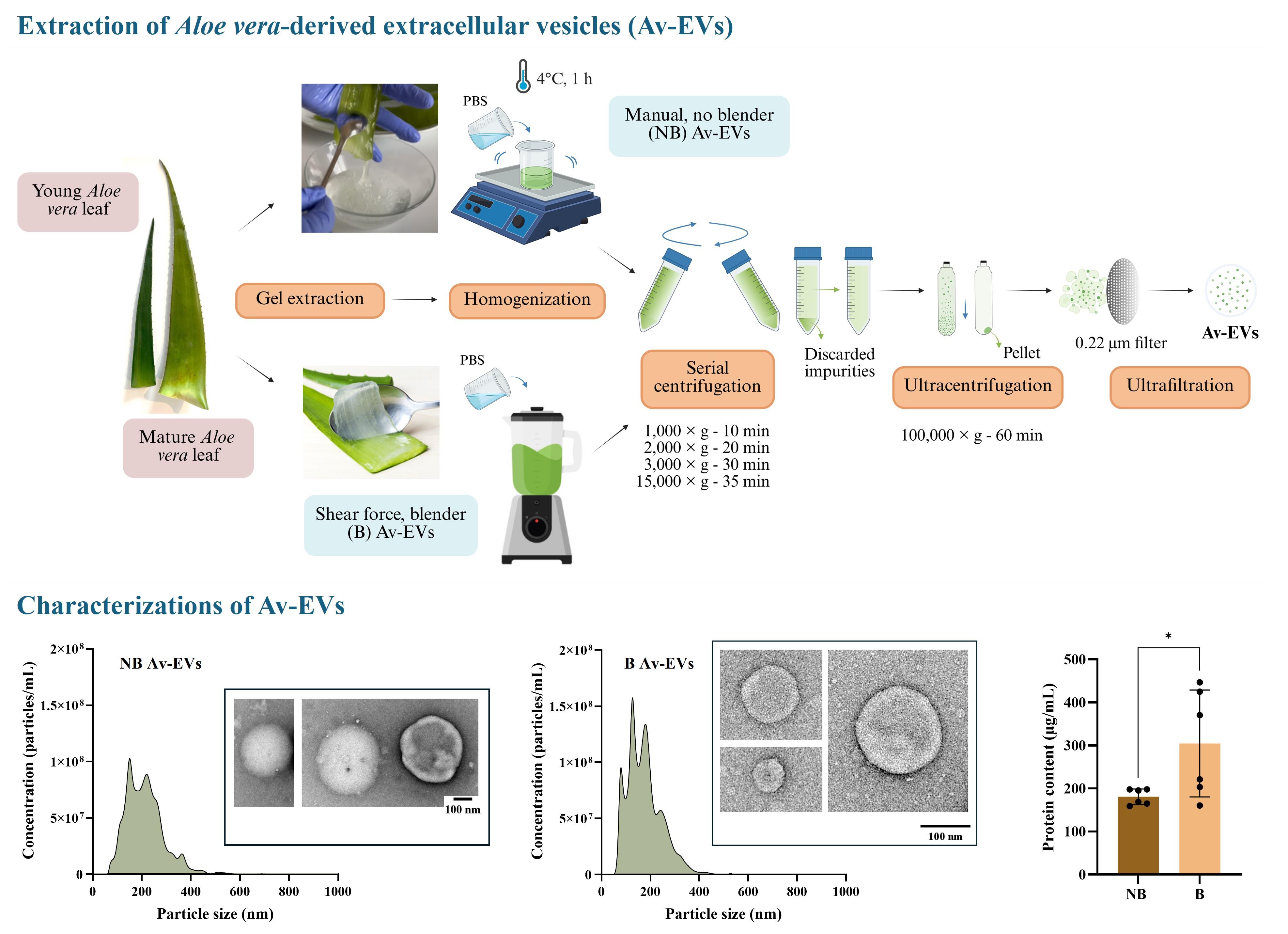
Aloe vera has long been used for its diverse pharmacological properties, motivating continued interest in isolating and preserving the bioactive molecules responsible for its therapeutic potential. More recently, Aloe vera–derived extracellular vesicles (Av-EVs) have emerged as nanoscale, cell-free carriers capable of retaining and delivering these properties, making them attractive for various biomaterials, nanomedicine, and regenerative medicine applications. Multiple techniques are available for extracellular vesicle isolation. These include ultracentrifugation, polymer-based precipitation, size-exclusion chromatography, immunoaffinity capture, ultrafiltration, density gradient separation, and emerging microfluidic platforms. Each method presents distinct trade-offs in purity, yield, scalability, and downstream compatibility. Despite this diversity, standardized workflows tailored to Av-EV isolation remain limited, and the influence of homogenization-induced shear forces and plant maturity on vesicle recovery and characterization has not been systematically addressed. Here, we present a reproducible protocol for isolating Av-EVs from Aloe vera gel employing two distinct homogenization strategies: manual, no-shear force (NB EVs), and blender-based shear-force homogenization (B EVs). The workflow covers gel preparation, serial centrifugation for debris removal, ultracentrifugation as the gold standard for vesicle enrichment, and final sterile filtration. This protocol enables consistent recovery of Av-EVs suitable for physicochemical characterization and functional analyses. It is simple and relies on commonly available laboratory equipment, facilitating broad adoption by ultracentrifugation users and offering adaptability to diverse research projects involving purified Aloe vera gel and Av-EVs, including studies focused on wound healing, fibrotic scarring, and regenerative processes, where coordinated antioxidant, anti-inflammatory, antimicrobial, immunomodulatory, and moisturizing responses are of interest.
Key features
• This protocol allows direct comparison of vesicle yield, size distribution, and protein content across extraction methods.
• This protocol yields ~1.4–2.0 × 10¹⁰ particles/mL per mature leaf for a total of ~8 × 10¹² particles per leaf.
• This protocol yields ~1.2–2.8 × 10¹⁰ particles/mL per young leaf for a total of ~2.8 × 10¹² per leaf.
• EVs from mature Aloe leaves yield protein concentrations of ~160–447 μg/mL, corresponding to ~3,840–10,728 μg of protein per leaf.
Keywords: Exosome (外泌体) EVs (细胞外囊泡)
Plant-derived EVs (PDEVs) (植物来源细胞外囊泡)
Ultracentrifugation (超速离心)
EV characterization (细胞外囊泡表征)
EVs (细胞外囊泡)
Plant-derived EVs (PDEVs) (植物来源细胞外囊泡)
Ultracentrifugation (超速离心)
EV characterization (细胞外囊泡表征)
Graphical overview
Background
Plant-derived extracellular vesicles (PDEVs) have emerged as a promising class of naturally occurring nanovesicles that retain the therapeutic benefits of the parent plant while functioning as biocompatible carriers of lipids, proteins, nucleic acids, and metabolites. Delivery of this molecular cargo supports intercellular communication, maintenance of tissue homeostasis, and regenerative processes [1]. Among PDEVs, Aloe vera–derived extracellular vesicles (Av-EVs) have attracted particular attention owing to the plant’s well-recognized pharmacological properties. Aloe vera contains a rich repertoire of bioactive molecules that collectively confer wound healing, antioxidant, anti-inflammatory, antimicrobial, immunomodulatory, and moisturizing effects [2–7]. Historically, these constituents have motivated extensive investigation of Aloe vera–based topical and oral formulations, with numerous in vitro studies, preclinical models, and clinical trials demonstrating high efficacy in skin-related applications [8–25]. Although such formulations remain widely used, recent interest has shifted toward Av-EVs, in which these bioactive molecules are enriched within a small vesicular fraction (~1% of the total gel volume) rather than being dispersed throughout the predominantly aqueous matrix [5].
Preliminary studies indicate that Av-EVs can modulate inflammatory signaling, enhance antioxidant defenses, and inhibit myofibroblast differentiation and contractile activity. Their versatility has also been demonstrated in applications such as drug encapsulation for targeted cancer therapy [26–29]. However, reported isolation methods vary widely, and to our knowledge, no prior study has provided a standardized workflow tailored to Av-EV isolation or systematically examined the combined influence of homogenization-induced shear forces and plant maturity on physicochemical characterization and functional analyses.
Multiple techniques have been described for the isolation of extracellular vesicles, including ultracentrifugation, polymer-based precipitation, size-exclusion chromatography, immunoaffinity capture, ultrafiltration, density gradient separation, and emerging microfluidic platforms. Each method presents distinct trade-offs in terms of purity, yield, scalability, and instrumentation requirements [30]. In this protocol, ultracentrifugation was selected as the gold-standard strategy due to its widespread adoption, reproducibility, and compatibility with downstream analyses. Here, we describe a reproducible workflow for isolating Av-EVs that incorporates two homogenization strategies: manual, no-shear force (NB EVs), and blender-based shear-force homogenization (B EVs). These steps are followed by serial centrifugation for debris removal, ultracentrifugation for vesicle enrichment, and final sterile filtration.
By enabling controlled comparison across extraction strategies and plant maturity stages, the protocol enables systematic evaluation of how processing conditions and source material impact Av-EV properties. The resulting vesicles can then be employed in experimental contexts related to pathological wound healing, such as fibrotic scarring, where coordinated antioxidant, anti-inflammatory, and pro-regenerative responses are relevant. In these settings, Av-EVs may contribute to restoring redox balance, modulating persistent inflammatory responses, and limiting excessive extracellular matrix deposition associated with fibrosis. In particular, our study demonstrated that the manual approach preserved vesicle integrity and bioactivity more effectively than shear-based homogenization, highlighting the importance of the extraction strategy for maintaining vesicle quality [31]. Additionally, EVs derived from mature leaves consistently outperformed those obtained from younger plants, showing stronger downregulation of inflammatory mediators and fibrotic markers [31]. Together, these findings suggest that both extraction strategies and plant maturity are crucial variables that must be carefully controlled to ensure the reproducibility and potency of EV-based therapeutics derived from Aloe sources.
While developed in the context of skin fibrosis research, this workflow can be broadly applied to studies investigating Av-EVs in biomaterials, nanomedicine, and regenerative medicine. For instance, future work may examine whether additional Aloe-associated properties, such as antimicrobial, analgesic, and moisturizing activities, are also mediated by its vesicles. Exploring these potential functions represents an important area for further broadening our understanding of the translational potential of Av-EVs in therapeutic delivery systems, bioactive materials, and tissue regeneration applications.
Materials and reagents
Biological materials
1. Aloe barbadensis miller leaves from mature plants and potted young plants
Note: Aloe barbadensis miller is commonly known as Aloe vera. Leaves from mature plants are commercially available for human consumption and can be obtained from supermarkets or grocery stores; in this study, they were sourced from Melissa’s/World Variety Produce, Inc. (USA). These leaves measured approximately 67 cm × 9 cm × 3 cm, yielded ~400 mL of gel per leaf, and resulted in ~8 × 10¹² particles per leaf. In contrast, Aloe vera potted plants sold in plant shops or nurseries typically represent young specimens. Young leaves measured approximately 45 cm × 5 cm × 1.5 cm, yielded ~100 mL of gel per leaf, and resulted in ~2.8 × 10¹² particles per leaf. These criteria were used to ensure consistency and comparability between maturity groups.
Reagents
1. Dulbecco’s phosphate-buffered saline (DPBS) (Cytiva, catalog number: SH30028.02)
2. Deionized (DI) water
3. PierceTM RIPA lysis buffer (Thermo Scientific, catalog number: 89901)
4. PierceTM BCA Protein Assay kit (Thermo Scientific, catalog number: 23225)
a. Bovine serum albumin (BSA) ampules, 2 mg/mL (Thermo Scientific, catalog number: 23209)
b. Reagent A (Thermo Scientific, catalog number: 23228)
c. Reagent B (Thermo Scientific, catalog number: 1859078)
5. Molecular biology–grade water (Cytiva, catalog number: SH30538)
6. Uranyl formate (Electron Microscopy Sciences, catalog number: 22450)
Solutions
1. BSA standards (see Recipes)
2. 0.75% uranyl formate (see Recipes)
Recipes
1. BSA standards
| Vial | Volume of diluent (RIPA) | Volume and source of BSA | Final BSA concentration |
|---|---|---|---|
| A | 0 | 300 μL of stock | 2,000 μg/mL |
| B | 125 μL | 375 μL of stock | 1,500 μg/mL |
| C | 325 μL | 325 μL of stock | 1,000 μg/mL |
| D | 175 μL | 175 μL of vial B dilution | 750 μg/mL |
| E | 325 μL | 325 μL of vial C dilution | 500 μg/mL |
| F | 325 μL | 325 μL of vial E dilution | 250 μg/mL |
| G | 325 μL | 325 μL of vial F dilution | 125 μg/mL |
| H | 400 μL | 100 μL of vial G dilution | 25 μg/mL |
| I | 400 μL | 0 | 0 = Blank |
2. 0.75% uranyl formate (10 mL)
a. Wear appropriate personal protective equipment, including an N95 respirator.
b. Boil 50 mL of deionized (DI) water in a 100 mL beaker on a hot plate.
c. In a fume hood, weigh 75 mg of uranyl formate into a 50 mL beaker and wrap the container with aluminum foil to protect it from light.
d. Add 10 mL of freshly boiled DI water, then stir until fully dissolved (~5 min) while keeping the solution protected from light.
e. Immediately before use, filter the 0.75% uranyl formate solution through a 0.22 μm syringe filter, placing filtered drops directly into your grid.
Laboratory supplies
1. Sterilized cutting surface
2. Sterilized tablespoon
3. Sterile tweezers
4. Aluminum foil
5. Nitrile examination gloves XS–XL (VWR, catalog number: 76518)
6. Sterilized knife (Victorinox, catalog number: 6.7833.6)
7. Sterile scissors (iBayam, 8" Multipurpose)
8. Sterilized fine-mesh sieve (Homquen)
9. Sterilized graduated containers with a capacity of 1 L
10. 50 mL conical tubes (Cellstar, catalog number: 227261)
11. 10 mL serological pipettes (Celltreat, catalog number: 229210)
12. 50 mL serological pipettes (Celltreat, catalog number: 229230)
13. Centrifuge tubes (Beckman Coulter, catalog number: 361625)
14. Sterile blades (Swann-Morton, catalog number: 0207)
15. 100 μm sterile cell strainer (Greiner Bio-One, catalog number: 542000)
16. 70 μm sterile cell strainer (Greiner Bio-One, catalog number: 542070)
17. 40 μm sterile cell strainer (Falcon, catalog number: 352340)
18. 0.22 μm sterile syringe filters (GVS, catalog number: FJ25BSCCA002AL01)
19. 10 mL sterile luer lock solo syringes (Norm-Ject, catalog number: NJ-4606728-02)
20. 1.5 mL Eppendorf tubes (Greiner Bio-One, catalog number: 616201)
21. 10 μL pipette tips (VWR, catalog number: 76323-394)
22. 20–200 μL pipette tips (VWR, catalog number: 76323-390)
23. 1,000 μL pipette tips (VWR, catalog number: 76323-454)
24. 1 mL sterile luer solo syringes (Norm-Ject, catalog number: NJ-9166017-02)
25. Formvar/carbon grid (200 mesh) (Electron Microscopy Science, catalog number: FCF200-Cu-50)
26. 50 mL glass beaker (Pyrex, catalog number: CLS100050)
27. 100 mL glass beaker (Pyrex, catalog number: CLS1000100)
28. 20 mm × 8 mm magnetic stir bar (Heathrow Scientific, catalog number: HS120548)
29. N95 respirator with valve (VWR, catalog number: 89201-510)
Equipment
1. Orbital shaker (Chemglass CLS-4021-100 Versa-Orb)
2. Commercial blender
3. Centrifuge with adapters for 50 mL conical tubes (Eppendorf, model: 5810)
4. Analytical balance (VWR, model: 124B2)
5. Ultracentrifuge with an MLA-50 rotor (Beckman Coulter, model: OptimaTM MAX-XP)
6. Nanoparticle tracking analysis (NTA) (Malvern Panalytical, model: NanoSight NS300)
7. Spectrophotometer (Molecular Devices, model: SpectraMax iD3®)
8. Talos L120C transmission electron microscopy (TEM, Thermo Fisher Scientific)
Software and datasets
1. NanoSight NTA 3.4 (Malvern Panalytical)
2. Softmax Pro 7 (Molecular Devices)
3. Microsoft Excel (Microsoft 365)
4. TIA (TEM imaging and analysis) (Thermo Fisher Scientific)
Procedure
登录/注册后免费查看全文
文章信息
稿件历史记录
提交日期: Feb 4, 2026
接收日期: Mar 19, 2026
在线发布日期: Mar 30, 2026
出版日期: Apr 20, 2026
版权信息
© 2026 The Author(s); This is an open access article under the CC BY license (https://creativecommons.org/licenses/by/4.0/).
如何引用
Ceballos-Santa, M. C., Sanchez-Ortiz, I., Gaborski, T. R. and Wuertz-Kozak, K. (2026). A Step-by-Step Protocol for the Isolation of Aloe vera–Derived Extracellular Vesicles via Manual and Shear-Force Homogenization. Bio-protocol 16(8): e5664. DOI: 10.21769/BioProtoc.5664.

 Download Citation in RIS Format
分类
植物科学 > 植物细胞生物学 > 细胞器分离
细胞生物学 > 细胞器分离 > 胞外囊泡
细胞生物学 > 细胞器分离 > 外来体
您对这篇实验方案有问题吗?
在此处发布您的问题,我们将邀请本文作者来回答。同时,我们会将您的问题发布到Bio-protocol Exchange,以便寻求社区成员的帮助。
 发布问题
发布问题


