


MDISCO: A High-Throughput Tissue-Clearing Protocol for Preservation of Endogenous Fluorescence in Whole Mouse Brains
MDISCO:一种用于全小鼠脑内源荧光保持的高通量组织透明化方法


发布: 2026年04月05日第16卷第7期 DOI: 10.21769/BioProtoc.5649 浏览次数: 695
评审: Marion HoggBaeksun KimAnonymous reviewer(s)
 PDF
PDF  Q&A
Q&A
Advertisement









Abstract
Organic solvent–based tissue clearing methods are widely used for whole-brain imaging but often compromise endogenous fluorescence. Existing protocols, such as iDISCO and fluorescence-preserving variants, have improved optical transparency but still present trade-offs between fluorescence retention, tissue stability, and workflow complexity. Here, we present MDISCO, a modified iDISCO-based clearing protocol designed to enhance preservation of endogenous fluorescence while maintaining high transparency and stable tissue morphology. MDISCO is directly compared with FDISCO+, an established fluorescence-preserving protocol, for the preservation of endogenous tdTomato and YFP. Performance across clearing steps is evaluated by measuring brain weight, anteroposterior and mediolateral dimensions, and optical transparency before and after solvent clearing and refractive index matching. Fluorescence preservation is assessed using whole-brain light-sheet microscopy with standardized imaging parameters to enable direct comparison. This protocol provides an accessible and high-throughput, reproducible workflow for solvent-based clearing with robust endogenous fluorescence preservation, offering clear advantages for whole-brain 3D imaging of genetically encoded fluorescent reporters.
Key features
• Preserves endogenous tdTomato and YFP fluorescence in whole mouse brains without signal amplification through immunolabeling.
• Improves optical clarity and cellular resolvability while maintaining anatomical integrity.
• Supports high-throughput “clearing” of whole-tissue samples.
Keywords: Tissue clearing (组织透明化) Whole-brain imaging (全脑成像)
Solvent-based clearing (基于溶剂的透明化)
Light-sheet microscopy (光片显微镜)
MDISCO (MDISCO)
Whole-brain imaging (全脑成像)
Solvent-based clearing (基于溶剂的透明化)
Light-sheet microscopy (光片显微镜)
MDISCO (MDISCO)
Graphical overview
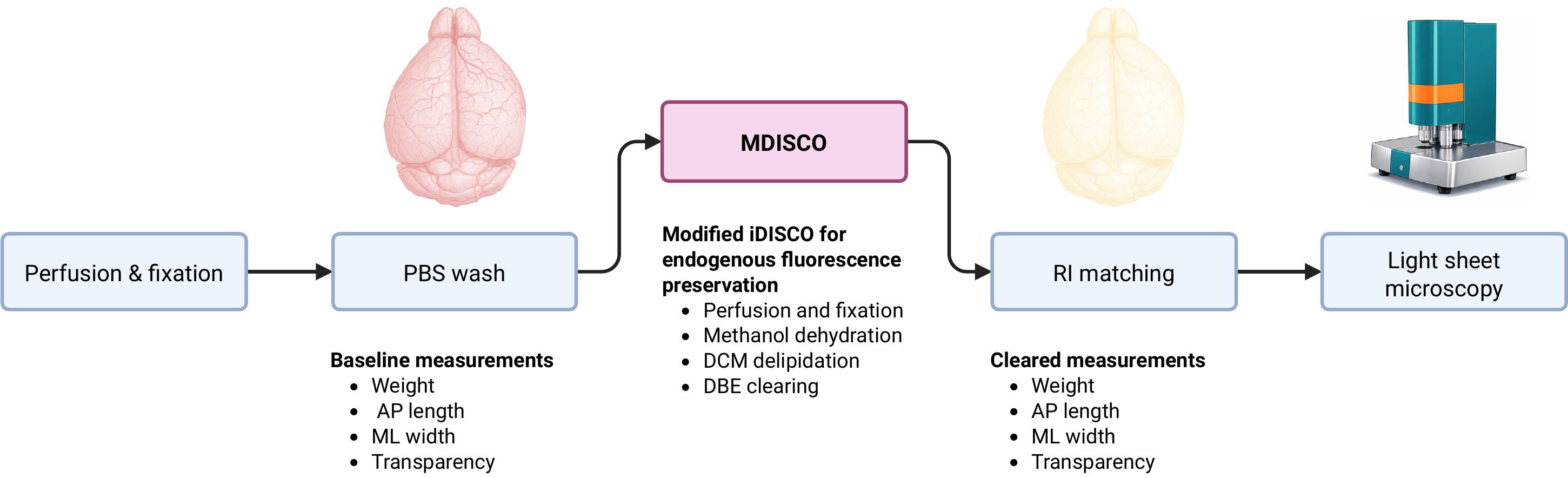
Experimental overview of MDISCO. Whole mouse brains are perfused, fixed, and stored in PBS prior to baseline measurements of weight, anteroposterior (AP) length, mediolateral (ML) width, and transparency. Samples are then processed using MDISCO via dichloromethane (DCM) delipidation and dibenzyl ether (DBE) clearing. Following refractive index (RI) matching, post-clearing measurements are collected, and brains are imaged by light-sheet microscopy.
Background
Tissue-clearing methods have enabled high-resolution imaging of intact biological samples by rendering tissue optically transparent while preserving structural organization. In rodent brain imaging, these approaches facilitate visualization of genetically encoded fluorescent reporters and large-scale neural circuits without the need for physical sectioning [1,2]. However, many organic solvent–based clearing protocols result in substantial quenching of endogenous fluorescent proteins, often necessitating additional immunolabeling steps that increase experimental complexity, cost, and processing time [2,3].
Organic solvent–based tissue clearing was first introduced with the 3DISCO protocol, which achieves optical transparency through graded dehydration in tetrahydrofuran (THF), followed by delipidation with dichloromethane (DCM) and refractive index matching in dibenzyl ether (DBE) [3]. This approach enables rapid and effective clearing of whole organs but is associated with substantial quenching of endogenous fluorescent proteins, limiting its utility for experiments relying on genetically encoded reporters [2,3]. The iDISCO protocol adapted this framework to support whole-mount immunolabeling by replacing THF with methanol-based dehydration, introducing hydrogen peroxide bleaching to reduce background, and incorporating rehydration and permeabilization steps to facilitate antibody penetration [3]. While iDISCO greatly expanded the applicability of solvent-based clearing for molecular labeling, it was not specifically optimized to preserve endogenous fluorescence and often results in diminished fluorescent protein signal in cleared tissue.
Several optimized clearing strategies, including uDISCO (ultimate DISCO) and FDISCO (fluorescence-preserving DISCO)-based methods, have been developed to improve fluorescence retention relative to earlier protocols [4,5]. While some of these approaches reduce regulatory and handling constraints by avoiding certain organic solvents, practical challenges related to incomplete clearing, background haze, and prolonged refractive index matching times can still limit imaging efficiency and data quality. In particular, residual scattering and bubble formation within incompletely cleared tissue can substantially increase imaging time and reduce effective cellular resolvability, even when protocols are followed precisely.
Immunolabeling is frequently used to visualize molecular targets in cleared tissue; however, this approach can introduce substantial variability due to incomplete antibody penetration, nonuniform labeling, off-target binding, and increased background signal, particularly in large or densely packed samples. Antibody-based labeling also substantially extends processing time and increases experimental cost, and labeling efficiency can vary across brain regions and experimental batches. In experimental contexts where genetically encoded fluorescent reporters are already present, preserving endogenous fluorescence avoids the additional cost, time, and variability associated with antibody-based signal amplification, including antibody optimization, staining condition adjustments, and reagent selection. While immunolabeling remains essential for targets that cannot be genetically encoded, these limitations motivate the continued development of clearing approaches that preserve endogenous fluorescence when reporter expression is available.
To address these limitations, we developed MDISCO (modified DISCO), a modified iDISCO (immunolabeling-enabled DISCO)-based clearing protocol designed to preserve endogenous fluorescence while maintaining optical clarity and anatomical integrity. Targeted adjustments to solvent handling and exposure conditions were implemented to minimize fluorophore quenching and improve clearing uniformity. These modifications enable consistent, high-quality whole-brain imaging without reliance on antibody labeling or proprietary reagents.
Subsequent DISCO-derived protocols have sought to balance optical transparency, tissue integrity, and preservation of endogenous fluorescence. uDISCO introduced solvent-mediated tissue shrinkage to improve imaging depth and resolution, but retained aggressive dehydration conditions that can compromise fluorescent protein stability [4]. More recent fluorescence-preserving approaches, such as FDISCO and FDISCO+, employ THF-based dehydration in combination with antioxidant additives to reduce fluorophore quenching, achieving improved fluorescence retention at the cost of increased workflow sensitivity and prolonged refractive index matching [5,6].
Building on these prior methods, MDISCO is derived from the iDISCO framework and retains methanol-based dehydration and peroxide bleaching while introducing targeted modifications to solvent exposure timing and post-clearing handling. In MDISCO, graded methanol dehydration is used in place of THF to reduce exposure to highly aggressive organic solvents known to destabilize fluorescent protein chromophores. DCM exposure is limited to short, defined delipidation steps rather than prolonged clearing, minimizing cumulative solvent-induced fluorescence loss while still achieving effective lipid removal. Equilibration in dibenzyl ether (DBE) is used primarily for uniform refractive index matching and to facilitate air bubble dissipation, followed by transfer to ethyl cinnamate (ECi) for long-term storage and imaging. ECi provides stable refractive index matching and has been reported to exhibit improved compatibility with endogenous fluorescent proteins. Together, these targeted modifications are designed to preserve fluorescent protein signal while maintaining optical clarity and tissue morphology, resulting in a practical and reproducible workflow that reduces variability and improves imaging readiness for whole-brain fluorescence microscopy.
In this protocol, MDISCO is evaluated in direct comparison with FDISCO+ using whole brains from tdTomato- and YFP-expressing transgenic mice. Tissue morphology, optical transparency, and endogenous fluorescence preservation are assessed across defined stages of the clearing workflow. Together, these analyses provide a practical framework for evaluating fluorescence-preserving tissue-clearing methods based not only on chemical composition but also on imaging performance, data interpretability, and experimental efficiency.
Materials and reagents
Biological materials
1. B6.Cg-Gt(ROSA)26Sortm9(CAG-tdTomato)Hze/J mice [Jackson Laboratory, strain #007909; common name: Ai9 or Ai9(RCL-tdT)] [7]
2. STOCK Fostm2.1(icre/ERT2)Luo/J mice [Jackson Laboratory, strain #030323, common name: Fos2A-iCreER (TRAP2)] [8]
3. TRAP2/Ai9 mice [TRAP2 (Fos2A-iCreER) and Rosa26LSL-tdTomato (Ai9) mice were crossed in-house mice] [9]
4. B6.129(Cg)-Oprl1tm1.1Mrbr/J mice (gifted from the Bruchas Lab at the University of Washington, Jackson Laboratory, strain #036308, common name: NOPRloxP/YFP) [10]
Reagents
1. Paraformaldehyde (PFA) powder, EM-grade (Fisher Scientific, catalog number: AC416785000)
2. PBS powder, pH 7.4 (Sigma-Aldrich, catalog number: P3813)
3. Methanol, ≥99.8% (Sigma-Aldrich, catalog number: 179957)
4. Dichloromethane (DCM), ≥99.9% (Applied BiosystemsTM, catalog number: 402152)
5. Hydrogen peroxide (H2O2) solution, 30% (Thermo Scientific, catalog number: 033323.AY)
6. Dibenzyl ether (DBE), ≥99% (Thermo Scientific, catalog number: A18447.36)
7. Ethyl cinnamate (ECi), ≥98% (Thermo Scientific, catalog number: A12906.36)
8. Double-distilled H2O (laboratory supply)
Laboratory supplies
1. Solvent-resistant gloves
2. Glass scintillation vials (e.g., Grainger, catalog number: 3LDT2)
Equipment
1. 37 °C incubator (e.g., Benchmark ScientificTM, SKU: RS7165)
2. Nutating mixer (Fisher Scientific, catalog number: 88-861-041)
3. Light-sheet microscope (e.g., UltraMicroscope Blaze, Miltenyi Biotec)
Software and datasets
1. ImSpector Pro (Miltenyi Biotec, version 8.0.3, requires a license)
2. MACS iQ View, 3D Large Volume Software (Miltenyi Biotec, version 1.2.3, requires a license)
3. Imaris (Oxford Instruments, Version 11.0, requires a license)
4. FIJI (ImageJ)
Procedure
登录/注册后免费查看全文
文章信息
稿件历史记录
提交日期: Jan 14, 2026
接收日期: Feb 26, 2026
在线发布日期: Mar 10, 2026
出版日期: Apr 5, 2026
版权信息
© 2026 The Author(s); This is an open access article under the CC BY license (https://creativecommons.org/licenses/by/4.0/).
如何引用
Martinez, M., Thornberry, J., Ozawa, A. and Toll, L. (2026). MDISCO: A High-Throughput Tissue-Clearing Protocol for Preservation of Endogenous Fluorescence in Whole Mouse Brains. Bio-protocol 16(7): e5649. DOI: 10.21769/BioProtoc.5649.

 Download Citation in RIS Format
分类
神经科学 > 神经解剖学和神经环路 > 荧光成像
细胞生物学 > 组织分析 > 组织成像
生物化学 > 蛋白质 > 荧光
您对这篇实验方案有问题吗?
在此处发布您的问题,我们将邀请本文作者来回答。同时,我们会将您的问题发布到Bio-protocol Exchange,以便寻求社区成员的帮助。
 发布问题
发布问题


