


Denaturing SUMO Immunoprecipitation From Mitotic Cells
有丝分裂细胞中变性条件下的SUMO免疫沉淀方法

发布: 2026年04月05日第16卷第7期 DOI: 10.21769/BioProtoc.5648 浏览次数: 390
评审: Ritu GuptaVipin Yadav YadavAnonymous reviewer(s)
 PDF
PDF  Q&A
Q&A
参见作者原研究论文
The authors used this protocol in:

Oct 2025
Advertisement








Abstract
Small ubiquitin-related modifiers (SUMOs) are covalently conjugated onto the proteome and serve as signaling molecules in many aspects of eukaryotic cell biology, from S. cerevisiae and C. elegans to H. sapiens. The conjugatable SUMO variants, SUMO1 and the almost identical SUMO2 and SUMO3 (designated SUMO2/3), are processed by an E1(SAE1:SAE2)-E2(UBC9)-E3 enzyme cascade to produce SUMO-modified proteins. The prerogative of the SUMO biology field is to identify and study the specific proteins undergoing SUMOylation, which grants us insights into the biological pathway of interest. This protocol was developed using the human osteosarcoma cell line U2OS to enable the investigation of SUMO conjugates in mitosis, the cell division phase of the cell cycle. We enrich the cell population for mitotic cells, which are isolated and subjected to stringent lysis conditions involving a high concentration of SDS and DTT in RIPA buffer, to promote complete protein denaturation. The lysates in high SDS RIPA buffer are diluted to reduce the overall SDS concentration and undergo conventional immunoprecipitation using SUMO1- or SUMO2/3-specific antibodies bound to protein A/G agarose beads. The samples are then compatible with downstream readouts such as western blots and mass spectrometry. This protocol detects endogenous SUMOylated proteins and avoids exogenous SUMO overexpression, which can alter SUMO conjugate formation. Furthermore, this denaturing protocol ensures only SUMOylated proteins are immunoprecipitated, and not their interactors.
Key features
• Purifies endogenous SUMO-modified proteins by building on Becker et al. [1].
• Enriches and isolates cells in mitosis using nocodazole and mitotic shake-off.
• 1% SDS RIPA lysis promotes robust denaturation ahead of SUMO-specific immunoprecipitation.
• Compatible with downstream readouts such as western blots and mass spectrometry.
Keywords: SUMO1 (SUMO1) SUMO2/3 (SUMO2/3)
Mitosis (有丝分裂)
Nocodazole (诺考达唑)
Denature (变性条件)
Immunoprecipitation (免疫沉淀)
Western blot (Western blot)
SUMO2/3 (SUMO2/3)
Mitosis (有丝分裂)
Nocodazole (诺考达唑)
Denature (变性条件)
Immunoprecipitation (免疫沉淀)
Western blot (Western blot)
Graphical overview
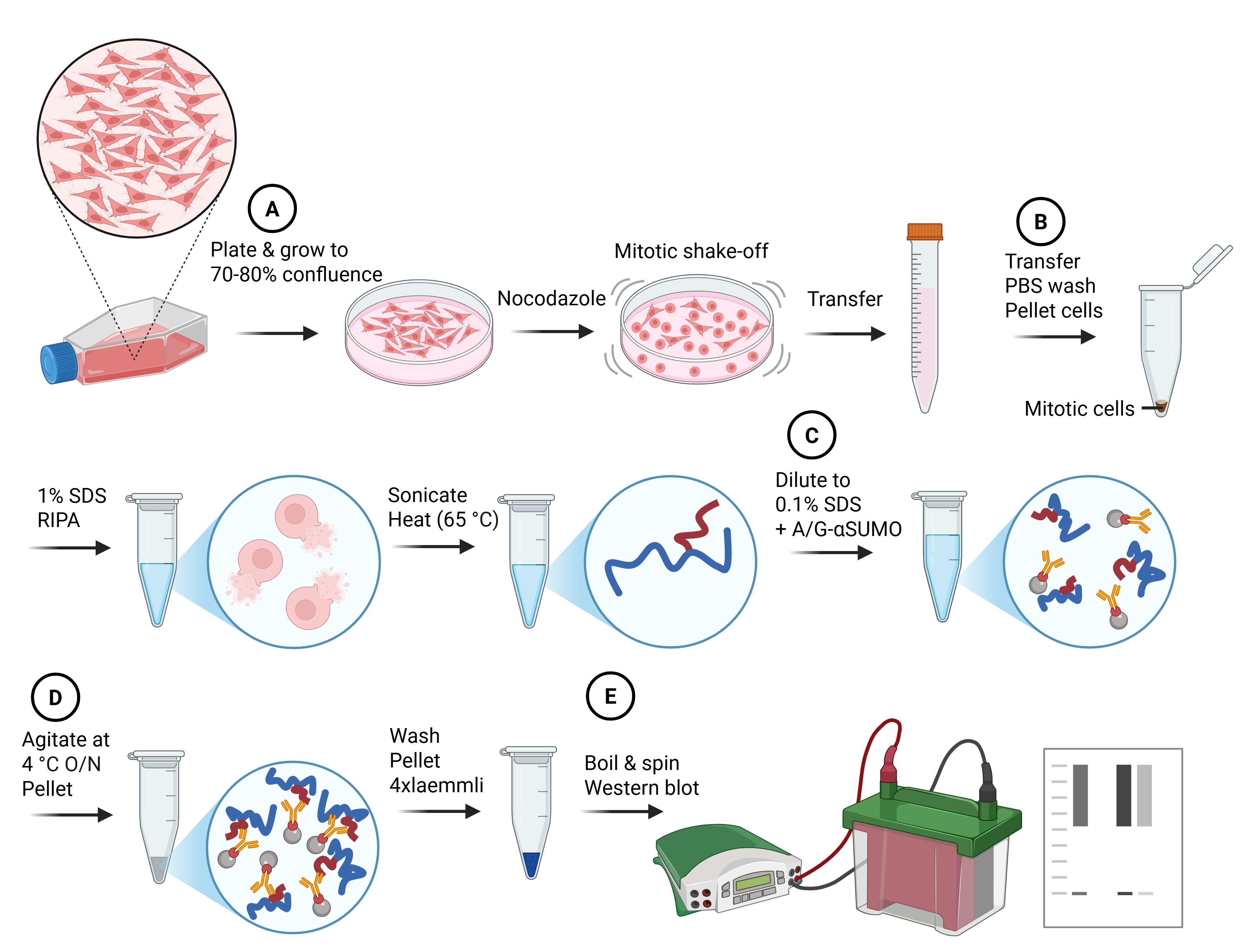
Denaturing small ubiquitin-related modifiers (SUMO) immunoprecipitation workflow. Major steps of the protocol and the impacts on cells and proteins are summarized in cartoon style. Letters in circles correspond to the protocol subsections (below). Blue lines indicate denatured proteins, and red lines indicate denatured SUMO protein conjugates.
Background
The small ubiquitin-related modifier (SUMO) can be covalently transferred onto proteins, altering their subcellular localization, protein–protein interactions, and/or activity. The conjugation of SUMO moieties onto substrates occurs via the processive actions of a unique E1 activation enzyme (composed of a heterodimer containing SAE1/AOS1 and SAE2/UBA2) and an E2 conjugating enzyme (UBC9), with many E3 ligases assisting [2]. Within higher eukaryotes, there are three variants of conjugatable SUMO: SUMO1, SUMO2, and SUMO3. SUMO2 and SUMO3 share a 97% sequence identity and are virtually indistinguishable [3]; as such, they are often designated SUMO2/3. SUMO1 and SUMO2/3 share many substrates, but proteomic studies have revealed that SUMO2/3-modified substrates do not exclusively overlap with those modified by SUMO1 [1,4]. The divergence between SUMO1 and SUMO2/3 is particularly evident in mitosis, where SUMO1 localizes to the mitotic spindle, whereas SUMO2/3 co-occupies areas associated with centromeres and condensed chromosomes [5].
SUMOylation has emerged as a vital regulator of mitosis, with defects in mitotic progression and chromosome segregation observed upon deficiency, depletion, or chemical inhibition of core components within the SUMO conjugation machinery [6–10]. While SUMO conjugation is required for efficient and accurate mitotic progression, it is notable that global SUMO conjugates are lower during this phase of the cell cycle [5], which can make identifying alterations in SUMOylation during mitosis challenging. Therefore, obtaining a large and relatively “pure” mitotic population is essential to accurately pinpoint mitotic SUMOylation events that are not skewed by asynchronous contaminants. Due to the short duration of this phase of the cell cycle (approximately 1 h in RPE, HeLa and MCF10A) [11], cell synchronization is commonly used to obtain sufficient cell numbers to study. This has classically been achieved using microtubule poisons such as nocodazole [12,13] and mitotic shake-off [14].
The identification of SUMO conjugates requires cell lysates to be prepared under denaturing conditions. This encourages the stability of the SUMO modification by inhibiting the actions of SUMO-specific proteases/isopeptidases, as well as reducing the co-precipitation of non-covalent SUMO interactors [15]. The simplest method of identifying SUMOylated proteins is through the detection of a mobility shift on denaturing gels that is sensitive to SUMO-specific proteases and/or disruption to SUMO conjugation machinery. Whilst this may be a valid starting point for identifying a SUMO substrate, it is indirect, biased toward abundant or highly modified targets, and only speaks to the modification of one protein. To increase the abundance of SUMO-modified substrates and to enable the identification of SUMOylated sites on target proteins, affinity purification with overexpressed SUMO constructs has frequently been employed. The use of 6xHis-tagged exogenous proteins and Ni-NTA resin is particularly used due to its compatibility with denaturing conditions [4,15–20]. However, the use of overexpression is an inherent issue, as it intrinsically alters the balance of SUMO variants (i.e., SUMO1 and SUMO2/3), which has the potential to alter their usage [7,21]. A further caveat to using overexpression is the prerequisite for cells to have an acceptable degree of transfectability. This can limit the cell lines and types of samples that can be used to identify SUMOylated substrates.
A number of methods have been developed to enrich SUMO conjugates under endogenous conditions [1,22–28]. Many of these techniques rely on mass spectrometry–based proteomics to identify SUMO moieties or remnants on peptides [22–26]. These methods are integral to our understanding of SUMO biology, enabling the site-specific identification of SUMOylation on substrates. However, the main limitation in adopting these approaches for more frequent use, among the wider molecular biology community, is the high degree of specialism they require. The level of information that these techniques are able to acquire may also lie beyond the scope of the specific research question under investigation.
The enrichment of endogenous SUMOylated proteins (as opposed to peptides) has been described using SUMO-traps [alternatively named SUMO binding entities (SUBEs)] [28] and antibodies [1]. Both approaches require less specialism to perform, and protein identification can occur through targeted western blotting or mass spectrometry. SUMO-traps can capture endogenous poly-SUMOylated proteins using a GST-fusion protein bearing tandem SUMO interacting motif repeats from the SUMO E3 RNF4. However, these are not suited to the identification of mono-SUMOylated proteins and consequently bias results toward SUMO2/3-containing substrates. Additionally, as it is currently presented in the literature, this assay is performed under non-denaturing conditions, and therefore, non-covalent SUMO interactors and interacting proteins can also be co-precipitated with SUMOylated substrates [28]. Becker et al. [1] described a procedure that used commercially available antibodies and denaturing conditions to enrich SUMOylated substrates. The main benefits of this technique derive from its ease of use, accessibility of reagents, and relative affordability. Due to the initial denaturation of lysates, misidentification of non-covalent interactors should be avoided. However, the reliance of this assay on antibodies is a major limitation. Antibodies are inherently limited by the specificity of their antigen and may not capture all SUMO substrates or bind other SUMO paralogs to those intended. Batch-to-batch variation within polyclonal antibodies can also contribute to variation in results. Lastly, if a significant enough enrichment is not achieved through immunoprecipitation, this assay may struggle to detect modifications on lowly expressed or modified proteins.
Here, we describe an optimized procedure to assess SUMO conjugation in mitosis. This assay was successfully employed to characterize an alteration in SUMO variant usage during mitosis when an acetyl mimic of the SUMO E1 component SAE2 (SAE2-K164Q) was expressed in cells [7]. Building on the assay described by Becker et al. [1], cells are enriched for mitosis using nocodazole and further isolated via a mitotic shake-off. Lysis and denaturation then proceed, using a high-percentage SDS buffer and treatment with DTT. The denatured lysate is diluted tenfold with a 0% SDS lysis buffer before a standard immunoprecipitation with commercial antibodies. This method bears the same underlying disadvantages as the Becker et al. technique, described above, with some additional caveats: the requirement for a large number of cells and the use of agents to synchronize cells before harvesting. The benefits of using this assay over other techniques include its ease of use, high adaptability to investigate specific research questions, lack of requirement for specialist equipment, and the absence of SUMO variant imbalance generated by overexpression.
Materials and reagents
Biological materials
1. U-2 OS, Homo sapiens bone osteosarcoma (ATCC, HTB-96)
Reagents
1. Dulbecco’s modified Eagle’s medium, high glucose (Sigma-Aldrich, catalog number: D5796-500ML)
2. Fetal bovine serum (FBS) (Gibco, catalog number: A5256801)
3. Penicillin-Streptomycin (Sigma-Aldrich, catalog number: P4333)
4. Phosphate-buffered saline (PBS) tablets (Sigma-Aldrich, catalog number: P4417)
5. Trypsin 10× (Sigma-Aldrich, catalog number: T4174-100ML)
6. Nocodazole (Sigma-Aldrich, catalog number: SML1665-1ML)
7. Sodium phosphate monobasic (Sigma-Aldrich, catalog number: 342483)
8. Sodium chloride (Fisher Scientific, catalog number: 10316943)
9. Sodium dodecyl sulfate (SDS) (Sigma-Aldrich, catalog number: 436143)
10. Sodium deoxycholate (Sigma-Aldrich, catalog number: D6750)
11. Ethylenediaminetetraacetic Acid (EDTA) (Fisher Scientific, catalog number: BP118-500)
12. Triton® X-100 (Promega, catalog number: H5141)
13. N-Ethylmaleimide (NEM) (Sigma-Aldrich, catalog number: 04260)
14. PhosSTOPTM (Roche, catalog number: 4906837001)
15. cOmpleteTM, Mini, EDTA-free protease inhibitor cocktail (Roche, catalog number: 11836170001)
16. 1,4-dithioTM threitol (DTT) (Roche, catalog number: 10197777001)
17. Tris-HCL (Trizma® hydrochloride) (Sigma-Aldrich, catalog number: T5941)
18. Urea (Promega, catalog number: V3171)
19. Glycerol (Fisher, catalog number: G/0650/08)
20. Bromophenol blue (Acros, catalog number: 403140050)
21. 2-Mercaptoethanol (Sigma-Aldrich, catalog number: M6250)
22. PierceTM protein A/G agarose (Thermo Scientific, catalog number: 20421)
23. SUMO1 antibody (Abcam, catalog number: ab32058)
24. SUMO2/3 antibody (Abcam, catalog number: ab81371)
25. Hydrochloric acid (HCl), 32% (required to adjust pH) (Fisher Scientific, catalog number: 10458980)
26. Sodium hydroxide (NaOH) (required to adjust pH) (Merck, catalog number: 1064621000)
27. Tris-Glycine-SDS PAGE buffer 10× (National diagnostics, catalog number: EC-869)
28. Novex Tris-Glycine Mini Protein Gels (Life Technologies Ltd., catalog number: XP04205BOX)
29. PageRulerTM Plus Prestained Protein Ladder (ThermoFisher Scientific, catalog number: 26619)
30. Tris-Glycine electroblotting buffer 10× (National diagnostics, catalog number: EC-880)
31. PVDF membrane 0.45 μm pore (VWR, catalog number: 10600023)
32. Methanol (VWR, catalog number: 20847.307)
33. Skimmed milk powder (Millipore, catalog number: 70166)
34. Tween 20 (VWR, catalog number: 663684B)
35. Polyclonal swine anti-rabbit immunoglobulins HRP (Agilent Dako, catalog number: P0217)
36. Polyclonal rabbit anti-mouse immunoglobulins HRP (Agilent Dako, catalog number: P0260)
37. ECL western blotting substrate (Promega, catalog number: W1015)
38. RANGAP1 polyclonal antibody (Invitrogen, catalog number: PA5-92435)
39. (Optional) PierceTM 660 nm protein assay (ThermoFisher Scientific, catalog number: 22660)
40. (Optional) ionic detergent compatibility reagent (ThermoFisher Scientific, catalog number: 22663)
41. (Optional) Cyclin B1 antibody (Cell Signaling, catalog number: 12231)
42. (Optional) α Tubulin antibody (Novus Biologicals, catalog number: NB100-690)
43. (Optional) Histone H3 (Cell Signaling, catalog number: 9715)
Solutions
1. Cell growth medium (see Recipes)
2. 1× PBS (see Recipes)
3. 1× Trypsin (see Recipes)
4. 10 M NaOH (see Recipes)
5. 1 M sodium phosphate, pH 7.4 (see Recipes)
6. 1% SDS lysis buffer (see Recipes)
7. 0% SDS lysis buffer (see Recipes)
8. 0.1% SDS buffer (see Recipes)
9. 1 M DTT (see Recipes)
10. 1 M Tris, pH 6.8 (see Recipes)
11. Loading buffer (see Recipes)
12. 1× running buffer (see Recipes)
13. 1× transfer buffer (see Recipes)
14. PBST 0.1% (see Recipes)
15. 5% milk (see Recipes)
Recipes
1. Cell growth medium
| Reagent | Final concentration | Quantity or volume |
|---|---|---|
| Dulbecco’s modified Eagle’s medium, high glucose | 445 mL | |
| FBS | 10% | 50 mL |
| Penicillin-streptomycin | 1% | 5 mL |
2. 1× PBS
| Reagent | Final concentration | Quantity or volume |
|---|---|---|
| PBS | 0.01 M phosphate buffer, 0.0027 M potassium chloride, and 0.137 M sodium chloride, pH 7.4, at 25 °C | 2 tablets |
| ddH2O | 400 mL |
Autoclave 1× PBS solution to sterilize.
3. 1× Trypsin
| Reagent | Final concentration | Quantity or volume |
|---|---|---|
| 10× trypsin | 1× | 5 mL |
| 1× PBS | 45 mL |
4. 10 M NaOH
| Reagent | Final concentration | Quantity or volume |
|---|---|---|
| NaOH | 10 M | 40 g |
| ddH2O | 100 mL |
5. 1 M sodium phosphate, pH 7.4
| Reagent | Final concentration | Quantity or volume |
|---|---|---|
| Sodium phosphate monobasic | 1 M | 69 g |
| ddH2O | Up to 500 mL |
Adjust pH to 7.4.
6. 1% SDS lysis buffer pH 7.4
| Reagent | Final concentration | Quantity or volume |
|---|---|---|
| 1 M sodium phosphate, pH 7.4 | 20 mM | 10 mL |
| Sodium chloride | 150 mM | 4.38 g |
| SDS | 1% w/v | 5 g |
| Sodium deoxycholate | 0.5% w/v | 2.5 g |
| EDTA | 5 mM | 730.6 mg |
| Triton X-100 | 1% | 5 mL |
| ddH2O | Up to 500 mL | |
| Directly before use, dissolve the following into 10 mL of the stock solution listed above | ||
| NEM | 10 mM | 12.5 mg |
| cOmpleteTM, Mini, EDTA-free protease inhibitor cocktail | 1× | 1 tablet |
| PhosSTOPTM | 1× | 1 tablet |
In aqueous solutions, NEM and inhibitor cocktails are unstable. Excess solution containing inhibitors can be aliquoted and stored at -20 °C for up to a month.
7. 0% SDS lysis buffer pH 7.4
| Reagent | Final concentration | Quantity or volume |
|---|---|---|
| 1 M sodium phosphate pH 7.4 | 20 mM | 10 mL |
| Sodium chloride | 150 mM | 4.38 g |
| Sodium deoxycholate | 0.5% | 2.5 g |
| EDTA | 5 mM | 730.6 mg |
| Triton X-100 | 1% | 5 mL |
| ddH2O | Up to 500 mL | |
| Directly before use, dissolve the following into 10 mL of the stock solution listed above | ||
| NEM | 10 mM | 12.5 mg |
| cOmpleteTM, Mini, EDTA-free protease inhibitor cocktail | 1× | 1 tablet |
| PhosSTOPTM | 1× | 1 tablet |
In aqueous solutions, NEM and inhibitor cocktails are unstable. Excess solution containing inhibitors can be aliquoted and stored at -20 °C for up to a month.
8. 0.1% SDS buffer pH 7.4
| Reagent | Final concentration | Quantity or volume |
|---|---|---|
| 1% SDS lysis buffer | 2 mL | |
| 0% SDS lysis buffer | 18 mL |
9. 1 M DTT
| Reagent | Final concentration | Quantity or volume |
|---|---|---|
| DTT | 1 M | 1.54 g |
| ddH2O | Up to 10 mL |
Store in dark conditions at -20 °C.
10. 1 M Tris, pH 6.8
| Reagent | Final concentration | Quantity or volume |
|---|---|---|
| Tris-HCl | 1 M | 157.60 g |
| ddH2O | Up to 1 L |
Adjust pH solution to 6.8.
11. Loading buffer
| Reagent | Final concentration | Quantity or volume |
|---|---|---|
| SDS | 8% w/v | 8 g |
| Urea | 6 M | 36 g |
| Glycerol | 40% | 40 mL |
| 1 M Tris-HCl, pH 6.8 | 0.2 M | 20 mL |
| 2-Mercaptoethanol | 5% | 5 mL |
| Bromophenol blue | 400 mg |
Open and dispense 2-mercaptoethanol in a chemical safety hood. To maximize the reducing capability of 2-mercaptoethanol, aliquots of loading buffer should be stored at -20 °C. Alternatively, 2-mercaptoethanol can be supplemented at 5% concentration into a volume of loading buffer directly before use.
12. 1× running buffer
| Reagent | Final concentration | Quantity or volume |
|---|---|---|
| Tris-Glycine-SDS PAGE buffer 10× | 1× | 100 mL |
| ddH2O | Up to 1 L |
13. 1× transfer buffer
| Reagent | Final concentration | Quantity or volume |
|---|---|---|
| Tris-glycine electroblotting buffer 10× | 1× | 100 mL |
| Methanol | 20% | 200 mL |
| ddH2O | Up to 1 L |
14. PBST 0.1%
| Reagent | Final concentration | Quantity or volume |
|---|---|---|
| PBS | 1× | 5 tablets |
| Tween 20 | 0.1% | 1 mL |
| ddH2O | 1 L |
15. 5% milk
| Reagent | Final concentration | Quantity or volume |
|---|---|---|
| Dried skimmed milk powder | 5% w/v | 2.5 g |
| PBST 0.1% | Up to 50 mL |
Laboratory supplies
1. 150 mm2 cell culture dish, tissue culture treated (Corning, catalog number: 430599)
2. 25 and 10 mL pipettes (Corning Costar, catalog numbers: CLS4250, CLS4488)
3. 50 mL centrifuge tubes (Corning, catalog number: CLS430921-500EA)
4. Cell counting chambers (Biosigma, catalog number: 390497)
5. 1.5 mL microtubes (Axygen, catalog number: MCT-150-C)
6. 10, 200, and 1,000 μL pipette tips (Starlab, catalog numbers: S1111-3700, S1111-1716, S1111-6811)
7. Whatman 3 MM chromatography paper (Cytiva, catalog number: 3030-917)
8. Cling film (Fisher Scientific, catalog number: 12872233)
9. X-ray film 18 × 24 cm (Scientific Laboratory Supplies, catalog number: MOL7016)
Equipment
1. CO2 incubator (Thermo Scientific, model: BB15)
2. Biosafety cabinet (Biopharma group, model: SafeFAST Classic 212 D)
3. Pipette filler (Thermo Scientific, catalog number: 9531)
4. Water bath (Grant Instruments, model: SAP18)
5. Light microscope (Olympus, model: CK30)
6. Large benchtop centrifuge (Thermo Scientific, model: Heraeus Megafuge 40)
7. Refrigerated benchtop centrifuge (Eppendorf, model: 5430 R)
8. Heat block (Labnet International, model: D1100)
9. Sonicator (Misonix, model: XL2000)
10. Vortex (Labnet International, model: S0200-230V-UK)
11. Roller (Stuart, model: SRT9D)
12. Minigel tank (Invitrogen, catalog number: A25977)
13. Powerpack (Fisherbrand, model: Powerpro-300)
14. Transfer tank (Hoefer, model: TE62)
15. Automatic X-ray film processor (Protec, model: Optimax)
16. 18 × 24 cm X-ray film cassette (Cytiva, model: RPN 11642)
17. pH meter (HANNA instruments, model: HI-2020-02)
18. Manual pipettes (0.5–10 µL, 20–200 µL, 100–1,000 µL) (Starlab, catalog numbers: S7100-0510, S7100-2200, S7110-1000)
19. (Optional) Multiskan SkyHigh microplate spectrophotometer (Thermo Scientific, model: A51119600C)
Software and datasets
1. ImageJ (National Institute of Health, 1.54i) [29]
Procedure
文章信息
稿件历史记录
提交日期: Dec 8, 2025
接收日期: Feb 19, 2026
在线发布日期: Mar 10, 2026
出版日期: Apr 5, 2026
版权信息
© 2026 The Author(s); This is an open access article under the CC BY license (https://creativecommons.org/licenses/by/4.0/).
如何引用
Walker, A. K., Lanz, A. J. and Morris, J. R. (2026). Denaturing SUMO Immunoprecipitation From Mitotic Cells. Bio-protocol 16(7): e5648. DOI: 10.21769/BioProtoc.5648.

 Download Citation in RIS Format
分类
生物化学 > 蛋白质 > 修饰 > SUMO化
细胞生物学 > 基于细胞的分析方法 > 蛋白互作
生物化学 > 蛋白质 > 免疫检测 > 免疫沉淀(IP)
您对这篇实验方法有问题吗?
在此处发布您的问题,我们将邀请本文作者来回答。同时,我们会将您的问题发布到Bio-protocol Exchange,以便寻求社区成员的帮助。
 发布问题
发布问题


