


Analysis of Developing Pollen Grains within Intact Arabidopsis thaliana Anthers by Olympus Two-Photon Laser Scanning Microscopy
使用Olympus双光子激光扫描显微镜分析完整的拟南芥花药中正在发育的花粉粒


发布: 2015年12月05日第5卷第23期 DOI: 10.21769/BioProtoc.1677 浏览次数: 10577
评审: Tie LiuNing LiuAnonymous reviewer(s)
 PDF
PDF  Q&A
Q&A
参见作者原研究论文
The authors used this protocol in:
Nov 2014
Advertisement








Abstract
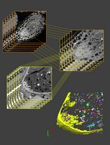
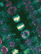
The method consists of imaging developing pollen grains as they form within intact, immature Arabidopsis thaliana anthers. Using two-photon excitation in the infrared wavelength range, the intrinsic fluorescence (autofluorescence) of developing pollen grains and surrounding sporophytic tissues of the anther wall, including the tapetum, middle layer, endothecium and epidermis, can be visualized in the three-dimensional space of an intact anther. In contrast to conventional confocal microscopy, the application of red-shifted light by two-photon microscopy improves depth penetration into specimens, while the scattering of light and subsequent phototoxicity is minimized, making this a superior method for imaging the developing pollen grains and tapetal cells enclosed within anthers. The technique described was optimized for the detection of autofluorescent components of the pollen wall, including sporopollenin and the pollen coat, and provided spatial and developmental data on the autofluorescent metabolites in anthers of wild-type and pollen wall mutant plants (Quilichini et al., 2014). The use of two-photon imaging of live, intact anthers holds potential for future studies aimed at understanding the spatial relationship between gametophytic and sporophytic tissues during pollen development and the distribution of metabolites or fluorescently-tagged proteins within developing anthers.
Materials and Reagents
- Ruler with millimetre divisions
- Glass bottom Petri dishes (MatTek Corporation, catalog number: P35G-1.5-14C )
- Coverslips (No.1.5, 0.17 mm thick, 22 x 22) (Thermo Fisher Scientific, catalog number 12-541B )
- Transfer pipettes 1.5 ml
- Arabidopsis thaliana plants in the flowering stage
- Non-toxic food-grade paraffin wax maintained in a liquid state at 40 ºC in a paraffin bath (Thera-Band, catalog number: 24050 )
- Distilled water
Equipment
- Spectra-Physics MaiTai HP Titanium:Sapphire mode-locked pulsed laser, with tuneable wavelengths spanning 690 to 1,040 nm.
- Olympus Fluoview 1000 scan head (Olympus America Inc., model: FV1000MPE ) modified by Olympus for two-photon imaging by the inclusion of a light path for the pulsed laser input and the housing for the non-descanned, multialkali side window photomultiplier tubes (PMTs).
- Olympus BX61WI upright microscope
- Olympus multiphoton microscope filter cube (440/40 bandpass filter) (OLYMPUS, model: FV10-MRVGR/XR 4CH NDD FILTER )
- Olympus multiphoton microscope dedicated objective lens optimized for transmitting IR light (25x, 1.05 numerical aperture, 2.0 mm working distance) (OLYMPUS, model: FV10-SNPXLU )
- Light tight, custom made multiphoton microscope enclosure. To build an enclosure, aluminum extruded components (Item North America) and flat-black painted fibre board can be used
- Forceps for fine dissections (Electron Microscopy Sciences, catalog number: 72700-D )
- Olympus stereomicroscope (magnification range of 6.3-63x) (OLYMPUS, model: SZX10 )
Software
- Olympus FluoView FV10-ASW 3.01
- Spectra-Physics MaiTai Control laser software
- Volocity version 6.1.1 software package
Note: ImageJ 1.47v can also be used to process images.
Procedure
文章信息
版权信息
© 2015 The Authors; exclusive licensee Bio-protocol LLC.
如何引用
Quilichini, T. D., Samuels, A. L. and Douglas, C. J. (2015). Analysis of Developing Pollen Grains within Intact Arabidopsis thaliana Anthers by Olympus Two-Photon Laser Scanning Microscopy . Bio-protocol 5(23): e1677. DOI: 10.21769/BioProtoc.1677.

 Download Citation in RIS Format
分类
植物科学 > 植物发育生物学 > 形态建成
植物科学 > 植物细胞生物学 > 细胞成像
细胞生物学 > 细胞成像 > 双光子显微镜
您对这篇实验方法有问题吗?
在此处发布您的问题,我们将邀请本文作者来回答。同时,我们会将您的问题发布到Bio-protocol Exchange,以便寻求社区成员的帮助。
 发布问题
发布问题


