往期刊物2023

卷册: 13, 期号: 16

left

Dec

20

Dec

5

Nov

20

Nov

5

Oct

20

Oct

5

Sep

20

Sep

5

Aug

20

Aug

5

Jul

20

Jul

5

Jun

20

Jun

5

May

20

May

5

Apr

20

Apr

5

Mar

20

Mar

5

Feb

20

Feb

5

Jan

20

Jan

5

right

生物信息学与计算生物学

利用单细胞RNA测序和参考图谱进行T细胞克隆分析

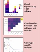
T Cell Clonal Analysis Using Single-cell RNA Sequencing and Reference Maps

利用单细胞RNA测序和参考图谱进行T细胞克隆分析

MA Massimo Andreatta
PG Paul Gueguen
NB Nicholas Borcherding
SC Santiago J. Carmona
4088 Views
Aug 20, 2023
T cells are endowed with T-cell antigen receptors (TCR) that give them the capacity to recognize specific antigens and mount antigen-specific adaptive immune responses. Because TCR sequences are distinct in each naïve T cell, they serve as molecular barcodes to track T cells with clonal relatedness and shared antigen specificity through proliferation, differentiation, and migration. Single-cell RNA sequencing provides coupled information of TCR sequence and transcriptional state in individual cells, enabling T-cell clonotype-specific analyses. In this protocol, we outline a computational workflow to perform T-cell states and clonal analysis from scRNA-seq data based on the R packages Seurat, ProjecTILs, and scRepertoire. Given a scRNA-seq T-cell dataset with TCR sequence information, cell states are automatically annotated by reference projection using the ProjecTILs method. TCR information is used to track individual clonotypes, assess their clonal expansion, proliferation rates, bias towards specific differentiation states, and the clonal overlap between T-cell subtypes. We provide fully reproducible R code to conduct these analyses and generate useful visualizations that can be adapted for the needs of the protocol user.Key features• Computational analysis of paired scRNA-seq and scTCR-seq data• Characterizing T-cell functional state by reference-based analysis using ProjecTILs• Exploring T-cell clonal structure using scRepertoire• Linking T-cell clonality to transcriptomic state to study relationships between clonal expansion and functional phenotypeGraphical overview

生物物理学

对成年小鼠体外脑切片进行穿孔膜片钳记录

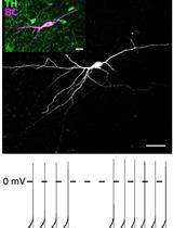
Perforated Patch Clamp Recordings in ex vivo Brain Slices from Adult Mice

对成年小鼠体外脑切片进行穿孔膜片钳记录

SH Simon Hess
HW Helmut Wratil
PK Peter Kloppenburg
1828 Views
Aug 20, 2023
Intracellular signaling pathways directly and indirectly regulate neuronal activity. In cellular electrophysiological measurements with sharp electrodes or whole-cell patch clamp recordings, there is a great risk that these signaling pathways are disturbed, significantly altering the electrophysiological properties of the measured neurons. Perforated-patch clamp recordings circumvent this issue, allowing long-term electrophysiological recordings with minimized impairment of the intracellular milieu. Based on previous studies, we describe a superstition-free protocol that can be used to routinely perform perforated patch clamp recordings for current and voltage measurements.

癌症生物学

哺乳动物细胞染色体畸变的定量分析

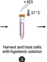
Quantification of Chromosomal Aberrations in Mammalian Cells

哺乳动物细胞染色体畸变的定量分析

IP Inés Paniagua
JJ Jacqueline J. L. Jacobs
2082 Views
Aug 20, 2023
Maintenance of genome integrity requires efficient and faithful resolution of DNA breaks and DNA replication obstacles. Dysfunctions in any of the processes orchestrating such resolution can lead to chromosomal instability, which appears as numerical and structural chromosome aberrations. Conventional cytogenetics remains as the golden standard method to detect naturally occurring chromosomal aberrations or those resulting from the treatment with genotoxic drugs. However, the success of cytogenetic studies depends on having high-quality chromosome spreads, which has been proven to be particularly challenging. Moreover, a lack of scoring guidelines and standardized methods for treating cells with genotoxic agents contribute to significant variability amongst different studies. Here, we report a simple and effective method for obtaining well-spread chromosomes from mammalian cells for the analysis of chromosomal aberrations. In this method, cells are (1) arrested in metaphase (when chromosome morphology is clearest), (2) swollen in hypotonic solution, (3) fixed before being dropped onto microscope slides, and (4) stained with DNA dyes to visualize the chromosomes. Metaphase chromosomes are then analyzed using high-resolution microscopy. We also provide examples, representative images, and useful guidelines to facilitate the scoring of the different chromosomal aberrations. This method can be used for the diagnosis of genetic diseases, as well as for cancer studies, by identifying chromosomal defects and providing insight into the cellular processes that influence chromosome integrity.Graphical overview

细胞生物学

在不同发育时间点立体定向递送依赖性腺病毒载体,对小鼠花萼突触进行年龄依赖性分子操作

Stereotactic Delivery of Helper-dependent Adenoviral Viral Vectors at Distinct Developmental Time Points to Perform Age-dependent Molecular Manipulations of the Mouse Calyx of Held

在不同发育时间点立体定向递送依赖性腺病毒载体,对小鼠花萼突触进行年龄依赖性分子操作

CK Christian Keine
MA Mohammed Al-Yaari
TR Tamara Radulovic
SY Samuel M. Young Jr.
1127 Views
Aug 20, 2023
Synapses are specialized structures that enable neuronal communication, which is essential for brain function and development. Alterations in synaptic proteins have been linked to various neurological and neuropsychiatric disorders. Therefore, manipulating synaptic proteins in vivo can provide insight into the molecular mechanisms underlying these disorders and aid in developing new therapeutic strategies. Previous methods such as constitutive knock-out animals are limited by developmental compensation and off-target effects. The current approach outlines procedures for age-dependent molecular manipulations in mice using helper-dependent adenovirus viral vectors (HdAd) at distinct developmental time points. Using stereotactic injection of HdAds in both newborn and juvenile mice, we demonstrate the versatility of this method to express Cre recombinase in globular bushy cells of juvenile Rac1fl/fl mice to ablate presynaptic Rac1 and study its role in synaptic transmission. Separately, we overexpress CaV2 α1 subunits at two distinct developmental time points to elucidate the mechanisms that determine presynaptic CaV2 channel abundance and preference. This method presents a reliable, cost-effective, and minimally invasive approach for controlling gene expression in specific regions of the mouse brain and will be a powerful tool to decipher brain function in health and disease.Key features• Virus-mediated genetic perturbation in neonatal and young adult mice.• Stereotaxic injection allows targeting of brain structures at different developmental stages to study the impact of genetic perturbation throughout the development.

医学

肺动脉和颈动脉导管同时测量肺动脉高压大鼠模型的平均肺动脉压和系统动脉压

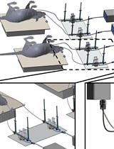
Catheterization of Pulmonary and Carotid Arteries for Concurrent Measurement of Mean Pulmonary and Systemic Arterial Pressure in Rat Models of Pulmonary Arterial Hypertension

肺动脉和颈动脉导管同时测量肺动脉高压大鼠模型的平均肺动脉压和系统动脉压

TS Tanoy Sarkar
AI Ayman Isbatan
SM Sakib M. Moinuddin
JC Jiwang Chen
FA Fakhrul Ahsan
1224 Views
Aug 20, 2023
Pulmonary hypertension (PH) is a group of pulmonary vascular disorders in which mean pulmonary arterial pressure (mPAP) becomes abnormally high because of various pathological conditions, including remodeling of the pulmonary arteries, lung and heart disorders, or congenital conditions. Various animal models, including mouse and rat models, have been used to recapitulate elevated mPAP observed in PH patients. However, the measurement and recording of mPAP and mean systemic arterial pressure (mSAP) in small animals require microsurgical procedures and a sophisticated data acquisition system. In this paper, we describe the surgical procedures for right heart catheterizations (RHC) to measure mPAP in rats. We also explain the catheterization of the carotid artery for simultaneous measurement of mPAP and mSAP using the PowerLab Data Acquisition system. We enumerate the surgical steps involved in exposing the jugular vein and the carotid artery for catheterizing these two blood vessels. We list the tools used for microsurgery in rats, describe the methods for preparing catheters, and illustrate the process for inserting the catheters in the pulmonary and carotid arteries. Finally, we delineate the steps involved in the calibration and setup of the PowerLab system for recording both mPAP and mSAP. This is the first protocol wherein we meticulously explain the surgical procedures for RHC in rats and the recording of mPAP and mSAP. We believe this protocol will be essential for PH research. Investigators with little training in animal handling can reproduce this microsurgical procedure for RHC in rats and measure mPAP and mSAP in rat models of PH. Further, this protocol is likely to help master RHC in rats that are performed for other conditions, such as heart failure, congenital heart disease, heart valve disorders, and heart transplantation.

微生物学

基于质谱的脂质组学、脂质生物能学和用于人类细胞中脂质分析和定量的网络工具

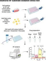
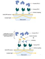
Mass Spectrometry-based Lipidomics, Lipid Bioenergetics, and Web Tool for Lipid Profiling and Quantification in Human Cells

基于质谱的脂质组学、脂质生物能学和用于人类细胞中脂质分析和定量的网络工具

LC Liang Cui
MY Meisam Yousefi
XY Xin Yap
CK Clara W.T. Koh
KT Kwan Sing Leona Tay
YO Yaw Shin Ooi
KC Kuan Rong Chan
2102 Views
Aug 20, 2023
Lipids can play diverse roles in metabolism, signaling, transport across membranes, regulating body temperature, and inflammation. Some viruses have evolved to exploit lipids in human cells to promote viral entry, fusion, replication, assembly, and energy production through fatty acid beta-oxidation. Hence, studying the virus–lipid interactions provides an opportunity to understand the biological processes involved in the viral life cycle, which can facilitate the development of antivirals. Due to the diversity and complexity of lipids, the assessment of lipid utilization in infected host cells can be challenging. However, the development of mass spectrometry, bioenergetics profiling, and bioinformatics has significantly advanced our knowledge on the study of lipidomics. Herein, we describe the detailed methods for lipid extraction, mass spectrometry, and assessment of fatty acid oxidation on cellular bioenergetics, as well as the bioinformatics approaches for detailed lipid analysis and utilization in host cells. These methods were employed for the investigation of lipid alterations in TMEM41B- and VMP1-deficient cells, where we previously found global dysregulations of the lipidome in these cells. Furthermore, we developed a web app to plot clustermaps or heatmaps for mass spectrometry data that is open source and can be hosted locally or at https://kuanrongchan-lipid-metabolite-analysis-app-k4im47.streamlit.app/. This protocol provides an efficient step-by-step methodology to assess lipid composition and usage in host cells.Graphical overview
酸水解-高效液相色谱法测定集胞藻PCC 6803中聚3-羟基丁酸酯的含量

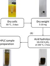
Determination of Poly(3-hydroxybutyrate) Content in Cyanobacterium Synechocystis sp. PCC 6803 Using Acid Hydrolysis Followed by High-performance Liquid Chromatography

酸水解-高效液相色谱法测定集胞藻PCC 6803中聚3-羟基丁酸酯的含量

JK Janine Kaewbai-ngam
Aran   Incharoensakdi Aran Incharoensakdi
TM Tanakarn Monshupanee
1258 Views
Aug 20, 2023
Various photoautotrophic cyanobacteria accumulate intracellular poly(3-hydroxybutyrate) (PHB) granules. This protocol can be used for determining the PHB contents of the cells as % PHB weight per dry cell weight using acid hydrolysis followed by high-performance liquid chromatography (HPLC). This HPLC analysis is rapid, with a running time of approximately 5 min per sample. The technique can accurately determine PHB concentrations in the range of 2–1,000 μg/mL PHB. However, this technique is not applicable for determining the contents of poly(3-hydroxybutyrate-co-3-hydroxyvalerate) in cyanobacteria.

分子生物学

利用嘌呤霉素结合对停滞的核糖体进行体外分析

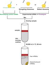
In Vitro Analysis of Stalled Ribosomes using Puromycin Incorporation

利用嘌呤霉素结合对停滞的核糖体进行体外分析

MS MaKenzie R. Scarpitti
MK Michael G. Kearse
2334 Views
Aug 20, 2023
Ribosome footprint profiling has demonstrated that ribosomes can be slowed or stalled on select mRNAs, often due to the presence of rare codons, stalling motifs, or via a ribosome-binding protein (e.g., FMRP). Stalled ribosomes can act as physical roadblocks for trailing ribosomes and ultimately can cause ribosome collisions that stimulate no-go mRNA decay. Detecting stalled or slowed ribosomes in cells by ribosome footprint profiling or classic polysome profiling is laborious, technically challenging, and low throughput. Here, we present a protocol to assay for stalled ribosomes on in vitro–transcribed reporter mRNAs using a robust, commercially available mammalian in vitro translation lysate and an optimized low-speed sucrose cushion. In short, we take advantage of the ability of puromycin to incorporate into the nascent polypeptide and cause the ribosome to dissociate from the mRNA during active elongation, as well as the ability to selectively pellet ribosomes through a low-speed sucrose cushion due to their large molecular weight. Stalled ribosomes are not actively elongating and do not incorporate puromycin, allowing the ribosome-bound mRNA to pellet in the low-speed sucrose cushion. RT-qPCR is used to quantify the amount of ribosome-bound reporter mRNA in the pellet. This workflow allows for direct assessment of stalled ribosomes and is fully amendable to insertion of putative stalling motifs in the target mRNA, as well as supplementation with recombinant proteins or small molecule inhibitors that target translation elongation.Key features• This protocol is optimized for cap-dependent in vitro translation in the dynamic linear range.• Details for generating capped reporter mRNA in one day are provided.• Requires as little as one day to complete if starting with in vitro–transcribed mRNA.• This protocol requires access to an ultracentrifuge and a real-time PCR system.Graphical overview

神经科学

小鼠视网膜研究中骨髓细胞特征的多色流式细胞术方案

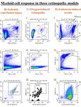
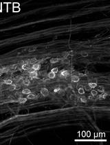
Multi-color Flow Cytometry Protocol to Characterize Myeloid Cells in Mouse Retina Research

小鼠视网膜研究中骨髓细胞特征的多色流式细胞术方案

WX Wei Xiao
RS Rami A. Shahror
CM Carol A. Morris
RC Ruth B. Caldwell
AF Abdelrahman Y. Fouda
2160 Views
Aug 20, 2023
Myeloid cells, specifically microglia and macrophages, are activated in retinal diseases and can improve or worsen retinopathy outcomes based on their inflammatory phenotype. However, assessing the myeloid cell response after retinal injury in mice remains challenging due to the small tissue size and the challenges of distinguishing microglia from infiltrating macrophages. In this protocol paper, we describe a flow cytometry–based protocol to assess retinal microglia/macrophage and their inflammatory phenotype after injury. The protocol is amenable to the incorporation of other markers of interest to other researchers.Key features• This protocol describes a flow cytometry–based method to analyze the myeloid cell response in retinopathy mouse models.• The protocol can distinguish between microglia- and monocyte-derived macrophages.• It can be modified to incorporate markers of interest.We show representative results from three different retinopathy models, namely ischemia-reperfusion injury, endotoxin-induced uveitis, and oxygen-induced retinopathy.
利用荧光报告蛋白和 ImageJ 软件检测和量化培养细胞内质网和细胞质中的钙离子

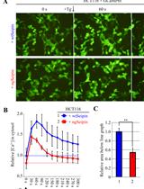
Detection and Quantification of Calcium Ions in the Endoplasmic Reticulum and Cytoplasm of Cultured Cells Using Fluorescent Reporter Proteins and ImageJ Software

利用荧光报告蛋白和 ImageJ 软件检测和量化培养细胞内质网和细胞质中的钙离子

SS Shunsuke Saito
KM Kazutoshi Mori
2130 Views
Aug 20, 2023
This protocol describes a method for detecting and quantifying calcium ions in the endoplasmic reticulum (ER) and cytoplasm of cultured cells using fluorescent reporter proteins and ImageJ software. Genetically engineered fluorescent reporter proteins, such as G-CEPIA1er and GCaMP6f, localize to intracellular regions of interest (i.e., ER and cytoplasm) and emit green fluorescence upon binding to calcium ions. In this way, the fluorescence brightness of cells transfected with expression vectors for these reporters reflects the calcium ion concentration in each intracellular region. Here, we describe procedures for observing cultured cells expressing these fluorescent reporters under a fluorescence microscope, analyzing the obtained image using the free image analysis software ImageJ (https://imagej.net/ij/index.html), and determining the average fluorescence brightness of multiple cells present in the image. The current method allows us to quickly and easily quantify calcium ions on an image containing multiple cells and to determine whether there are relative differences in intracellular calcium ion concentration among experiments with different conditions.Key features• Detection and quantification of calcium ions in the ER and cytoplasm using fluorescent reporter proteins• Quick and easy verification of measurement results using ImageJ• Simultaneous comparison between various experimental conditions (drug treatment, mutants, etc.)
Harpegnathos saltator蚂蚁群落中的种姓转换和回归

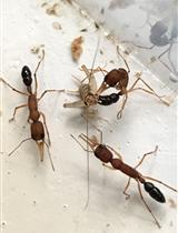
Caste Transition and Reversion in Harpegnathos saltator Ant Colonies

Harpegnathos saltator蚂蚁群落中的种姓转换和回归

CO Comzit Opachaloemphan
FC Francisco Carmona-Aldana
HY Hua Yan
977 Views
Aug 20, 2023
Living organisms possess the ability to respond to environmental cues and adapt their behaviors and physiologies for survival. Eusocial insects, such as ants, bees, wasps, and termites, have evolved advanced sociality: living together in colonies where individuals innately develop into reproductive and non-reproductive castes. These castes exhibit remarkably distinct behaviors and physiologies that support their specialized roles in the colony. Among ant species, Harpegnathos saltator females stand out with their highly plastic caste phenotypes that can be easily manipulated in a laboratory environment. In this protocol, we provide detailed instructions on how to generate H. saltator ant colonies, define castes based on behavioral and physiological phenotypes, and experimentally induce caste switches, including the transition from a non-reproductive worker to a reproductive gamergate and vice versa (known as reversion). The unusual features of H. saltator make it a valuable tool to investigate cellular and molecular mechanisms underlying phenotypic plasticity in eusocial organisms.Key features• H. saltator is one of few ant species showing remarkable caste plasticity with striking phenotypic changes, being a useful subject for studying behavioral plasticity.• Caste switches in H. saltator can be easily manipulated in a controlled laboratory environment by controlling the presence of reproductive females in a colony.• The relatively large size of H. saltator females allows researchers to dissect various tissues of interest and conduct detailed phenotypic analyses.
蜜蜂摇摆舞社会信号学习的研究方法

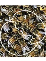
A Method for Studying Social Signal Learning of the Waggle Dance in Honey Bees

蜜蜂摇摆舞社会信号学习的研究方法

SD Shihao Dong
TL Tao Lin
JN James C. Nieh
KT Ken Tan
932 Views
Aug 20, 2023
Honey bees use a complex form of spatial referential communication. Their waggle dance communicates to nestmates the direction, distance, and quality of a resource by encoding celestial cues, retinal optic flow, and relative food value into motion and sound within the nest. This protocol was developed to investigate the potential for social learning of this waggle dance. Using this protocol, we showed that correct waggle dancing requires social learning. Bees (Apis mellifera) that did not follow any dances before they first danced produced significantly more disordered dances, with larger waggle angle divergence errors, and encoded distance incorrectly. The former deficits improved with experience, but distance encoding was set for life. The first dances of bees that could follow other dancers had none of these impairments. Social learning, therefore, shapes honey bee signaling, as it does communication in human infants, birds, and multiple other vertebrate species. However, much remains to be learned about insects’ social learning, and this protocol will help to address knowledge gaps in the understanding of sophisticated social signal learning, particularly in understanding the molecular bases for such learning.Key features• It was unclear if honey bees (Apis mellifera) could improve their waggle dance by following experienced dancers before they first waggle dance.• Honey bees perform their first waggle dances with more errors if they cannot follow experienced waggle dancers first.• Directional and disorder errors improved over time, but distance error was maintained. Bees in experimental colonies continued to communicate longer distances than control bees.• Dancing correctly, with less directional error and disorder, requires social learning.• Distance encoding in the honey bee dance is largely genetic but may also include a component of cultural transmission.

植物科学

拟南芥中硝酸盐的荧光生物传感器成像

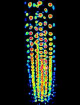
Fluorescent Biosensor Imaging of Nitrate in Arabidopsis thaliana

拟南芥中硝酸盐的荧光生物传感器成像

YC Yen-Ning Chen
CH Cheng-Hsun Ho
2583 Views
Aug 20, 2023
Nitrate (NO3–) is an essential element and nutrient for plants and animals. Despite extensive studies on the regulation of nitrate uptake and downstream responses in various cells, our knowledge of the distribution of nitrogen forms in different root cell types and their cellular compartments is still limited. Previous physiological models have relied on in vitro biochemistry and metabolite level analysis, which limits the ability to differentiate between cell types and compartments. Here, to address this, we report a nuclear-localized, genetically encoded fluorescent biosensor, which we named nlsNitraMeter3.0, for the quantitative visualization of nitrate concentration and distribution at the cellular level in Arabidopsis thaliana. This biosensor was specifically designed for nitrate measurements, not nitrite. Through genetic engineering to create and select sensors using yeast, Xenopus oocyte, and Arabidopsis expression systems, we developed a reversible and highly specific nitrate sensor. This method, combined with fluorescence imaging systems such as confocal microscopy, allows for the understanding and monitoring of nitrate transporter activity in plant root cells in a minimally invasive manner. Furthermore, this approach enables the functional analysis of nitrate transporters and the measurement of nitrate distribution in plants, providing a valuable tool for plant biology research. In summary, we provide a protocol for sensor development and a biosensor that can be used to monitor nitrate levels in plants.Key features• This protocol builds upon the concept of FRET biosensors for in vivo visualization of spatiotemporal nitrate levels at a cellular resolution.• Nitrate levels can be quantified utilizing the biosensor in conjunction with either a plate reader or a fluorescence microscope.Graphical overview
拟南芥灰葡萄孢生长的定量研究

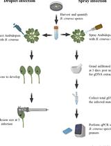
Quantification of Botrytis cinerea Growth in Arabidopsis thaliana

拟南芥灰葡萄孢生长的定量研究

PS Patricia Scholz
KC Kent D. Chapman
TI Till Ischebeck
AG Athanas Guzha
1978 Views
Aug 20, 2023
Yield losses attributed to plant pathogens pose a serious threat to plant productivity and food security. Botrytis cinerea is one of the most devastating plant pathogens, infecting a wide array of plant species; it has also been established as a model organism to study plant–pathogen interactions. In this context, development of different assays to follow the relative success of B. cinerea infections is required. Here, we describe two methods to quantify B. cinerea development in Arabidopsis thaliana genotypes through measurements of lesion development and quantification of fungal genomic DNA in infected tissues. This provides two independent techniques that are useful in assessing the susceptibility or tolerance of different Arabidopsis genotypes to B. cinerea.Key features• Protocol for the propagation of the necrotrophic plant pathogen fungus Botrytis cinerea and spore production.• Two methods of Arabidopsis thaliana infection with the pathogen using droplet and spray inoculation.• Two readouts, either by measuring lesion size or by the quantification of fungal DNA using quantitative PCR.• The two methods are applicable across plant species susceptible the B. cinerea.Graphical overviewA simplified overview of the droplet and spray infection methods used for the determination of B. cinerea growth in different Arabidopsis genotypes
拟南芥果胶单糖的GC–MS分析

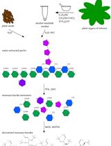
Analysis of Pectin-derived Monosaccharides from Arabidopsis Using GC–MS

拟南芥果胶单糖的GC–MS分析

PS Patricia Scholz
KC Kent D. Chapman
TI Till Ischebeck
AG Athanas Guzha
1269 Views
Aug 20, 2023
Pectin is a complex polysaccharide present in the plant cell wall, whose composition is constantly remodelled to adapt to environmental or developmental changes. Mutants with altered pectin composition have been reported to exhibit altered stress or pathogen resistance. Understanding the link between mutant phenotypes and their pectin composition requires robust analytical methods to detect changes in the relative monosaccharide composition. Here, we describe a quick and efficient gas chromatography–mass spectrometry (GC–MS)-based method that allows the differential analysis of pectin monosaccharide composition in plants under different conditions or between mutant plants and their respective wild types. Pectin is extracted from seed mucilage or from the alcohol-insoluble residue prepared from leaves or other organs and is subsequently hydrolysed with trifluoracetic acid. The resulting acidic and neutral monosaccharides are then derivatised and measured simultaneously by GC–MS.Key features• Comparative analysis of monosaccharide content in Arabidopsis-derived pectin between different genotypes or different treatments.• Procedures for two sources of pectin are shown: seed coat mucilage and alcohol-insoluble residue.• Allows quick analyses of neutral and acidic monosaccharides simultaneously.Graphical overview
使用平板阅读器测定植物 NADP-苹果酸脱氢酶活性的半通量程序

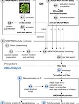
A Semi-throughput Procedure for Assaying Plant NADP-malate Dehydrogenase Activity Using a Plate Reader

使用平板阅读器测定植物 NADP-苹果酸脱氢酶活性的半通量程序

KB Kevin Baudry
EI Emmanuelle Issakidis-Bourguet
902 Views
Aug 20, 2023
Chloroplast NADP-dependent malate dehydrogenase (NADP-MDH) is a redox regulated enzyme playing an important role in plant redox homeostasis. Leaf NADP-MDH activation level is considered a proxy for the chloroplast redox status. NADP-MDH enzyme activity is commonly assayed spectrophotometrically by following oxaloacetate-dependent NADPH oxidation at 340 nm. We have developed a plate-adapted protocol to monitor NADP-MDH activity allowing faster data production and lower reagent consumption compared to the classic cuvette format of a spectrophotometer. We provide a detailed procedure to assay NADP-MDH activity and measure the enzyme activation state in purified protein preparations or in leaf extracts. This protocol is provided together with a semi-automatized data analysis procedure using an R script.
用醋酸洋红和苏木精染色法观察丝状绿藻Zynema(轮藻门)染色体的改进方法

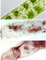
Improved Methods for Acetocarmine and Haematoxylin Staining to Visualize Chromosomes in the Filamentous Green Alga Zygnema (Charophyta)

用醋酸洋红和苏木精染色法观察丝状绿藻Zynema(轮藻门)染色体的改进方法

NR Nina Rittmeier
AH Andreas Holzinger
890 Views
Aug 20, 2023
Genome sizes of Zygnema spp. vary greatly, being unknown whether polyploidization occurred. The exact number of chromosomes in this genus is unknown since counting methods established for higher plants cannot be applied to green algae. The massive presence of pectins and arabinogalactan proteins in the cell wall interferes with the uptake of staining solutions; moreover, cell divisions in green algae are not restricted to meristems as in higher plants, which is another limiting factor. Cell divisions occur randomly in the thallus, due to the intercalary growth of algal filaments. Therefore, we increased the number of cell divisions via synchronization by changing the light cycle (10:14 h light/dark). The number of observed mitotic stages peaked at the beginning of the dark cycle. This protocol describes two methods for the visualization of chromosomes in the filamentous green alga Zygnema. Existing protocols were modified, leading to improved acetocarmine and haematoxylin staining methods as investigated by light microscopy. A freeze-shattering approach with liquid nitrogen was applied to increase the accessibility of the haematoxylin dye. These modified protocols allowed reliable chromosome counting in the genus Zygnema.Key features• Improved method for chromosome staining in filamentous green algae.• Optimized for the Zygnema strains SAG 698-1a (Z. cylindricum), SAG 698-1b (Z. circumcarinatum), and SAG 2419 (Zygnema ‘Saalach’).• This protocol builds upon the methods of chromosomal staining in green algae developed by Wittmann (1965), Staker (1971), and Fujii and Guerra (1998).• Cultivation and synchronization: 14 days; fixation and permeabilization: 24 h; staining: 1 h; image analysis and chromosome number quantification: up to 20 h.

干细胞

人肾脏祖细胞培养物的制备及其向足细胞的分化

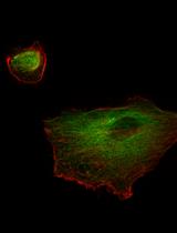
Preparation of Human Kidney Progenitor Cultures and Their Differentiation into Podocytes

人肾脏祖细胞培养物的制备及其向足细胞的分化

MM Maria Elena Melica
MA Maria Lucia Angelotti
GA Giulia Antonelli
AP Anna J. Peired
CC Carolina Conte
LD Letizia De Chiara
BM Benedetta Mazzinghi
EL Elena Lazzeri
LL Laura Lasagni
PR Paola Romagnani
1610 Views
Aug 20, 2023
Kidney diseases are a global health concern. Modeling of kidney disease for translational research is often challenging because of species specificities or the postmitotic status of kidney epithelial cells that make primary cultures, for example podocytes. Here, we report a protocol for preparing primary cultures of podocytes based on the isolation and in vitro propagation of immature kidney progenitor cells subsequently differentiated into mature podocytes. This protocol can be useful for studying physiology and pathophysiology of human kidney progenitors and to obtain differentiated podocytes for modeling podocytopathies and other kidney disorders involving podocytes.Graphical overview

更正

更正:基于表面等离子体共振(SPR)生物传感器的人PD-1/PD-L1阻断检测方法的建立

Correction Notice: Establishment of Human PD-1/PD-L1 Blockade Assay Based on Surface Plasmon Resonance (SPR) Biosensor

更正:基于表面等离子体共振(SPR)生物传感器的人PD-1/PD-L1阻断检测方法的建立

TP Tess Puopolo
hL Huifang Li
JG Justin Gutkowski
AC Ang Cai
NS Navindra P Seeram
HM Hang Ma
CL Chang Liu
541 Views
Aug 20, 2023