往期刊物2023

卷册: 13, 期号: 2

left

Dec

20

Dec

5

Nov

20

Nov

5

Oct

20

Oct

5

Sep

20

Sep

5

Aug

20

Aug

5

Jul

20

Jul

5

Jun

20

Jun

5

May

20

May

5

Apr

20

Apr

5

Mar

20

Mar

5

Feb

20

Feb

5

Jan

20

Jan

5

right

生物物理学

裂解液到网格:从出芽酵母中快速分离天然复合物用于低温冷冻电镜成像

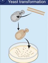
Lysate-to-grid: Rapid Isolation of Native Complexes from Budding Yeast for Cryo-EM Imaging

裂解液到网格:从出芽酵母中快速分离天然复合物用于低温冷冻电镜成像

IC Ian Cooney
DM Deirdre C. Mack
AF Aaron J. Ferrell
MS Michael G. Stewart
SW Shuxin Wang
HD Helen M. Donelick
DT Daniela Tamayo-Jaramillo
DG Dakota L. Greer
DZ Danyang Zhu
WL Wenyan Li
PS Peter S. Shen
1914 Views
Jan 20, 2023
Single-particle electron cryo-microscopy (cryo-EM) is an effective tool to determine high-resolution structures of macromolecular complexes. Its lower requirements for sample concentration and purity make it an accessible method to determine structures of low-abundant protein complexes, such as those isolated from native sources. While there are many approaches to protein purification for cryo-EM, attaining suitable particle quality and abundance is generally the major bottleneck to the typical single-particle project workflow. Here, we present a protocol using budding yeast (S. cerevisiae), in which a tractable immunoprecipitation tag (3xFLAG) is appended at the endogenous locus of a gene of interest (GOI). The modified gene is expressed under its endogenous promoter, and cells are grown and harvested using standard procedures. Our protocol describes the steps in which the tagged proteins and their associated complexes are isolated within three hours of thawing cell lysates, after which the recovered proteins are used directly for cryo-EM specimen preparation. The prioritization of speed maximizes the ability to recover intact, scarce complexes. The protocol is generalizable to soluble yeast proteins that tolerate C-terminal epitope tags. Graphical abstract Overview of lysate-to-grid workflow. Yeast cells are transformed to express a tractable tag on a gene of interest. Following cell culture and lysis, particles of interest are rapidly isolated by co-immunoprecipitation and prepared for cryo-EM imaging (created with BioRender.com).

发育生物学

一种可靠的和一致的方法来量化黑腹果蝇的致死率和寿命

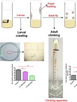
A Reliable and Consistent Method to Quantify Percent Lethality and Life Span in Drosophila melanogaster

一种可靠的和一致的方法来量化黑腹果蝇的致死率和寿命

PK Priyanka Kumari
UA Ushashi Ain
HF Hena Firdaus
1399 Views
Jan 20, 2023
Drosophila melanogaster is a classic model organism to study gene function as well as toxicological effects. To study gene function, the expression of a particular gene of interest is disrupted by using the widely explorable Drosophila genetic toolkit, whereas to study toxicological effects the flies are exposed to a particular toxicant through diet. These experiments often require the quantification of lethality from embryonic to adult stages, as well as the assessment of the life span in order to check the role of the gene/toxicant of interest in Drosophila. Here, we propose an experimental protocol that enables a consistent and rigorous assessment of lethality and life span of cadmium chloride (CdCl2)–exposed or genetically perturbed flies [downregulation and overexpression of the cytosolic Cu, Zn superoxide dismutase (SOD1) gene], consecutively. The protocol insists upon the requirement of one single experimental setup that is unique, distinctive, and cost-effective as it engages minimal laboratory equipment and resources. The described methods lead to the smooth observation of the embryos, their successive stagewise transition, and life span of the adult flies post eclosion. Additionally, these methods also facilitate the assessment of crawling and climbing behavioral parameters of the larvae and adults, respectively, and allow the calculation of lethal concentration (LC50) for the mentioned toxicant as well as median survival of the flies, which can be a determining factor in proceeding with further stages of experiments.Graphical abstract

药物发现

 在tau蛋白病变中检测自噬-靶向嵌合体(AUTOTAC)介导的靶向蛋白降解的方法

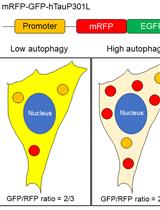
Methods to Detect AUTOphagy-Targeting Chimera (AUTOTAC)-mediated Targeted Protein Degradation in Tauopathies

在tau蛋白病变中检测自噬-靶向嵌合体(AUTOTAC)介导的靶向蛋白降解的方法

ML Min Ju Lee
SK Su Bin Kim
HK Hee Yeon Kim
SL Su Jin Lee
JL Ji Su Lee
YK Yong Tae Kwon
CJ Chang Hoon Ji
1859 Views
Jan 20, 2023
Targeted protein degradation (TPD) facilitates the selective elimination of unwanted and pathological cellular cargoes via the proteasome or the lysosome, ranging from proteins to organelles and pathogens, both within and outside the cell. Currently, there are several in vitro and in vivo protocols that assess the degradative potency of a given degrader towards a myriad of targets, most notably soluble, monomeric oncoproteins. However, there is a clear deficiency of methodologies to assess the degradative potency of heterobifunctional chimeric degraders, especially those in the autophagy space, against pathological, mutant tau species, such as detergent-insoluble oligomers and high-molecular aggregates. The protocol below describes both in vitro and in vivo biochemical assays to induce tau aggregation, as well as to qualitatively and quantitatively measure the degradative potency of a given degrader towards said aggregates, with specific applications of the AUTOTAC (AUTOphagy-TArgeting Chimera) platform provided as an example. A well-defined set of methodologies to assess TPD-mediated degradation of pathological tau species will help expand the scope of the TPD technology to neurodegeneration and other proteinopathies, in both the lab and the clinic.Graphical abstractOverview of assays observing elimination of tauP301L aggregates with AUTOTAC. (A) Description of the biological working mechanism of heterobifunctional chimeric AUTOTAC degraders. (B) Schematic illustration of assays described in this paper.

分子生物学

DNA组装的GRS-DNA-CuS纳米蒲公英的制备和表征

Preparation and Characterization of DNA-assembled GRS-DNA-CuS Nanodandelions

DNA组装的GRS-DNA-CuS纳米蒲公英的制备和表征

HW Haoze Wang
YC Yanna Cui
YZ Yongming Zhang
ZX Zeyu Xiao
965 Views
Jan 20, 2023
In this study, we introduce a detailed protocol for the preparation of DNA-assembled GRS-DNA-copper sulfide (CuS) nanodandelion, a multifunctional theranostics nanoparticle. Using transmission electron microscope (TEM) and dynamic light scattering techniques, we characterize the physicochemical property of DNA-assembled GRS-DNA-CuS nanodandelions and their dissociation property after the first near-infrared (NIR) light irradiation. In addition, we systematically monitor the processes of tumor accumulation and uniform intratumoral distribution (UITD) of ultrasmall CuS photothermal agents (PAs), which are dissociated from GRS-DNA-CuS nanodandelions, by Raman imaging and photoacoustic imaging, respectively. The UITD of the dissociated ultrasmall CuS PAs can enhance the therapeutic efficiency of photothermal treatment under the second NIR light irradiation. Overall, this protocol provides a powerful tool to achieve UITD of PAs by explosively breaking the hydrogen bonds of DNA in GRS-DNA-CuS nanodandelions under NIR light irradiation. We expect DNA-assembled nanotheranostics to serve as a robust platform for a variety of biomedical applications related to photothermal therapy in the oncology field. This protocol can increase experimental reproducibility and contribute to efficient theranostics nanomedicine.

神经科学

束缚应激诱导的厌食症和社会隔离诱导的厌食症小鼠模型的建立

Establishment of Restraint Stress–induced Anorexia and Social Isolation–induced Anorexia Mouse Models

束缚应激诱导的厌食症和社会隔离诱导的厌食症小鼠模型的建立

IP Ingrid Camila Possa-Paranhos
KC Kerem Catalbas
JB Jared Butts
KO Kyle O’Berry
PS Patrick Sweeney
1243 Views
Jan 20, 2023
Anorexia nervosa (AN) is a devastating neuropsychiatric disease with a prevalence rate of approximately 0.3%–1% among women and morbidity and mortality rates among the highest of all neuropsychiatric disorders. The disease etiology is complex but primarily characterized by reduced food intake and body weight, and intense anxiety and fear associated with gaining weight. Existing rodent models of AN are useful and capture features of the disease, but either require specialized genetic mouse models or are difficult to implement in mice. Here, we describe two simple mouse models of stress-induced anorexia that are easy to implement in basic research labs, and capture core features associated with AN, such as reduced food intake in the context of social/physical stress and increased anxiety-related behavior. These protocols provide reproducible and robust assays for stress-induced anorexia and may be implemented with additional assays to probe the neural circuitry mediating the effects of psychological stress on feeding in mice.Graphical abstract
利用共聚焦荧光显微镜对果蝇大脑溶酶体压力的体内评估

In vivo Assessment of Lysosomal Stress in the Drosophila Brain Using Confocal Fluorescence Microscopy

利用共聚焦荧光显微镜对果蝇大脑溶酶体压力的体内评估

FM Felipe Martelli
1082 Views
Jan 20, 2023
Lysosomes play a central role in signaling, nutrient sensing, response to stress, and the degradation and recycling of cellular content. Defects in lysosomal digestive enzymes or structural components can impair lysosomal function with dire consequences to the cell, such as neurodegeneration. A number of methods exist to assess lysosomal stress in the model Drosophila, such as specific driver and reporter strains, transmission electron microscopy, and the investigation of gene expression. These methods, however, can be time consuming and, in some cases, costly. The procedure described here provides a quick, reliable, and low-cost approach to measure lysosomal stress in the Drosophila brain. Using fluorescence confocal microscopy and the LysoTracker staining, this protocol allows for the direct measurement of lysosome size and number. This method can be used to assess lysosomal stress under a number of different genetic and environmental scenarios in the Drosophila brain.

植物科学

用树莓派和延时摄影评估植物抗旱性

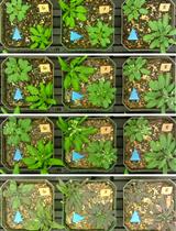
Evaluating Plant Drought Resistance with a Raspberry Pi and Time-lapse Photography

用树莓派和延时摄影评估植物抗旱性

Daniel N. Ginzburg Daniel N. Ginzburg
SR Seung Y. Rhee
1968 Views
Jan 20, 2023
Identifying genetic variations or treatments that confer greater resistance to drought is paramount to ensuring sustainable crop productivity. Accurate and reproducible measurement of drought stress symptoms can be achieved via automated, image-based phenotyping. Many phenotyping platforms are either cost-prohibitive, require specific technical expertise, or are simply more complex than necessary to effectively evaluate drought resistance. Certain mutations, allelic variations, or treatments result in plants that constitutively use less water. To accurately identify genetic differences or treatments that confer a drought phenotype, plants from all experimental groups must be subjected to equal levels of drought stress. This can be easily achieved by growing and imaging plants that are grown in the same pot. Here, we provide a detailed protocol to configure a Raspberry Pi computer and camera module to image seedlings of multiple genotypes growing in shared pots and to transfer images and metadata via the cloud for downstream analyses. Also detailed is a method to calculate percent soil water content of pots while being imaged to allow for comparison of stress symptoms with water availability. This protocol was recently used to uncouple differential water usage from drought resistance in a dwarf Arabidopsis thaliana mutant chiquita1-1/cost1 compared to the wild-type control. It is cost effective, suitable for any plant species, customizable to various biological questions, and requires no prior experience with electronics or basic software programming.
用相关光电子显微镜的方法确定拟南芥嫁接界面的超微结构单元

Targeting Ultrastructural Events at the Graft Interface of Arabidopsis thaliana by A Correlative Light Electron Microscopy Approach

用相关光电子显微镜的方法确定拟南芥嫁接界面的超微结构单元

CC Clément Chambaud
SC Sarah J. Cookson
NO Nathalie Ollat
AB Amélie Bernard
LB Lysiane Brocard
1793 Views
Jan 20, 2023
Combining two different plants together through grafting is one of the oldest horticultural techniques. In order to survive, both partners must communicate via the formation of de novo connections between the scion and the rootstock. Despite the importance of grafting, the ultrastructural processes occurring at the graft interface remain elusive due to the difficulty of locating the exact interface at the ultrastructural level. To date, only studies with interfamily grafts showing enough ultrastructural differences were able to reliably localize the grafting interface at the ultrastructural level under electron microscopy. Thanks to the implementation of correlative light electron microscopy (CLEM) approaches where the grafted partners were tagged with fluorescent proteins of different colors, the graft interface was successfully and reliably targeted. Here, we describe a protocol for CLEM for the model plant Arabidopsis thaliana, which unambiguously targets the graft interface at the ultrastructural level. Moreover, this protocol is compatible with immunolocalization and electron tomography acquisition to achieve a three-dimensional view of the ultrastructural events of interest in plant tissues.Graphical abstract

干细胞

来自骨髓增生性肿瘤患者的造血祖细胞的血小板生成素不依赖性巨核细胞分化

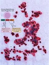
Thrombopoietin-independent Megakaryocyte Differentiation of Hematopoietic Progenitor Cells from Patients with Myeloproliferative Neoplasms

来自骨髓增生性肿瘤患者的造血祖细胞的血小板生成素不依赖性巨核细胞分化

CT Chloe A. L. Thompson-Peach
JF Johannes Foßelteder
AR Andreas Reinisch
DT Daniel Thomas
1299 Views
Jan 20, 2023
Primary hematopoietic stem and progenitor cell (HSPC)-derived megakaryocytes are a valuable tool for translational research interrogating disease pathogenesis and developing new therapeutic avenues for patients with hematologic disorders including myeloproliferative neoplasms (MPNs). Thrombopoietin (TPO)-independent proliferation and megakaryocyte differentiation play a central role in the pathogenesis of essential thrombocythemia and myelofibrosis, two MPN subtypes that are characterized by increased numbers of bone marrow megakaryocytes and somatic mutations in either JAK2, CALR, or MPL. However, current culture strategies generally use healthy HSPCs for megakaryocyte production and are not optimized for the investigation of TPO-independent or TPO-hypersensitive growth and megakaryocyte-directed differentiation of primary patient–derived HSPCs. Here, we describe a detailed protocol covering all necessary steps for the isolation of CD34+ HSPCs from the peripheral blood of MPN patients and the subsequent TPO-independent differentiation into CD41+ megakaryocytes using both a collagen-based colony assay and a liquid culture assay. This protocol provides a novel, reproducible, and cost-effective approach for investigating megakaryocyte growth and differentiation properties from primary MPN patient cells that can be easily adapted for research on other megakaryocyte-related disorders.Graphical abstractSchematic representation of the isolation of CD34+ progenitor cells and subsequent TPO-independent megakaryocyte differentiation

系统生物学

组织培养中CRISPRi sgRNAs的快速多通道流式细胞术验证

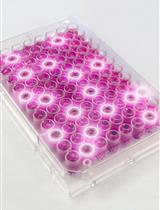
Rapid Multiplexed Flow Cytometric Validation of CRISPRi sgRNAs in Tissue Culture

组织培养中CRISPRi sgRNAs的快速多通道流式细胞术验证

JC John S. Chorba
VX Vivian Q. Xia
GS Geoffrey A. Smith
AP Arun Padmanabhan
1494 Views
Jan 20, 2023
Genome-wide CRISPR-based screening is a powerful tool in forward genetics, enabling biologic discovery by linking a desired phenotype to a specific genetic perturbation. However, hits from a genome-wide screen require individual validation to reproduce and accurately quantify their effects outside of a pooled experiment. Here, we describe a step-by-step protocol to rapidly assess the effects of individual sgRNAs from CRISPR interference (CRISPRi) and CRISPR activation (CRISPRa) systems. All steps, including cloning, lentivirus generation, cell transduction, and phenotypic readout, can be performed entirely in 96-well plates. The system is highly flexible in both cell type and selection system, requiring only that the phenotype(s) of interest be read out via flow cytometry. We expect that this protocol will provide researchers with a rapid way to sift through potential screening hits, and prioritize them for deeper analysis in more complex in vitro or even in vivo systems. Graphical abstract

更新

更新:一种综合的实验方法:利用免疫组织化学染色和分析进行蛋白表达的半定量测定

Update Notice: Semi-quantitative Determination of Protein Expression Using Immunohistochemistry Staining and Analysis

更新:一种综合的实验方法:利用免疫组织化学染色和分析进行蛋白表达的半定量测定

Alexandra R Crowe Alexandra R Crowe
Wei Yue Wei Yue
1650 Views
Jan 20, 2023