往期刊物2018

卷册: 8, 期号: 8

left

Dec

20

Dec

5

Nov

20

Nov

5

Oct

20

Oct

5

Sep

20

Sep

5

Aug

20

Aug

5

Jul

20

Jul

5

Jun

20

Jun

5

May

20

May

5

Apr

20

Apr

5

Mar

20

Mar

5

Feb

20

Feb

5

Jan

20

Jan

5

right

生物化学

海洋微生物酶法定量微藻中的昆布多糖

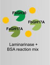
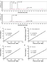
Laminarin Quantification in Microalgae with Enzymes from Marine Microbes

海洋微生物酶法定量微藻中的昆布多糖

SB Stefan Becker
Jan-Hendrik  Hehemann Jan-Hendrik Hehemann
7421 Views
Apr 20, 2018
The marine beta-glucan laminarin is an abundant storage polysaccharide in microalgae. High production rates and rapid digestion by heterotrophic bacteria turn laminarin into an ideal carbon and energy source, and it is therefore a key player in the marine carbon cycle. As a main storage glucan laminarin also plays a central role in the energy metabolism of the microalgae (Percival and Ross, 1951; Myklestad, 1974; Painter, 1983). We take advantage of enzymes that digest laminarin selectively and can thereby quantify only this polysaccharide in environmental samples. These enzymes hydrolyze laminarin into glucose and oligosaccharides, which are measured with a standard reducing sugar assay to obtain the laminarin concentration. Prior to this assay, the three enzymes need to be produced via heterologous expression and purification. The assay can be used to monitor laminarin concentrations in environmental microalgae, which were concentrated from seawater by filtering, or in samples derived from algal lab cultures.

癌症生物学

表达萤光素酶的肿瘤细胞系的生成

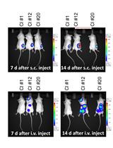
Generation of Luciferase-expressing Tumor Cell Lines

表达萤光素酶的肿瘤细胞系的生成

TB Todd V. Brennan
LL Liwen Lin
XH Xiaopei Huang
YY Yiping Yang
19072 Views
Apr 20, 2018
Murine tumor models have been critical to advances in our knowledge of tumor physiology and for the development of effective tumor therapies. Essential to these studies is the ability to both track tumor development and quantify tumor burden in vivo. For this purpose, the introduction of genes that confer tumors with bioluminescent properties has been a critical advance for oncologic studies in rodents. Methods of introducing bioluminescent genes, such as firefly luciferase, by viral transduction has allowed for the production of tumor cell lines that can be followed in vivo longitudinally over long periods of time. Here we describe methods for the production of stable luciferase expressing tumor cell lines by lentiviral transduction.
使用2-NBDG探针并基于FACS的小鼠胚胎成纤维细胞和乳腺癌细胞的葡萄糖摄取测定

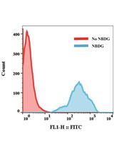
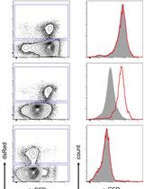
FACS-based Glucose Uptake Assay of Mouse Embryonic Fibroblasts and Breast Cancer Cells Using 2-NBDG Probe

使用2-NBDG探针并基于FACS的小鼠胚胎成纤维细胞和乳腺癌细胞的葡萄糖摄取测定

SD Shengli Dong
Suresh K Alahari Suresh K Alahari
10821 Views
Apr 20, 2018
This is a flow cytometry-based protocol to measure glucose uptake of mouse embryonic fibroblasts (MEFs) and breast cancer cells in vitro. The method is a slightly modified and updated version as previously described (Dong et al., 2017). Briefly, the target cells are incubated with the fluorescently tagged 2-(N-(7-nitrobenz-2-oxa-1,3-diazol-4-yl)amino)-2-deoxyglucose (2-NBDG) for 2 h or 30 min, and the efficiency of glucose uptake is examined using a flow cytometer. This method can be adapted to measure a variety of adipocytes, immune cells, MEFs and cancer cells.

细胞生物学

一种从染色质网络中提取核骨架的方法

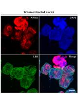
A Method for Extracting the Nuclear Scaffold from the Chromatin Network

一种从染色质网络中提取核骨架的方法

JC Junjie Chen
BT Boon Heng Dennis Teo
JL Jinhua Lu
7085 Views
Apr 20, 2018
Each cell contains many large DNA polymers packed in a nucleus of approx. 10 μm in diameter. With histones, these DNA polymers are known to form chromatins. How chromatins further compact in the nucleus is unclear but it inevitably depends on an extensive non-chromatin nuclear scaffold. Imaging of endogenous chromatin network and the complementary scaffold that support this network has not been achieved but biochemical and proteomic investigations of the scaffold can still provide important insights into this chromatin-organizing network. However, this demands highly inclusive and reproducible extraction of the nuclear scaffold. We have recently developed a simple protocol for releasing the scaffold components from chromatins. The inclusiveness of the extract was testified by the observation that, upon its extraction from the nuclei, the remaining nuclear chromatins were liberated into extended and often parallel chromatin fibers. Basically, this protocol includes the generation of pure nuclei, treatment of the nuclei with Triton X-100 to generate envelope-depleted nuclei (TxN), and extraction of the nuclei at 500 mM NaCl in a sucrose-containing buffer. This combined extract of TxN is known as TxNE.

免疫学

肿瘤相关巨噬细胞和卵巢癌细胞的3D共培养系统

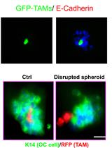
3D Co-culture System of Tumor-associated Macrophages and Ovarian Cancer Cells

肿瘤相关巨噬细胞和卵巢癌细胞的3D共培养系统

LL Lingli Long
MY Mingzhu Yin
Wang  Min Wang Min
11082 Views
Apr 20, 2018
Ovarian cancer is fairly unique in that ovarian carcinoma cells can detach and spread directly through peritoneal cavity. It has been unclear, however, how detached cancer cells survive in the peritoneum and form spheroid structure. We have recently reported that there is a strong correlation between Tumor-associated macrophages (TAMs)-associated spheroid and clinical pathology of ovarian cancer, and that TAMs promote spheroid formation and tumor growth at early stages of transcoelomic metastasis in orthotopic mouse models. We have established an in vitro spheroid formation assay using a 3D co-culture system in which mouse GFP+F4/80+CD206+ TAMs isolated from spheroids of ovarian cancer-bearing donor tomatolysM-cre mice were mixed with ID8 cells (TAM:ID8 at a ratio of 1:10) in medium containing 2% Matrigel and seeded onto the 24-well plate precoated with Matrigel. As transcoelomic metastasis is also associated with many other cancers such as pancreatic and colon cancers, TAM-mediated spheroid formation assay would provide a useful approach to define the molecular mechanism and therapeutic targets for ovarian cancer and other transcoelomic metastasis cancers.

微生物学

白色念珠菌的CRISPR/Cas9诱变方法

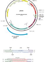
Method for CRISPR/Cas9 Mutagenesis in Candida albicans

白色念珠菌的CRISPR/Cas9诱变方法

ND Neta Dean
HN Henry Ng
12075 Views
Apr 20, 2018
Candida albicans is the most prevalent and important human fungal pathogen. The advent of CRISPR as a means of gene editing has greatly facilitated genetic analysis in C. albicans. Here, we describe a detailed step-by-step procedure to construct and analyze C. albicans deletion mutants. This protocol uses plasmids that allow simple ligation of synthetic duplex 23mer guide oligodeoxynucleotides for high copy gRNA expression in C. albicans strains that express codon-optimized Cas9. This protocol allows isolation and characterization of deletion strains within nine days.
从粪便样本中提取小分子并检测其对微生物的生理学活性

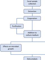
Extraction of Small Molecules from Fecal Samples and Testing of Their Activity on Microbial Physiology

从粪便样本中提取小分子并检测其对微生物的生理学活性

EA Eduardo S. Alves
RF Rosana B. R. Ferreira
L. Caetano M. Antunes L. Caetano M. Antunes
7334 Views
Apr 20, 2018
The human body is colonized by vast communities of microbes, collectively known as microbiota, or microbiome. Although microbes colonize every surface of our bodies that is exposed to the external environment, the biggest collection of microbes colonizing humans and other mammals can be found in the gastrointestinal tract. Given the fact that the human gut is colonized by several hundred microbial species, our group hypothesized that the chemical diversity of this environment should be significant, and that many of the molecules present in that environment would have important signaling roles. Therefore, we devised a protocol to extract these molecules from human feces and test their signaling properties. Potentially bioactive extracts can be tested through addition to culture medium and analyses of bacterial growth and gene expression, among other properties. The protocol described herein provides an easy and rapid method for the extraction and testing of metabolites from fecal samples using Salmonella enterica as a model organism. This protocol can also be adapted to the extraction of small molecules from other matrices, such as cultured mammalian cells, tissues, body fluids, and axenic microbial cultures, and the resulting extracts can be tested against various microbial species.
利用透射光学显微镜技术和计算机图像定量分析密集菌落中的细菌蹭行运动

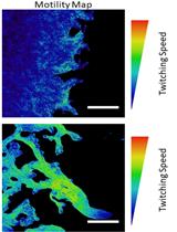
Quantification of Bacterial Twitching Motility in Dense Colonies Using Transmitted Light Microscopy and Computational Image Analysis

利用透射光学显微镜技术和计算机图像定量分析密集菌落中的细菌蹭行运动

BS Benjamin Smith
Jianfang  Li Jianfang Li
MM Matteo Metruccio
Stephanie  Wan Stephanie Wan
David Evans David Evans
SF Suzanne Fleiszig
6992 Views
Apr 20, 2018
A method was developed to allow the quantification and mapping of relative bacterial twitching motility in dense samples, where tracking of individual bacteria was not feasible. In this approach, movies of bacterial films were acquired using differential interference contrast microscopy (DIC), and bacterial motility was then indirectly quantified by the degree to which the bacteria modulated the intensity of light in the field-of-view over time. This allowed the mapping of areas of relatively high and low motility within a single field-of-view, and comparison of the total distribution of motility between samples.
使用金属标签透射电子显微技术定位酵母菌中番茄丛矮病毒复制区

Metal-tagging Transmission Electron Microscopy for Localisation of Tombusvirus Replication Compartments in Yeast

使用金属标签透射电子显微技术定位酵母菌中番茄丛矮病毒复制区

Isabel Fernández de Castro Isabel Fernández de Castro
CR Cristina Risco
6638 Views
Apr 20, 2018
Positive-stranded (+) RNA viruses are intracellular pathogens in humans, animals and plants. To build viral replicase complexes (VRCs) viruses manipulate lipid flows and reorganize subcellular membranes. Redesigned membranes concentrate viral and host factors and create an environment that facilitates the formation of VRCs within replication organelles. Therefore, efficient virus replication depends on the assembly of specialized membranes where viral macromolecular complexes are turned on and hold a variety of functions. Detailed characterization of viral replication platforms in cells requires sophisticated imaging approaches. Here we present a protocol to visualize the three-dimensional organization of the tombusvirus replicase complex in yeast with MEtal-Tagging Transmission Electron Microscopy (METTEM). This protocol allowed us to image the intracellular distribution of the viral replicase molecules in three-dimensions with METTEM and electron tomography. Our study showed how viral replicase molecules build replication complexes within specialized cell membranes.
宿主调控的乙型肝炎病毒衣壳在无哺乳动物细胞系统中的组装

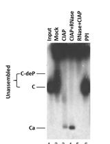
Host-regulated Hepatitis B Virus Capsid Assembly in a Mammalian Cell-free System

宿主调控的乙型肝炎病毒衣壳在无哺乳动物细胞系统中的组装

KL Kuancheng Liu
JH Jianming Hu
6400 Views
Apr 20, 2018
The hepatitis B virus (HBV) is an important global human pathogen and represents a major cause of hepatitis, liver cirrhosis and liver cancer. The HBV capsid is composed of multiple copies of a single viral protein, the capsid or core protein (HBc), plays multiple roles in the viral life cycle, and has emerged recently as a major target for developing antiviral therapies against HBV infection. Although several systems have been developed to study HBV capsid assembly, including heterologous overexpression systems like bacteria and insect cells, in vitro assembly using purified protein, and mammalian cell culture systems, the requirement for non-physiological concentrations of HBc and salts and the difficulty in manipulating host regulators of assembly presents major limitations for detailed studies on capsid assembly under physiologically relevant conditions. We have recently developed a mammalian cell-free system based on the rabbit reticulocyte lysate (RRL), in which HBc is expressed at physiological concentrations and assembles into capsids under near-physiological conditions. This system has already revealed HBc assembly requirements that are not anticipated based on previous assembly systems. Furthermore, capsid assembly in this system is regulated by endogenous host factors that can be readily manipulated. Here we present a detailed protocol for this cell-free capsid assembly system, including an illustration on how to manipulate host factors that regulate assembly.
肠凝聚型大肠埃希杆菌对HEK293细胞的粘附测定

Adhesion of Enteroaggregative E. coli Strains to HEK293 Cells

肠凝聚型大肠埃希杆菌对HEK293细胞的粘附测定

JA Jorge Luis Ayala-Lujan
FR Fernando Ruiz-Perez
6295 Views
Apr 20, 2018
Enteroaggregative Escherichia coli (EAEC) is a recognized cause of acute diarrhea among both children and adults worldwide. EAEC strains are characterized by the presence of aggregative adherence fimbriae (AAF), which play a key role in pathogenesis by mediating attachment to the intestinal mucosa and by triggering host inflammatory responses. The aggregative adherence fimbria II (AAF/II) is the most important adherence factor of EAEC prototype strain 042 (EAEC042) to intestinal cells. Multiple receptors for AAF/II on epithelial cells have been identified including the transmembrane signaling mucin Muc1. This protocol describes a method to measure adherence of EAEC strains to HEK293 cells expressing the Muc1 glycoprotein.
蓝藻细胞内渗透调节物质的测定

Determination of Intracellular Osmolytes in Cyanobacterial Cells

蓝藻细胞内渗透调节物质的测定

XT Xiaoming Tan
KS Kuo Song
CQ Cuncun Qiao
XL Xuefeng Lu
6096 Views
Apr 20, 2018
Most of the cyanobacteria accumulate osmolytes including sucrose, glucosylglycerol, in their cells in response to salt stress. Here we describe a protocol of our laboratory for extraction and quantification of cyanobacterial intracellular sucrose and glucosylglycerol. We have confirmed this protocol was applicable to at least four kinds of cyanobacteria, filamentous cyanobacterium Anabaena sp. PCC 7120, unicellular cyanobacterium Synechocystis sp. PCC 6803, Synechococcus elongatus PCC 7942 and halotolerant unicellular cyanobacterium Synechococcus sp. PCC 7002.

分子生物学

microRNA海绵文库的生成

Generation of microRNA Sponge Library

microRNA海绵文库的生成

Sebastian   Herzog Sebastian Herzog
7417 Views
Apr 20, 2018
This protocol describes the generation and functional validation of microRNA (miRNA) sponge or decoy constructs. When expressed from a strong promoter, these transcripts can sequester specific miRNA:RISC complexes, thereby resulting in a derepression of endogenous target mRNA. Hence, cells expressing such sponges display a partial or full miRNA loss-of-function phenotype.Depending on the sponge sequence, the activity of any miRNA of choice can be inhibited by sponge sequestration, but it should be noted that these constructs do not seem to be specific for one particular miRNA. Rather, all miRNAs of the same family as defined by the seed sequence will be affected, albeit to a different degree.

神经科学

电生理记录小鼠神经肌肉突触制备物中诱发的终板电位

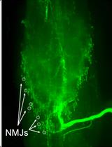
Electrophysiological Recordings of Evoked End-Plate Potential on Murine Neuro-muscular Synapse Preparations

电生理记录小鼠神经肌肉突触制备物中诱发的终板电位

Giulia Zanetti Giulia Zanetti
SN Samuele Negro
AM Aram Megighian
Marco Pirazzini Marco Pirazzini
12128 Views
Apr 20, 2018
Neuromuscular junction (NMJ) is the specialized chemical synapse that mediates the transmission of the electrical impulse running along motor neuron axons to skeletal muscle fibers. NMJ is the best characterized chemical synapse and its study along many years of research has provided most of the general knowledge of synapse development, structure and functionality.Electrophysiology is the most accurate experimental procedure to study NMJ physiology and it largely contributed to the elucidation of synaptic transmission basic principles. Many electrophysiological techniques have been developed to study NMJ physiology and physiopathology. In this paper, we describe an ex vivo tissue preparation for electrophysiology that can be applied to investigate nerve-muscle transmission functionality in mice. It is routinely used in our laboratory to study presynaptic neurotoxins, antitoxins, and to monitor NMJ degeneration and regeneration. This is a broadly applicable technique which can also be adopted to investigate alterations of NMJ activity in mouse models of neuromuscular diseases, including peripheral neuropathies, motor neuron disorders and myasthenic syndromes.
人类硬脑膜淋巴管的磁共振成像及组织病理学观察

Magnetic Resonance Imaging and Histopathological Visualization of Human Dural Lymphatic Vessels

人类硬脑膜淋巴管的磁共振成像及组织病理学观察

SH Seung-Kwon Ha
GN Govind Nair
MA Martina Absinta
NL Nicholas J. Luciano
DR Daniel S. Reich
11540 Views
Apr 20, 2018
In this protocol, we describe a method to visualize and map dural lymphatic vessels in-vivo using magnetic resonance imaging (MRI) and ex-vivo using histopathological techniques. While MRI protocols for routine imaging of meningeal lymphatics include contrast-enhanced T2-FLAIR and T1-weighted black-blood imaging, a more specific 3D mapping of the lymphatic system can be obtained by administering two distinct gadolinium-based MRI contrast agents on different days (gadofosveset and gadobutrol) and subsequently processing images acquired before and after administration of each type of contrast. In addition, we introduce methods for optimal immunostaining of lymphatic and blood vessel markers in human dura mater ex-vivo.
小鼠胚胎纹状体神经元的分离与维持

Isolation and Maintenance of Murine Embryonic Striatal Neurons

小鼠胚胎纹状体神经元的分离与维持

LN Luana  Naia
AR A. Cristina Rego
10091 Views
Apr 20, 2018
Primary cultures of murine striatal neurons are widely used to explore cellular mechanisms in neurobiology, including brain diseases. Here we describe a detailed and standardized protocol to dissect and culture embryonic murine striatal neurons GABA-positive/DARPP-32-positive for 12 days in vitro, when they show good neuronal cell connectivity and the presence of dendritic spines, which reflects the maturation of the network.
间接量热法测定慢性化学遗传神经元刺激对能量平衡的影响

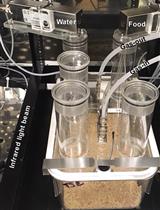
Testing Effects of Chronic Chemogenetic Neuronal Stimulation on Energy Balance by Indirect Calorimetry

间接量热法测定慢性化学遗传神经元刺激对能量平衡的影响

Sangho   Yu Sangho Yu
HM Heike Münzberg
5510 Views
Apr 20, 2018
The fundamental of neuroscience is to connect the firing of neurons to physiological and behavioral outcomes. Chemogenetics enables researchers to control the activity of a genetically defined population of neurons in vivo through the expression of designer receptor exclusively activated by designer drug (DREADD) in specific neurons and the administration of its synthetic ligand clozapine N-oxide (CNO) (Sternson and Roth, 2014). Using stimulatory Gq-coupled DREADD (hM3Dq) in mice, we showed that leptin receptor (LepRb)-expressing neurons in the preoptic area (POA) of the hypothalamus are warm-sensitive neurons that mediate warm-responsive metabolic and behavioral adaptations by reducing energy expenditure and food intake (Yu et al., 2016). We also used DREADD technology to test effects of chronic stimulation of POA LepRb neurons on energy expenditure, food intake, and body weight with the TSE indirect calorimetry system. Here we describe the detailed protocol of how we used indirect calorimetry to study the outcome of chronic stimulation of POA LepRb neurons. This protocol can be adapted to study long-term metabolic and behavioral consequences of other neuronal modulations, with possible modifications to the type of DREADD, duration of CNO treatment, or method of CNO delivery.

植物科学

植物多重应激下脂质过氧化作用的希夫试剂定性分析:组织化学方法

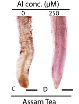
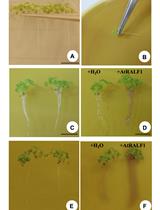
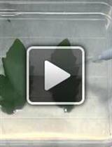
Qualitative Analysis of Lipid Peroxidation in Plants under Multiple Stress Through Schiff’s Reagent: A Histochemical Approach

植物多重应激下脂质过氧化作用的希夫试剂定性分析:组织化学方法

JA Jay Prakash Awasthi
BS Bedabrata Saha
Bhaben  Chowardhara Bhaben Chowardhara
Sanjenbam Sanjibia Devi Sanjenbam Sanjibia Devi
Pankaj  Borgohain Pankaj Borgohain
SP Sanjib Kumar Panda
14018 Views
Apr 20, 2018
Lipid peroxidation is a physiological indicator of both biotic and abiotic stress responses, hence is often used as a biomarker to assess stress-induced cell damage or death. Here we demonstrate an easy, quick and cheap staining method to assess lipid peroxidation in plant tissues. In this methodology, Schiff’s reagent, is used to assay for membrane degradation. Histochemical detection of lipid peroxidation is performed in this protocol. In brief, Schiff’s reagent detects aldehydes that originate from lipid peroxides in stressful condition. Schiff’s reagent is prepared and applied to plants tissue. After the reaction, plant tissue samples are rinsed with a sulfite solution to retain the staining color. From this analysis, qualitative visualization of lipid peroxidation in plant tissue is observed in the form of magenta coloration. This reagent is useful for visualization of stress induced lipid peroxidation in plants. In this protocol, Indica rice root, Assam tea root and Indian mustard seedlings are used for demonstration.
番茄离体及活体灰葡萄孢菌接种试验

In-vitro and in-planta Botrytis cinerea Inoculation Assays for Tomato

番茄离体及活体灰葡萄孢菌接种试验

JL Jiajie Lian
HH Hongyu Han
JZ Jiuhai Zhao
Chuanyou  Li Chuanyou Li
13432 Views
Apr 20, 2018
Botrytis cinerea (B. cinerea) attacks many crops of economic importance, represents one of the most extensively studied necrotrophic pathogens. Inoculation of B. cinerea and phenotypic analysis of plant resistance are key procedures to investigate the mechanism of plant immunity. Here we describe a protocol for B. cinerea inoculation on medium and planta based on our study using the tomato-B. cinerea system.
用Ilastik及ImageJ Fiji定量蓟马危害

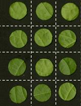
Quantification of Thrips Damage Using Ilastik and ImageJ Fiji

用Ilastik及ImageJ Fiji定量蓟马危害

IV Isabella G. S. Visschers
Nv Nicole M. van Dam
JP Janny L. Peters
10208 Views
Apr 20, 2018
Quantification of insect damage is an essential measurement for identifying resistance in plants. In screening for host plant resistance against thrips, the total damaged leaf area is used as a criterion to determine resistance levels. Here we present an objective novel method for analyzing thrips damage on leaf disc using the freely available software programs Ilastik and ImageJ. The protocol was developed in order to screen over 40 Capsicum lines for resistance against Frankliniella occidentalis (Western Flower Thrips) and Thrips tabaci (Onion thrips).

更正

更正:通过含有pH敏感指示剂溴甲酚紫的凝胶评估根部pH值的变化

Correction Notice: Evaluation of Root pH Change Through Gel Containing pH-sensitive Indicator Bromocresol Purple

更正:通过含有pH敏感指示剂溴甲酚紫的凝胶评估根部pH值的变化

Aparecida L. Silva Aparecida L. Silva
KD Keini Dressano
Paulo H. O. Ceciliato Paulo H. O. Ceciliato
Juan Carlos Guerrero-Abad Juan Carlos Guerrero-Abad
Daniel S. Moura Daniel S. Moura
3625 Views
Apr 20, 2018