- Submit a Protocol
- Receive Our Alerts
- Log in
- /
- Sign up
- My Bio Page
- Edit My Profile
- Change Password
- Log Out
- EN
- EN - English
- CN - 中文
- Protocols
- Articles and Issues
- For Authors
- About
- Become a Reviewer
- EN - English
- CN - 中文
- Home
- Protocols
- Articles and Issues
- For Authors
- About
- Become a Reviewer
pERK Detection Assays Using the Surefire AlphaScreen® Kit (TGR Biosciences and PerkinElmer)
Published: Vol 3, Iss 13, Jul 5, 2013 DOI: 10.21769/BioProtoc.806 Views: 10582
Reviewed by: Cheng Zhang

Protocol Collections
Comprehensive collections of detailed, peer-reviewed protocols focusing on specific topics
Related protocols

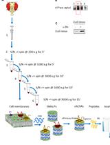
Enrichment of Membrane Proteins for Downstream Analysis Using Styrene Maleic Acid Lipid Particles (SMALPs) Extraction
Benedict Dirnberger [...] Kathryn S. Lilley
Aug 5, 2023 2833 Views

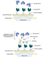
Establishment of Human PD-1/PD-L1 Blockade Assay Based on Surface Plasmon Resonance (SPR) Biosensor
Tess Puopolo [...] Chang Liu
Aug 5, 2023 2750 Views

A Computational Workflow for Membrane Protein–Ligand Interaction Studies: Focus on α5-Containing GABA (A) Receptors
Syarifah Maisarah Sayed Mohamad [...] Ahmad Tarmizi Che Has
Nov 20, 2025 1917 Views
Abstract
Extracellular signal-regulated kinase 1 and 2 (ERK1/2) are serine/threonine protein kinases that are phosphorylated on Thr202/Tyr204 (ERK1) and Thr185/Tyr187 (ERK2). Phosphorylation of ERK1/2 (pERK1/2) arises from multiple stimuli, resulting in physiological responses that include cell growth, proliferation and differentiation. This protocol has been optimized for the detection of ligand-mediated pERK1/2 in adherent immortal cell lines expressing G protein-coupled receptors (GPCRs).
Materials and Reagents
- Dulbecco’s modified eagle medium (DMEM) (Life Technologies, Gibco®, catalog number: 11995-065 )
- Bovine serum albumin (BSA) (Sigma-Aldrich, catalog number: A7906 )
- SureFire® Reagents (Includes Lysis, Activation and Reaction buffers) (TGR BioSciences, catalog number: TGRES500 )
- AlphaScreen® General IgG (Protein A) detection kit (PerkinElmer, catalog number: 6760617 )
- White ProxiPlate 384-well microplate (PerkinElmer, catalog number: 6008280 )
- TopSeal (PerkinElmer, catalog number: 6005250 )
- NaCl
- KCl
- Na2HPO4
- KH2PO4
- Phosphate buffered saline (PBS) (see Recipes)
- Detection buffer (see Recipes)
Equipment
- Fusion-α plate reader or Envision plate reader with appropriate Alphascreen detection modules (PerkinElmer)
- Sterile 96-well clear flat bottom plates (BD Biosciences, Falcon®, catalog number: 353072 )
- Humidified incubator
- Multichannel pipettes
- Micropipettes
- Orbital shaker
Procedure
Notes:
1) Cells can either be stably or transiently expressing receptor of interest.
2) It is recommended to first perform a timecourse analysis to determine the time at which ligand-mediated pERK1/2 is maximal. For this, follow the same protocolusing a single concentration of ligand (recommended concentration 100x Kd).
Recommended initial timecourse (min): 90, 60, 45, 30, 15, 10, 8, 6, 4, 2, 1, 0.
3) Subsequent timecourses can then be refined to determine the precise time at which maximum ligand-induced pERK1/2 occurs.
I. Cell preparation
Seed cells in suitable nutrient media (e.g. DMEM, 10% FBS, no antibiotics) into a sterile 96-well plate and incubate in a humidified environment at 37 °C, 5% CO2 to be ~90% confluent the following day (~24 h).
Note: Optimization for cell number depending on the cell line used will be necessary (we suggest a starting range of 10,000-50,000 cells per well). Recommended density for CHO FlpIN cells is 30,000 cells/ well.
II. Stimulation
- The day following seeding, aspirate nutrient media, rinse once with 100 μl PBS, and replace with 90 μl prewarmed DMEM (no FBS).
- Incubate in a humidified environment at 37 °C, 5% CO2 for a minimum of 4 h (recommended 6 h, up to overnight (O/N)).
- Prepare serial dilutions of ligands at 10x final concentration in DMEM, enough for 10 μl/well, to be performed in duplicate (minimum), and enough for the number of timepoints if doing a timecourse.
Note: Concentration range to use will depend on ligand affinity for receptor. For initial timecourse test, select a concentration 100x Kd of ligand and a DMEM control. The concentrations can then be refined in subsequent experiments. If using a peptide or ‘sticky’ ligand, prepare serial dilutions in DMEM with 0.1% BSA.
- Prepare a suitable concentration of FBS in DMEM, enough for 10 μl/well, to be performed in duplicate (minimum), and enough for the number of timepoints if doing a timecourse.
Note: This is the internal control for the experiment – FBS promotes pERK1/2. Recommended final concentration of FBS in a CHO FlpIN cell line is 3-10%.
- Following preincubation in DMEM, add 10 μl of 10x prepared ligands or FBS to cells for a total volume of 100 μl, 1x final concentration.
Note: For initial timecourse, begin at 90 min, and add ligand or FBS to cells at each timepoint until time 0 (no addition). For concentration response, add ligand or FBS at time of maximal induced pERK1/2 as determined through timecourse.
- After completion of stimulation, rapidly remove ligand-containing media from cells.
Note: Depending on the cell type, this may involve flicking or gentle aspiration.
- Add 50 μl 1x Surefire® Lysis buffer.
Note: Optimization for lysis volume will be necessary, and depends on the cell type, expression level of the receptor and efficiency of coupling to pERK1/2 pathways. Recommended starting lysis volume in a CHO FlpIN cell line is 30-100 μl.
- Incubate lysates at room temperature (RT) for 5-10 min on an orbital shaker.
III. Detection
- In reduced lighting conditions, prepare detection buffer.
- Transfer 5 μl of cell lysate from each well to a 384-well ProxiPlate.
- In reduced lighting conditions, add 8.5 μl Detection buffer to each sample.
- Seal the plate with TopSeal and wrap in foil.
Note: Small volumes are subject to evaporation, TopSeal is essential.
- Incubate at RT for 2 h or 37 °C for 1 h in reduced lighting conditions.
Note: If incubating at 37 °C, ensure the plate has returned to RT before measuring luminescence (~15 min at RT following 37 °C incubation should suffice). Detection beads are temperature sensitive.
- Analyse luminescence on a Fusion-α or Envision plate reader using standard α-screen settings.
IV. Data analysis
Data should be normalized to the response elicited by the FBS control.
Recipes
- Phosphate Buffered Saline (PBS)
137 mM NaCl, 2.7 mM KCl, 10 mM Na2HPO4, 1.8 mM KH2PO4, pH 7.4.
- Detection buffer
85.0% SureFire® reaction buffer
14.2% SureFire® activation buffer*
0.4% Acceptor beads
0.4% Donor beads
* Activation buffer should be stored at 4 °C, however, precipitation will occur at this temperature. Before use, heat to 37 °C to ensure all is dissolved.
Prepare Detection buffer immediately before use. Discard unused detection buffer. Mix detection buffer gently. Do not vortex.
Additional note: Lysates may be stored at -20 °C and pERK1/2 detected at a later time, but no longer than 2 weeks following stimulation.
References
- Koole, C., Wootten, D., Simms, J., Valant, C., Sridhar, R., Woodman, O. L., Miller, L. J., Summers, R. J., Christopoulos, A. and Sexton, P. M. (2010). Allosteric ligands of the glucagon-like peptide 1 receptor (GLP-1R) differentially modulate endogenous and exogenous peptide responses in a pathway-selective manner: implications for drug screening. Mol Pharmacol 78(3): 456-465.
Article Information
Copyright
© 2013 The Authors; exclusive licensee Bio-protocol LLC.
How to cite
Koole, C., Wootten, D. and Sexton, P. M. (2013). pERK Detection Assays Using the Surefire AlphaScreen® Kit (TGR Biosciences and PerkinElmer). Bio-protocol 3(13): e806. DOI: 10.21769/BioProtoc.806.
Category
Cell Biology > Cell signaling > Phosphorylation
Biochemistry > Protein > Modification
Biochemistry > Protein > Interaction > Protein-ligand interaction
Do you have any questions about this protocol?
Post your question to gather feedback from the community. We will also invite the authors of this article to respond.
Share
Bluesky
X
Copy link


