- Protocols

- Articles and Issues
- For Authors
- About
- Become a Reviewer

Past Issue in 2013
Volume: 3, Issue: 14
Cancer Biology
Measurement of Endogenous MALT1 Activity
Estradiol Receptor (ER) Chromatin Immunoprecipitation in MCF-7 Cells


Immunology
Isolation of Phagosomes from Dendritic Cells by Using Magnetic Beads

Endosomal pH Measurement in Bone Marrow Derived Dendritic Cells

Microbiology
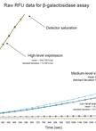
High-throughput β-galactosidase and β-glucuronidase Assays Using Fluorogenic Substrates

Extraction and Quantification of Cyclic Di-GMP from Pseudomonas aeruginosa


Analyzing Inhibitory Effects of Reagents on Mycoplasma Gliding and Adhesion


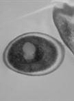
Preparation of Candida albicans Biofilms for Transmission Electron Microscopy

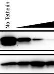
HIV-1 Virus-like Particle Budding Assay
Preparation of Candida albicans Biofilms Using an in vivo Rat Central Venous Catheter Model
Molecular Biology
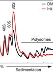
Polysome Profiling Analysis

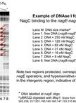
DNase I Footprinting to Identify Protein Binding Sites
Plant Science
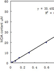
Analysis of Malondialdehyde, Chlorophyll Proline, Soluble Sugar, and Glutathione Content in Arabidopsis seedling


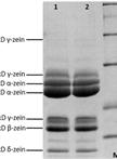
Maize Endosperm Protein Extraction and Analysis
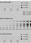
Determination of Enzyme Kinetic Parameters of UDP-glycosyltransferases

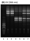
Extraction and Reglucosylation of Barbarea vulgaris Sapogenins