- Protocols

- Articles and Issues
- For Authors
- About
- Become a Reviewer

Past Issue in 2026
Volume: 16, Issue: 3
Biochemistry
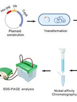
A One-Step Method for Efficient Purification of Functional Cas9 Protein

Biochemical Reconstitution and FRAP Analysis of Membrane-Associated Condensates on Supported Lipid Bilayers
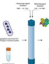
On-Column Dual-Gradient Refolding for Efficient Recovery of Insoluble Affinity-Tagged Recombinant Proteins
Bioinformatics and Computational Biology
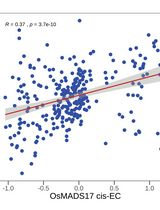
Identifying Causal Genes and Building Regulatory Networks in Crops Using the CisTrans-ECAS Method
Cancer Biology
A Quantitative DNA Fiber Assay to Monitor Replication Fork Progression, Protection, and Restart
The Generation of Tissue-Specific ECM Hydrogels From Melanoma and Associated Organs to Study Cancer Biology
Developmental Biology
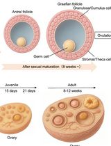
Monitoring of Sperm-Independent Calcium Oscillations in Immature Oocytes of Mice
Environmental science
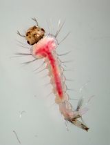
Qualitative Detection of Lipid Peroxidation in Mosquito Larvae Using Schiff’s Reaction: A Simple Histochemical Tool for In Situ Assessment of Oxidative Damage

Immunology
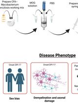
Advancing EAE Modeling: Establishment of a Non-Pertussis Immunization Protocol for Multiple Sclerosis
Mechanobiology
Quantifying Mechanical Strain–Induced Membrane Damage in Early Neuronal Cells Using an In Vitro Traumatic Brain Injury Model
Microbiology
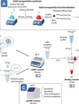
Visual Nanoprobe-Enhanced Loop-Mediated Isothermal Amplification Protocol for Rapid Detection of Infectious Laryngotracheitis Virus from Avian Respiratory Swabs
Neuroscience
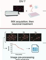
High Content In Vitro Survival Assay of Cortical Neurons
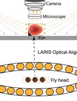
Low Angle Ring Illumination Stereomicroscopy (LARIS) Method for High-Contrast Imaging of Drosophila Compound Eyes
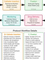
A Low-Stress, Long-Duration Stable Tail Vein Catheterization and Precise Drug Delivery Protocol for Awake, Freely Moving Mice
Plant Science
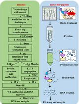
Turbo-RIP: A Protocol for TurboID-based RNA Immunopurification to Map RNA Landscapes in Plant Biomolecular Condensates
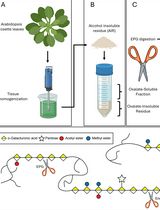
Detailed Method for the Purification of Rhamnogalacturonan-I (RG-I) in Arabidopsis thaliana
Isolation and Transfection of Protoplasts From Maize Mesophyll Cells
Stem Cell
Simple and Rapid Model to Generate Differentiated Endometrial Floating Organoids

