- EN - English
- CN - 中文
Measurement of Endogenous H2O2 and NO and Cell Viability by Confocal Laser Scanning Microscopy
内源H2O2和NO以及保卫细胞活力的定量分析-共聚焦激光扫描显微镜联合荧光染料法
发布: 2013年10月05日第3卷第19期 DOI: 10.21769/BioProtoc.920 浏览次数: 14308
评审: Tie Liu
Abstract
Recently, there is compelling evidence that hydrogen peroxide (H2O2) and nitric oxide (NO) function as signaling molecules in plants, mediating a range of responses including stomatal movement. Thus, the choice of sensitive methods for detection of endogenous H2O2 and NO in guard cells are very important for understanding the role of H2O2 and NO in guard cell signaling. In addition, besides stomatal closure caused by interfering guard cell signaling, it can also be caused by widespread, nonspecific damage to guard cells. To determine whether stomatal movement is caused by damage to guard cells, sensitive methods for detection of guard cell viability are often required.
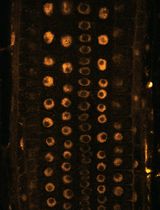
The oxidatively sensitive fluorophore 2′,7′-dichlorofluorecin (H2DCF) is commonly employed to measure changes in intracellular H2O2 level directly. The non-polar diacetate ester (H2DCFDA) of H2DCF enters the cell and is hydrolysed into the more polar, non-fluorescent compound H2DCF, which, therefore, is trapped. Subsequent oxidation of H2DCF by H2O2, catalysed by peroxidases, yields the highly fluorescent DCF. Similarly, the cell-permeable, NO-sensitive fluorescent probe 4,5-diaminofluorescein diacetate (DAF-2DA) is widely used for the direct detection of NO presence in both animal and plant cells. The non-polar DAF-2DA enters the cell and is hydrolyzed by cytosolic esterase into the more polar, non-fluorescent compound DAF-2, which in the presence of NO is converted to the highly fluorescent triazole derivative DAF-2T. The fluorescent indicator dyes fluorescein diacetate (FAD) and propidium iodide (PI) are widely used for detection of cell viability. FAD passes through cell membranes and is hydrolyzed by intracellular esterase to produce a polar compound that passes slowly through a living cell membrane but fast through a damaged or dead cell membrane, and thus accumulates inside the viable cells and exhibits green fluorescence when excited by blue light. In contrast, PI passes through damaged or dead cell membranes and intercalates with DNA and RNA to form a bright red fluorescent complex seen in the nuclei of dying or dead cells but not living cells. Based on the above analysis, the fluorescent indicator dyes H2DCFDA, DAF-2DA, FAD and PI load readily into guard cells, and their optical properties make them amenable to analysis by confocal laser scanning microscopy.
This protocol describes how to combine confocal laser scanning microscopy with fluorescent indicator dyes H2DCFDA, DAF-2DA, FAD and PI respectively for measurement of H2O2 and NO and viability of guard cell in leaves of Arabidopsis (Arabidopsis thaliana).
Materials and Reagents
- Leaves of Arabidopsis (Arabidopsis thaliana)
- Ethanesulfonic acid (MES)
- 2′,7′-dichlorofluorescin diacetate (H2DCFDA) (Sigma-Aldrich, catalog number: D6883 )
- 4, 5-diaminofluorescein diacetate (DAF-2DA) (Sigma-Aldrich, catalog number: D2813 )
- Propidium iodide (PI) (Sigma-Aldrich, catalog number: P4170 )
- Fluorescein diacetate (FDA) (Sigma-Aldrich, catalog number: F7378 )
- DMSO (Sigma-Aldrich, catalog number: D8418 )
- 10 mM MES/KCl buffer pH 6.15 (see Recipes)
- Tris/KCl buffer pH 7.2 (see Recipes)
- 10 mM H2DCFDA (see Recipes)
- 10 mM DAF-2DA (stock solution) (see Recipes)
- 1 mg/ml PI (stock solution) (see Recipes)
- 5 mg/ml FDA (stock solution) (see Recipes)
Equipment
- TCS-SP2 Confocal Laser Scanning Microscopy (Leica Lasertechnik Gmbh3, Heidelberg)
- 25 °C incubator
- Glass slide and cover glass
- Eyelbrow brush (Yellow wolf hair, length of hair: 0.8 cm, width of hair: 0.8 cm)
- Tweezers
- 6-cm diameter Petri plate
Software
- Leica confocal software (Leica Lasertechnik Gmbh3, Heidelberg)
- Photoshop software
Procedure
文章信息
版权信息
© 2013 The Authors; exclusive licensee Bio-protocol LLC.
如何引用
Readers should cite both the Bio-protocol article and the original research article where this protocol was used:
- Wu, M., Ma, X. and He, J. (2013). Measurement of Endogenous H2O2 and NO and Cell Viability by Confocal Laser Scanning Microscopy. Bio-protocol 3(19): e920. DOI: 10.21769/BioProtoc.920.
- He, J. M., Ma, X. G., Zhang, Y., Sun, T. F., Xu, F. F., Chen, Y. P., Liu, X. and Yue, M. (2013). Role and interrelationship of Galpha protein, hydrogen peroxide, and nitric oxide in ultraviolet B-induced stomatal closure in Arabidopsis leaves. Plant Physiol 161(3): 1570-1583.
分类
植物科学 > 植物生物化学 > 其它化合物
细胞生物学 > 细胞成像 > 共聚焦显微镜
生物化学 > 其它化合物 > 活性氧
您对这篇实验方法有问题吗?
在此处发布您的问题,我们将邀请本文作者来回答。同时,我们会将您的问题发布到Bio-protocol Exchange,以便寻求社区成员的帮助。
Share
Bluesky
X
Copy link


