

Simultaneous Immunofluorescence-Based In Situ mRNA Expression and Protein Detection in Bone Marrow Biopsy Samples
发布: 2026年05月05日第16卷第9期 DOI: 10.21769/BioProtoc.5612 浏览次数: 12
评审: Anonymous reviewer(s)


Abstract
Fluorescence in situ hybridization (FISH) can be employed to study the expression and subcellular localization of nucleic acids by using labeled antisense strands that hybridize with the target RNA or DNA molecules. Likewise, immunofluorescence antibody staining (IF) takes advantage of the specific interaction between a fluorophore-labeled antibody and its corresponding antigen. This protocol reports the combination of RNA-FISH and IF antibody staining for simultaneous detection of both RNA transcripts and proteins of interest in routine formalin-fixed paraffin-embedded (FFPE) bone marrow biopsy samples. Herein, we provide a detailed description of the methodology that we have developed and optimized to study the spatial expression of two transcripts—TGFB1 and PDGFA1—in human hematopoietic (CD45+) and non-hematopoietic (CD271+) cells in the bone marrow of patients with acute lymphoblastic leukemia (ALL).
Key features
• The protocol describes the simultaneous visualization of RNA target transcripts and protein expression.
• In situ RNA and surface marker analysis was established for routine human bone marrow biopsies.
Keywords: Multi-color immunofluorescence stainingGraphical overview
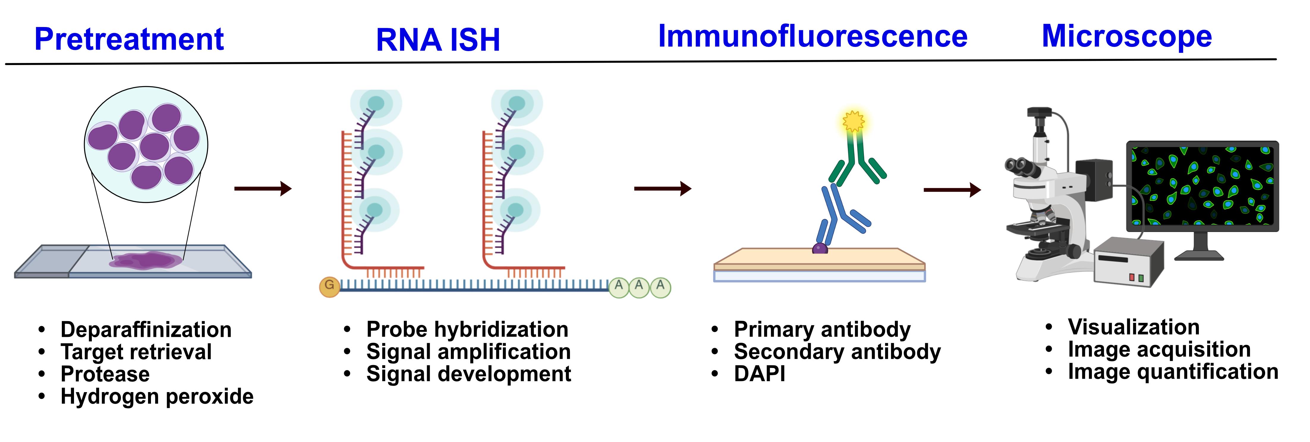
Dual RNA fluorescence in situ hybridization and protein immunofluorescence workflow
Background
Acute lymphoblastic leukemia (ALL) is a blood cancer defined by the aberrant production of lymphoid blast cells. The presence of bone marrow (BM) fibrosis in ALL is correlated with poorer prognosis [1]. However, the mechanisms leading to BM fibrosis in ALL remain unknown. We therefore studied the spatial expression of two cytokines, TGFB1 and PDGFA1, which are known to play a major role in primary myelofibrosis (PMF), in bone marrows of ALL patients in comparison with PMF to assess whether the two diseases share common mechanisms of fibrosis initiation and progression [2].
Real-time RT-PCR is widely used for the detection and quantification of target mRNA, being extremely sensitive. However, the RNA extraction step destroys the tissue, which makes it impossible to spatially assess tissue gene expression. On the other hand, traditional antibody-based fluorescence protein detection preserves tissue architecture but does not provide information about possible transcriptional changes.
In order to simultaneously assess gene and protein expression in bone marrow tissues, we therefore combined the RNAscope® in situ hybridization assay for TGFB1 and PDGFA1 transcripts with antibody-based immunofluorescence (IF) staining using hematopoietic and stromal cell surface markers. The main advantage of using RNAscopeTM technology is its high sensitivity and specificity compared to traditional in situ RNA hybridization methods. This is due to the use of double Z probes that allow the detection of lowly expressed or partially degraded RNA while maintaining target-specific signal. Combining the in situ mRNA detection with specific cell marker analysis allowed us to study the expression of our target genes in defined cell types, providing information about cellular heterogeneity and gene expression patterns throughout the tissue. This protocol, however, is currently restricted to the detection of up to three different RNA targets at a time, and the use of additional cell surface markers clearly depends on the spectral compatibility of the fluorophores and the number of channels available on the imaging platform.
Materials and reagents
Biological materials
1. FFPE bone marrow biopsy sections (5 μm thick) from the iliac crest were obtained from the Division of Pathology, Laboratory Medicine Skåne, Lund, Sweden. Sections were routine pathology specimens from nine Philadelphia-negative B-cell ALL (B-ALL) patients (three females and six males, ages ranging from 2 to 7 years old, with fibrosis grades 0–2) and three adult patients with primary myelofibrosis (one male and two females, with ages ranging from 32 to 50 years old). Samples from hematologically normal patients were included as negative controls.
Reagents
1. RNAscope® probes
a. RNAscope® probe for TGFB1 (Hs-TGFB1-no-XMm-C2) (Advanced Cell Diagnostics, catalog number: 443481-C2)
b. RNAscope® probe for PDGFA1 (Hs-PDGFA-no-XMm-C1) (Advanced Cell Diagnostics, catalog number: 441061)
c. Positive control probe against the human POLA2A PPIB UBC transcripts (Advanced Cell Diagnostics, catalog number: 320861)
d. Negative control probe against the DapB transcript (accession #EF191515) of the Bacillus subtilis strain SMY ± TSA VividTM Fluorophore 570 and TSA VividTM 650 (Advanced Cell Diagnostics, catalog number: 3200871)
2. RNAscope® Multiplex Fluorescent Reagent kit v2 (Advanced Cell Diagnostics, catalog number: 323100)
a. Pretreatment reagents
i. RNAscope® hydrogen peroxide (Advanced Cell Diagnostics, catalog number: 322381)
ii. RNAscope® protease plus (Advanced Cell Diagnostics, catalog number: 322381)
iii. RNAscope® 10× target retrieval (Advanced Cell Diagnostics, catalog number: 322000)
b. RNAscope® Multiplex fluorescent detection reagents v2 (Advanced Cell Diagnostics, catalog number: 323110)
i. RNAscope® Multiplex FL v2 AMP 1
ii. RNAscope® Multiplex FL v2 AMP 2
iii. RNAscope® Multiplex FL v2 AMP 3
iv. RNAscope® Multiplex FL v2 HRP-C1
v. RNAscope® Multiplex FL v2 HRP-C2
vi. RNAscope® Multiplex FL v2 HRP blocker
vii. RNAscope® Multiplex FL v2 DAPI
c. RNAscope® Scope Multiplex TSA buffer (Advanced Cell Diagnostics, catalog number: 322809)
d. RNAscope® 50× wash buffer (Advanced Cell Diagnostics, catalog number: 310091)
3. Tyramide signal amplification (TSA) Plus fluorophores
a. TSA VividTM fluorophore 570 (Advanced Cell Diagnostics, catalog number: 323272)
b. TSA VividTM fluorophore 650 (Advanced Cell Diagnostics, catalog number: 323273)
4. Primary antibodies
a. Mouse anti-human 271 (R&D Systems, catalog number: MAB367, RRID: AB_2152546)
b. Rabbit anti human CD45 (Sigma-Aldrich, catalog number: HPA000440, RRID: AB_611377)
5. Secondary antibodies
a. AlexaFluor® 488 goat anti-rabbit IgG1 (Jackson ImmunoResearch Labs, catalog number: 111-545-003, RRID: AB_2338046)
b. AlexaFluor® 700 goat anti-rabbit IgG (Thermo Fisher Scientific, catalog number: A-21038, RRID: AB_2535709)
6. HyCloneTM Dulbecco’s phosphate buffered saline (DPBS, Cytiva, catalog number: SH30028.02)
7. Tween 20 (Sigma-Aldrich, catalog number: P1379)
8. PBS tablets (GibcoTM, catalog number: 18912014)
9. Neo-ClearTM, xylene substitute (Sigma-Aldrich, catalog number: 1.09843.5000)
10. Absolute ethanol [Apoteket (Swedish pharmacy), catalog number: 910345]
11. Distilled water
12. Sodium azide (Merck, catalog number: 71289)
13. ProLong Gold antifade mountant (Thermo Fisher Scientific, catalog number: P36930)
14. Cover glass 24 mm × 50 mm (Thermo Fisher Scientific, catalog number: 12-545-F)
15. Carboy >3 L (any supplier)
16. Eppendorf tubes, 0.5, 1.5, and 2 mL (any supplier)
17. Serological pipettes, 5, 10, and 25 mL (any supplier)
18. Paper towel or absorbent paper
19. Normal goat serum (Jackson ImmunoResearch Labs, catalog number: 005-000-121, RRID: AB_2336990)
20. Sodium citrate tribasic dihydrate (Sigma-Aldrich, catalog number: S4641-25G)
21. Sodium chloride (Thermo Fisher Scientific, catalog number: 447302500)
Solutions
1. RNAscope® wash buffer 1× (see Recipes)
2. Saline sodium citrate (SSC) (see Recipes)
3. Blocking buffer (see Recipes)
4. Primary antibody dilution buffer (see Recipes)
5. Secondary antibody dilution buffer (see Recipes)
6. IF wash buffer (see Recipes)
Recipes
1. RNAscope® wash buffer 1×
| Reagent | Concentration | Volume |
|---|---|---|
| RNAscope® wash buffer | 50× | 120 mL |
| Distilled water | n/a | 5.88 L |
Warm RNAscope 50× wash buffer up to 40 °C for 10–20 min before preparation of RNAscope® wash buffer 1×. 1× wash buffer may be prepared ahead of time and stored at room temperature for up to one month.
2. Saline sodium citrate (SSC)
| Reagent | Quantity |
|---|---|
| NaCl | 175.3 g |
| Sodium citrate | 88.2 g |
| Distilled water | 1 L |
Note: Dissolve the NaCl and sodium citrate in 800 mL of distilled water, then adjust the pH to 7.0 with HCl and adjust the volume to 1 L with additional H2O. Sterilize by autoclaving.
3. Blocking buffer
| Reagent | Concentration | Volume |
|---|---|---|
| PBS | 1× | 10 mL |
| Normal goat serum | 1 mL | |
| Sodium azide | 100 μL |
4. Primary antibody dilution buffer
| Reagent | Concentration | Quantity |
|---|---|---|
| PBS | 1× | 10 mL |
| Normal goat serum | 500 μL | |
| Sodium azide | 100 μL |
5. Secondary antibody dilution buffer
| Reagent | Concentration | Quantity |
|---|---|---|
| PBS | 1× | 10 mL |
| Normal goat serum | 200 μL | |
| Sodium azide | 100 μL |
6. IF wash buffer
| Reagent | Concentration | Quantity |
|---|---|---|
| PBS | 1× | 2 tablets |
| Distilled water | 1 L | |
| Tween 20 | 500 μL |
Equipment
1. HybEZTM Hybridization System
a. HybEZTM II Hybridization System oven 110V (Advanced Cell Diagnostics, catalog number: 321711)
b. HybEZTM Humidity Control Tray with lid (Advanced Cell Diagnostics, catalog number: 310012)
c. RNAscope® EZ-BatchTM Slide Rack (Advanced Cell Diagnostics, catalog number: 310017)
d. HybEZTM Humidifying Paper (Advanced Cell Diagnostics, catalog number: 310015)
2. Olympus VS120-S6-W slide scanner (Olympus)
3. Leica Stellaris 5 Confocal laser-scanning inverted microscope (Leica Microsystems)
4. Steamer (Russell Hobbs)
5. Incubator/water bath (any supplier)
6. Dry oven (any supplier)
7. P1000, P200, P10 pipettors (any supplier)
8. Plastic Coplin jars (any supplier)
Software and datasets
1. ZEISS Arivis Pro software (ver. 4.0, Carl Zeiss Microscopy Software Center Rostock GmbH, Germany)
2. Prism software version 10.4.2, GraphPad
3. Microsoft Office Excel
Procedure
文章信息
稿件历史记录
提交日期: Nov 17, 2025
接收日期: Jan 18, 2026
在线发布日期: Feb 4, 2026
出版日期: May 5, 2026
版权信息
© 2026 The Author(s); This is an open access article under the CC BY license (https://creativecommons.org/licenses/by/4.0/).
如何引用
Sierras, A. L., Bräunig, S., Li, H. and Scheding, S. (2026). Simultaneous Immunofluorescence-Based In Situ mRNA Expression and Protein Detection in Bone Marrow Biopsy Samples. Bio-protocol 16(9): e5612. DOI: 10.21769/BioProtoc.5612.
分类
分子生物学
您对这篇实验方法有问题吗?
在此处发布您的问题,我们将邀请本文作者来回答。同时,我们会将您的问题发布到Bio-protocol Exchange,以便寻求社区成员的帮助。