

In Vitro Model of Cytokine-Induced Inflammatory 3T3-L1 Adipocytes Mimicking Obesity
模拟肥胖的细胞因子诱导炎症性 3T3-L1 脂肪细胞体外模型
发布: 2026年02月20日第16卷第4期 DOI: 10.21769/BioProtoc.5609 浏览次数: 318
评审: Komuraiah MyakalaShun Yu Jasemine YangAnonymous reviewer(s)
Advertisement


Abstract
Obesity is a risk factor for many diseases. The 3T3-L1 cell line is often used to obtain mature adipocytes, but these lack the inflammatory phenotype observed in obesity. Using a cocktail of cytokines that mimics the secretome of macrophages found in the inflammatory adipose tissue, we developed a protocol for obtaining mature inflammatory adipocytes. This model was validated at gene (RT-qPCR) and protein levels (multiplex adipokine array) as we found a decrease of adipogenic markers (C/EBPα, PPARУ, adiponectin, and CD36) and an increase of pro-inflammatory cytokines (IL-6, IL-1β, CXCL1, CXCL10, TNF-α, ICAM-1, and lipocalin-2). We provide a relevant in vitro model for studying the impact of low-grade chronic inflammation caused by obesity and its downstream effects on metabolic disorders and tumor microenvironments.
Key features
• Currently available protocols of adipocyte differentiation are not relevant for studying obesity in vitro.
• We developed a simple and reproducible method to generate inflammatory adipocytes in vitro using a cytokine cocktail.
• Gene expression analysis (qPCR) confirms the downregulation of adipogenic and protective markers (e.g., adiponectin, CD36) and the upregulation of pro-inflammatory cytokines (e.g., IL-6, IL-1β, TNF-α).
• Adipokine array reveals decreased secretion of anti-inflammatory molecules (adiponectin, IGFBPs, FGF-21, HGF) and increased release of pro-inflammatory adipokines (serpin E1, IGF-1, lipocalin-2, IL-6, ICAM-1).
• This protocol provides a relevant and versatile method for investigating obesity-related inflammation and its role in disease progression.
Keywords: Adipocyte (脂肪细胞)Graphical overview
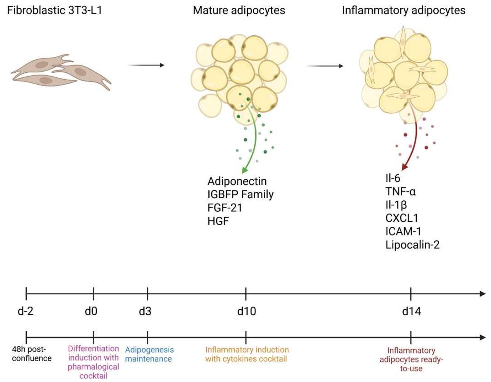
From differentiation to inflammation: Establishment of the inflammatory adipocyte model
Background
Adipose tissue plays key metabolic roles by secreting proteins known as adipokines. Under physiological conditions, adipose tissue secretes protective molecules such as adiponectin [1], the insulin-like growth factor–binding protein family (IGFBPs) [2], and fibroblast growth factor 1(FGF-1) [3], which contribute to insulin sensitivity and adipose tissue homeostasis. In contrast, obese adipose tissue shows decreased secretion of these protective factors and increased release of pro-inflammatory adipokines such as lipocalin-2 [4] and intercellular adhesion molecule-1 (ICAM-1) [5]. This change in phenotype is associated with a dedifferentiation of mature adipocytes by decreasing major adipogenic transcription factors like CCAAT/enhancer-binding protein alpha (C/EBPα) and peroxisome proliferator-activated receptor gamma (PPARУ).
One reason for adipose tissue remodeling during obesity is the development and exacerbation of chronic inflammation. During weight gain, excessive lipid accumulation in adipocytes leads to cell hypertrophy, hypoxia, and eventually necrosis. In addition to adipokines, this environment promotes the secretion of pro-inflammatory molecules such as tumor necrosis factor alpha (TNF-α), interleukin-6 (IL-6), interleukin-8 (IL-8), and interleukin-1 beta (IL-1β), which recruit immune cells, particularly circulating pro-inflammatory monocytes. Accumulating macrophages organize in crown-like structures (CLS) and amplify the inflammatory signaling [6].
This inflamed adipose tissue induces a chronic state of low-grade inflammation, which is crucial to investigate further, as it contributes not only to metabolic dysfunction but also to the development and progression of numerous diseases [7]. Indeed, individuals with obesity (BMI > 30) or individuals with BMI > 25 are well recognized to have an increased risk and worsened outcomes for cancer, cardiovascular diseases, diabetes, or hypertension [8].
Due to their ability to differentiate into adipocytes, 3T3-L1 cells—one of the most used fibroblast cell lines—are widely employed to study adipogenesis and adipocyte biology. This model is also frequently used in obesity research. However, inducing 3T3-L1 cell differentiation remains technically challenging due to the well-documented limited reproducibility of this cell line [9]. There is currently no standardized, fast, and cost-effective method to generate inflammatory adipocytes that accurately mimic obesity-associated adipocytes in vitro. Here, we describe a new in vitro approach to produce inflammatory adipocytes that avoids the need for specialized equipment such as a hypoxic chamber or additional conditioned media from other cells [10,11]. This innovative protocol provides a practical, accessible, and valuable tool for investigating the role of adipocytes in various pathologies, while limiting the use of animals.
Materials and reagents
Biological materials
1. 3T3-L1 cell line from mouse (Merck, catalog number: 86052701-1VL)
Reagents
1. Dulbecco’s modified Eagle medium 4.5 g/L glucose with sodium pyruvate and stabilized L-Glutamine (DMEM) (Grosseron, Biosera, catalog number: 0153043)
2. Fetal bovine serum (FBS) (Dutscher, Hyclone, catalog number: SV30160.03)
3. Dulbecco’s phosphate-tamponed saline 10× without calcium/magnesium (PBS) (Grosseron, Biosera, catalog number: 0153264)
4. Trypsin 1× with EDTA (Grosseron, Biosera, catalog number: 0153112)
5. Rosiglitazone >98% (HPLC), powder, PPARУ agonist (Merck, Sigma-Aldrich, catalog number: R2408-10MG)
6. 3-isobutyl-1-methyxanthine (IBMX) 250 mg (Merck, Sigma-Aldrich, catalog number: 410957-250MG)
7. Dexamethasone 100 mg (MP Biomedicals Germany GMBH, catalog number: 0219004080)
8. Insulin solution human 10 mg/mL (Merck, Sigma-Aldrich, catalog number: I9278-5mL)
9. Oil red O 0.5 L × 4 (DIAPATH France, catalog number: C0512/U)
10. 2-Propanol (isopropanol) >99.5% (Merck, Sigma-Alrich, catalog number: 190764-500ML)
11. Aqueous mounting medium (Abcam, catalog number: ab64230)
12. Dimethyl sulfoxide (DMSO) 250 mL (Corning, catalog number: 25-950-CQC)
13. Recombinant mouse IL-1β 10 μg (BioLegend, catalog number: 575102)
14. Recombinant mouse TNF-α 10 μg (BioLegend, catalog number: 575202)
15. Recombinant mouse IL-6 10 μg (BioLegend, catalog number: 575702)
16. Recombinant mouse CXCL1 (KC) 10 μg (BioLegend, catalog number: 573702)
17. Trypan blue solution 0.4% in PBS 100 mL (Fisher Scientific, catalog number: SV30084.01)
18. Maxwell RSC simplyRNA tissue (Promega, catalog number: AS1340 containing homogenization solution and thioglycerol)
19. Maxima First-Strand cDNA Synthesis kit for RT-qPCR (Thermo Fisher Scientific, catalog number: K1642)
20. PowerUp SYBR Green (Thermo Fisher Scientific, catalog number: A25778)
21. Proteome Profiler Mouse Adipokine Array kit (Bio-techne, catalog number: ARY013)
Solutions
1. Pre-adipocyte expansion medium (PM) (see Recipes)
2. Oil red O solution for lipid staining (see Recipes)
3. Adipocyte differentiation medium (DM) (see Recipes)
4. Adipocyte maintenance medium (MM) (see Recipes)
5. Adipocyte pro-inflammatory medium (see Recipes)
6. RT-PCR (see Recipes)
7. qPCR (see Recipes)
Recipes
1. Pre-adipocyte expansion medium (PM)
| Reagent | Final concentration | Quantity or volume |
|---|---|---|
| DMEM | 90% | 45 mL |
| FBS | 10% | 5 mL |
| Total | 100% | 50 mL |
Heat DMEM and FBS in a water bath at 37 °C. Prepare 50 mL of DMEM 10% FBS solution in a 50 mL Falcon tube by mixing 45 mL of DMEM and 5 mL of FBS. PM can be stored at 2–8 °C for up to four weeks. Bring the PM solution to 37 °C before use.
2. Oil red O solution for lipid staining
| Reagent | Final concentration | Quantity or volume |
|---|---|---|
| Oil red O | 60% | 6 mL |
| Distilled water | 40% | 4 mL |
| Total | 100% | 10 mL |
Prepare a staining solution by mixing 6 mL of Oil red O with 4 mL of distilled water. Pass this solution through a filter into a bottle. Then, let the solution settle for 30 min before use. This solution is stable for 24 h at room temperature.
3. Adipocyte differentiation medium (DM)
| Reagent | Final concentration | Quantity or volume |
|---|---|---|
| PM | 100% | 50 mL |
| Rosiglitazone | 2 μM | 5 μL |
| Dexamethasone | 1 μM | 50 μL |
| Insulin | 10 μg/mL | 50 μL |
| IBMX | 0.5 mM | 50 μL |
a. First, prepare a stock solution for each pharmacological inducer in powder. Thaw rosiglitazone and IBMX on ice before use:
Dexamethasone (1 mM): weigh 7.8 mg of dexamethasone and dissolve in 20 mL of DMSO.
Rosiglitazone (20 mM): weigh 3.57 mg of rosiglitazone and dissolve in 500 μL of DMSO.
IBMX (500 mM): weigh 55.56 mg of IBMX and dissolve in 500 μL of DMSO.
Mix well by vortexing. Each stock solution can be stored at -20 °C for one month and thawed on ice before use.
b. Prepare DM solution by mixing PM, dexamethasone, rosiglitazone, IBMX, and insulin in a 50 mL Falcon tube. DM can be stored at 2–8 °C for 2 weeks. Bring the DM solution to 37 °C before use.
4. Adipocyte maintenance medium (MM)
| Reagent | Final concentration | Quantity or volume |
|---|---|---|
| PM | 100% | 50 mL |
| Insulin | 10 μg/mL | 50 μL |
Prepare MM solution by mixing PM and insulin in a 50 mL Falcon tube. MM can be stored at 2–8 °C for 4 weeks. Bring MM solution to 37 °C before use.
5. Adipocyte pro-inflammatory medium (InfM)
Each cytokine (IL-6, IL-1β, CXCL1, TNF-α) is at 200 μg/mL (10 μg in 50 μL) and should be thawed on ice before use. Prepare InfM by mixing 20 mL of PM with each cytokine in a 50 mL Falcon tube. Final cytokine concentrations vary from 10 to 50 ng/mL according to technical requirements (please refer to the validation section). Below is the recipe for three different concentrations: 10, 25, and 50 ng/mL.
a. For cytokine concentration of 10 ng/mL
| Reagent | Final concentration | Quantity or volume |
|---|---|---|
| PM | n/a | 19.9996 μL |
| IL-6 | 10 ng/mL | 1 μL |
| IL-1β | 10 ng/mL | 1 μL |
| CXCL1 | 10 ng/mL | 1 μL |
| TNF-α | 10 ng/mL | 1 μL |
b. Protocol for cytokine concentration of 25 ng/mL
| Reagent | Final concentration | Quantity or volume |
|---|---|---|
| PM | n/a | 19.990 μL |
| IL-6 | 25 ng/mL | 2.5 μL |
| IL-1β | 25 ng/mL | 2.5 μL |
| CXCL1 | 25 ng/mL | 2.5 μL |
| TNF-α | 25 ng/mL | 2.5 μL |
c. Protocol for cytokine concentration of 50 ng/mL
| Reagent | Final concentration | Quantity or volume |
|---|---|---|
| PM | n/a | 19.980 μL |
| IL-6 | 50 ng/mL | 5 μL |
| IL-1β | 50 ng/mL | 5 μL |
| CXCL1 | 50 ng/mL | 5 μL |
| TNF-α | 50 ng/mL | 5 μL |
InfM is stable for 3 days at 2–8 °C. If adipocytes need to be treated for more than 24 h, InfM must be changed every day. Bring InfM to 37 °C before use.
6. RT-PCR
The reverse transcription-polymerase chain reaction for one sample is as follows:
| Reagent | Final concentration | Quantity or volume |
|---|---|---|
| 5× Reaction mix | n/a | 4 μL |
| Maxima enzyme mix | n/a | 2 μL |
| RNA | 1 μg | Defined by RNA concentration |
| Nuclease-free water | n/a | Up to 20 μL |
7. qPCR
| Reagent | Final concentration | Quantity or volume (for one sample) |
|---|---|---|
| Syber Green mix | n/a | 10 μL |
| Forward primer (see Table 1) | 0.5 pmol/μL | 0.1 μL |
| Reverse primer (see Table 1) | 0.5 pmol/μL | 0.1 μL |
| Nuclease-free water | n/a | 5.8 μL |
Table 1. Primer sequence for adipogenic transcription factors and inflammatory cytokines
| Target name | Forward primer (5′-3′) | Melting temperature (°C) | Reverse primer (3′-5′) | Melting temperature (°C) |
|---|---|---|---|---|
| PPARy | GTACTGTCGGTTTCAGAAGTGCC | 61.65 | ATCTCCGCCAACAGCTTCTCCT | 63.73 |
| Adiponectin | TCCCAATGTACCCATTCGCT | 59.08 | AGAGTCCCGGAATGTTGCAG | 60.04 |
| C/EBPα | GCAAAGCCAAGAAGTCGGTG | 60.04 | TCTCCACGTTGCGTTGTTTG | 59.62 |
| CXCL10 | GATGACGGGCCAGTGAGAAT | 59.82 | CTCAACACGTGGGCAGGATA | 59.75 |
| CD36 | TGGGTTAAAACAGGCACCACT | 60.06 | CTGCTGTTCTTTGCCACGTC | 60.04 |
| CXCL1 | AGACCATGGCTGGGATTCAC | 59.74 | CGCGACCATTCTTGAGTGTG | 59.56 |
| Il-1β | TGGACCTTCCAGGATGAGGACA | 62.52 | GTTCATCTCGGAGCCTGTAGTG | 60.48 |
| Il-6 | TACCACTTCACAAGTCGGAGGC | 62.24 | CTGCAAGTGCATCATCGTTGTTC | 61.21 |
| TNF-α | GTAGCCCACGTCGTAGCAAA | 60.39 | TTGAGATCCATGCCGTTGGC | 61.03 |
| GAPDH | CATCACTGCCACCCAGAAGACTG | 63.28 | ATGCCAGTGAGCTTCCCGTTCAG | 65.15 |
Laboratory supplies
1. Cell culture flask, 75 cm2, for adherent cells with vent cap (with breathable 0.22 μm membrane) (Grosseron, Nest, catalog number: 0999076)
2. Cell culture flask, 175 cm2, for adherent cells with vent cap (with breathable 0.22 μm membrane) (Grosseron, Nest, catalog number: 0999077)
3. 12-well cell culture-treated plate with flat bottom (Grosseron, Nest, catalog number: 0999051)
4. Sterile 15 mL conical Falcon tubes (Dutsher, catalog number: 352095)
5. Sterile 50 mL conical Falcon tubes (Dutsher, catalog number: 352070)
6. 5 mL serological pipettes, individually packaged (Grosseron, iSample, catalog number: GR00501)
7. 10 mL serological pipettes, individually packaged (Grosseron, iSample, catalog number: GR01001)
8. 25 mL serological pipettes, individually packaged (Grosseron, iSample, catalog number: GR02501)
9. 10 μL pipette tips with filter (Grosseron, Brand, catalog number: 9.409724)
10. 100 μL pipette tips with filter (Grosseron, Brand, catalog number: 9.409727)
11. 200 μL pipette tips with filter (Grosseron, Brand, catalog number: 9.409728)
12. 1000 μL pipette tips with filter (Grosseron, Brand, catalog number: 9.409729)
13. Qualitative filter papers (23–40 μm) (VWR, catalog number: 516-0287)
14. Corning counting chamber for Corning cell counter (Grosseron, Corning, catalog number: CG480200)
15. Coverslide diameter 18 mm (Grosseron, catalog number: 9.161062)
Equipment
1. Biological safety cabinet Faster (Labotherm, model: cytosafe 2004)
2. MCO170AIC CO2 incubator (Dutsher, PHCBI, catalog number: 099433)
3. CX43 Olympus microscope (objectives: 4×, 10×, 20×, 40×, and 60×) (Grosseron, Olympus, catalog number: S236200)
4. Corning automatic cell counter (Grosseron, Corning, catalog number: CG6749)
5. Laboratory centrifuge with rotors for 15- and 50-mL conical tubes (Rotofix 32A, Dutsher, Hettich, catalog number: 472112)
6. Water bath VWB2 (Avantor, catalog number: 462-0554)
7. Pipetboy (Grosseron, catalog number: P016500)
8. Liquid nitrogen (N2) tank
9. Freezer (-20 °C and -80 °C)
10. Refrigerator (2–8 °C)
11. Pacific 7 TII (UV) for pure water (Thermo Fisher Scientific, catalog number: 50132123)
12. Chemical safety storage cabinet K-UB 90 (Grosseron, Asecos, catalog number: A005306)
13. Veriti Thermal Cycler, 96-well Fast (Thermo Fisher Scientific, Applied Biosystems, catalog number: 4375305)
14. 7500 Real-Time PCR system (Thermo Fisher Scientific, Applied Biosystems, catalog number: 4351105)
15. iBright CL1500 imaging system (Thermo Fisher Scientific, Invitrogen, catalog number: A44114)
Software and datasets
1. 7500 Software (Applied Biosystems, Version 2.3)
2. iBright Analysis Software (Invitrogen, Version 5.2.1)
3. GraphPad Prism (GraphPad, Version 10)
Procedure
文章信息
稿件历史记录
提交日期: Oct 23, 2025
接收日期: Jan 16, 2026
在线发布日期: Jan 30, 2026
出版日期: Feb 20, 2026
版权信息
© 2026 The Author(s); This is an open access article under the CC BY-NC license (https://creativecommons.org/licenses/by-nc/4.0/).
如何引用
Cartier, L., Fournet, R., De Boni, M., Kotaich, N., Laassilii, C., Merrouche, Y. and Potteaux, S. (2026). In Vitro Model of Cytokine-Induced Inflammatory 3T3-L1 Adipocytes Mimicking Obesity. Bio-protocol 16(4): e5609. DOI: 10.21769/BioProtoc.5609.
分类
免疫学 > 炎症性疾病
细胞生物学 > 基于细胞的分析方法 > 炎症反应
免疫学 > 免疫机理 > 体外模型
您对这篇实验方法有问题吗?
在此处发布您的问题,我们将邀请本文作者来回答。同时,我们会将您的问题发布到Bio-protocol Exchange,以便寻求社区成员的帮助。