

Time-Lapse Into Immunofluorescence Imaging Using a Gridded Dish
利用带网格培养皿实现活细胞延时成像到免疫荧光成像的衔接观察
发布: 2026年02月20日第16卷第4期 DOI: 10.21769/BioProtoc.5606 浏览次数: 249
评审: Anonymous reviewer(s)
参见作者原研究论文
The authors used this protocol in:
Nov 2025
Advertisement


Abstract
Time-lapse into immunofluorescence (TL into IF) imaging combines the wealth of information acquired during live-cell imaging with ease of access for static immunofluorescence markers. In the field of mechanobiology, connecting live and static imaging to visualize cell biology dynamics is often troublesome. For instance, nuclear blebs are deformations of the nucleus that often rupture spontaneously, leading to changes in the molecular composition of the nucleus and the nuclear bleb. Current techniques to connect cellular dynamics and their downstream effects via live-cell imaging, followed by immunofluorescence, often require third-party analysis programs or stage position measurements to accurately track cells. This protocol simplifies the connection between live and static imaging by utilizing a gridded imaging dish. In our protocol, cells are plated on a dish with an engraved coordinate plane. Individual cells are then matched from when the time-lapse ends to the immunofluorescence images simply by their known coordinate location. Overall, TL into IF offers a straightforward method for connecting dynamic live-cell with static immunofluorescence imaging, in an easy and accessible tool for cell biologists.
Key features
• This protocol directly links live-cell imaging to immunofluorescence imaging.
• The only special equipment required for this protocol is gridded imaging dishes.
• This protocol does not require third-party applications.
Keywords: Live-cell imaging (活细胞成像)Graphical overview
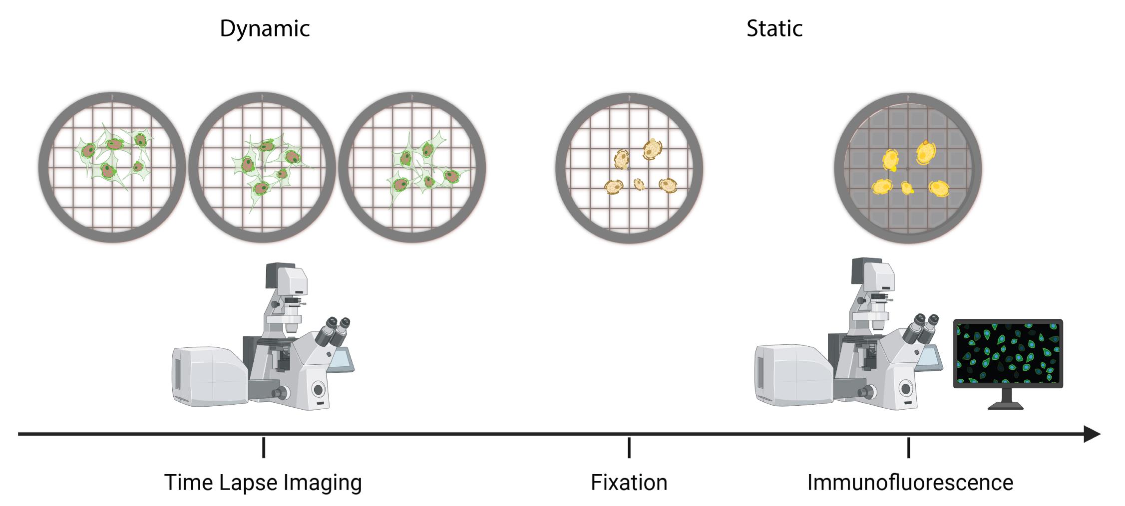
Overview of time-lapse into immunofluorescence imaging using a gridded dish
Background
Both time-lapse and immunofluorescence imaging are essential to understanding cell biology dynamics and their effects. However, by performing live cell and static experiments independently of one another, information is lost. Particularly in the field of mechanobiology, it has been difficult to connect nuclear shape fluctuations and ruptures to their causes and consequences due to the disconnect between these two types of experiments. We recently overcame this barrier through the development of TL into IF [1,2]. For instance, it is known that abnormal nuclear morphology is associated with numerous human diseases, including cancer [3]. Nuclear blebs are a type of nuclear deformation that is prone to spontaneous rupture and known to cause cellular dysfunctions, including DNA damage [4–9]. However, how nuclear blebs form and their molecular composition before and after rupture has yet to be fully elucidated. It was recently noted that decreased DNA density is the best indicator of a nuclear bleb, and that loss of lamin B1 in the bleb indicates previous nuclear rupture [1,2,10,11]. Enrichment or loss of other proteins in the bleb, such as lamin A/C, emerin, and cGAS, has also demonstrated an association with nuclear rupture [1,12,13]. These experiments, as well as numerous cell biology experiments, require a direct connection between dynamic live-cell and immunofluorescence imaging to understand cellular dynamics and their downstream effects. Current methods of tracking live-cell imaging into immunofluorescence imaging often require third-party applications such as MATLAB or matching microscope stage position coordinates [14,15]. The primary advantage of our method is the elimination of these extra steps; while the use of third-party applications allows for automated tracking, our protocol does not require any external applications. Additionally, matching stage position coordinates may prove difficult if different microscopes are used for live-cell and immunofluorescence experiments. The only special equipment needed for our protocol is a gridded imaging dish (D35-14-1.5GI, Cellvis), which has a coordinate plane engraved on the glass coverslip that is visible in transmitted light. By seeding cells on the gridded dish, cells are simply matched from time-lapse imaging to immunofluorescence imaging via their location on the coordinate plane. Gridded coverslips have been used in the past for similar imaging techniques [16,17]. Our protocol decreases the complexity of the experiment by confining it to a single imaging dish, which in turn increases its accessibility and utility for cell biology.
Materials and reagents
Biological materials
1. Mouse embryonic fibroblast wild-type NLS-GFP (MEF WT NLS-GFP) described in previous works [15,18,19]
Reagents
1. Dulbecco’s modified Eagle’s medium (DMEM) [+] 4.5 g/L glucose, L-glutamine, sodium pyruvate (Corning, catalog number: 45000-304)
2. Phosphate-buffered saline (PBS) 1× [-] calcium, magnesium (Corning, catalog number: 45000-446)
3. Characterized fetal bovine serum (FBS) (U.S.) (Cytiva HyCloneTM, catalog number: SH3007103)
4. Penicillin-streptomycin (pen/strep) solution (Corning, catalog number: MT30002CI)
5. 16% paraformaldehyde (PFA) aqueous solution, EM grade (Electron Microscopy Sciences, catalog number: 50-980-487)
6. Bovine serum albumin (BSA) fraction V (Fisher BioReagents, catalog number: BP1605-100)
7. Triton X-100 (VWR Life Science, catalog number: 9002-93-01)
8. Tween 20 molecular biology grade (Promega, catalog number: H5152)
9. 0.25% Trypsin, 0.1% EDTA in HBSS, no calcium, magnesium, and sodium bicarbonate (Corning, catalog number: MT25053CI)
10. Primary and secondary antibodies of choice
11. Hoechst (Invitrogen, catalog number: H3570)
Solutions
1. DMEM medium complete (see Recipes)
2. 4% PFA solution (see Recipes)
3. Triton X-100 solution (see Recipes)
4. Tween 20 solution (see Recipes)
5. BSA solution (see Recipes)
Recipes
1. DMEM medium complete
| Reagent | Final concentration | Quantity or volume |
|---|---|---|
| DMEM | 90% | 500 mL |
| FBS | 9% | 50 mL |
| Pen/strep | 1% | 5.5 mL |
| Total | 100% | 555.5 mL |
Thaw the frozen FBS and pen/strep in a 37 °C bead bath. Add 5.5 mL of pen/strep and 50 mL of FBS to 500 mL of DMEM. Mix by inverting. Store at 4 °C.
2. 4% PFA solution
| Reagent | Final concentration | Quantity or volume |
|---|---|---|
| 16% PFA | 25% | 3 mL |
| PBS | 75% | 9 mL |
| Total | 100% | 12 mL |
Add 3 mL of 16% PFA to 9 mL of PBS in a conical tube. Ensure the conical tube is covered in aluminum foil to avoid light exposure. Mix by inverting. Store at room temperature for up to one month.
Note: Handle PFA with gloves in a fume hood and dispose of PFA in a hazardous waste container.
3. Triton X-100 solution
| Reagent | Final concentration | Quantity or volume |
|---|---|---|
| Triton X-100 | 0.1% | 400 μL |
| PBS | 99.9% | 400 mL |
| Total | 100% | 400 mL |
Add 400 μL of Triton X-100 to 400 mL of PBS. Mix using a stir bar. Store at room temperature for up to one month.
4. Tween 20 solution
| Reagent | Final concentration | Quantity or volume |
|---|---|---|
| Tween 20 | 0.06% | 240 μL |
| PBS | 99.04% | 400 mL |
| Total | 100% | 400 mL |
Add 240 μL of Tween 20 to 400 mL of PBS. Mix using a stir bar. Store at room temperature for up to one month.
5. BSA solution
| Reagent | Final concentration | Quantity or volume |
|---|---|---|
| BSA | 2% | 500 mg |
| PBS | 98% | 25 mL |
| Total | 100% | 25 mL |
Add 500 mg of BSA to 25 mL of PBS. Mix by inverting. The BSA solution can be stored at 4 °C for up to one week.
Laboratory supplies
1. 60 mm surface-treated tissue culture dishes (Fisher, catalog number: FB012921)
2. 35 mm glass bottom dish with 14 mm micro-well #1.5 gridded cover glass (Cellvis, catalog number: D35-14-1.5GI)
3. 15 mL conical tubes (VWR International, catalog number: 470225-000)
4. 50 mL conical tubes (VWR International, catalog number: 470225-004)
5. 2 mL serological pipettes (Fisher, catalog number: 13-678-11C)
6. 5 mL serological pipettes (Fisher, catalog number: 13-678-11D)
7. 10 mL serological pipettes (Fisher, catalog number: 13-678-11E)
8. 2 μL pipette tips (Gilson, catalog number: 76178-284)
9. 20 μL pipette tips (Gilson, catalog number: 76178-282)
10. 1,000 μL pipette tips (Gilson, catalog number: 76180-360)
11. Parafilm (Amcor, catalog number: 13-374-5)
Equipment
1. Incubator (Heracell VIOS 160i Tri-Gas CO2 Incubator) (Thermo Scientific, catalog number: 51033720)
2. Pipet-Aid XP2 (Drummond Scientific Company, catalog number: 4-000-501)
3. Nutating mixer (VWR International, catalog number: 82007-202)
4. Light microscope [Nikon Instruments Ti-2E microscope, Orca Fusion Gen III Camera, Lumencor Aura III light engine, Perfect Focus System, TMC CleanBench air table, with 40× air objective (N.A 0.75, W.D. 0.66, MRH00401) or use with Crest V3 Spinning Disk Confocal]
5. Stage heater (Okolab Stage Heater, model: H401-T-Controller)
6. Stage top incubator (Okolab Stage Top Incubator, model: H301)
7. Refrigerator (4 °C)
Procedure
文章信息
稿件历史记录
提交日期: Dec 3, 2025
接收日期: Jan 15, 2026
在线发布日期: Jan 26, 2026
出版日期: Feb 20, 2026
版权信息
© 2026 The Author(s); This is an open access article under the CC BY license (https://creativecommons.org/licenses/by/4.0/).
如何引用
Lang, N., Chu, C. G. and Stephens, A. D. (2026). Time-Lapse Into Immunofluorescence Imaging Using a Gridded Dish. Bio-protocol 16(4): e5606. DOI: 10.21769/BioProtoc.5606.
分类
力学生物学
细胞生物学 > 细胞成像 > 活细胞成像
生物化学 > 蛋白质 > 免疫检测 > 免疫染色法
您对这篇实验方法有问题吗?
在此处发布您的问题,我们将邀请本文作者来回答。同时,我们会将您的问题发布到Bio-protocol Exchange,以便寻求社区成员的帮助。