

Protocol for In Utero Fetal-to-Fetal Kidney Transplantation in Rats
大鼠宫内胎间肾脏移植实验方案
发布: 2026年01月20日第16卷第2期 DOI: 10.21769/BioProtoc.5565 浏览次数: 340
评审: Komuraiah MyakalaAnonymous reviewer(s)
参见作者原研究论文
The authors used this protocol in:

Mar 2025
Advertisement


Abstract
Congenital renal disorders, such as the Potter sequence, result from renal dysgenesis. To explore a prenatal therapeutic approach for fetuses with kidney insufficiency, we established an in utero transplantation protocol using donor fetal kidneys. Although numerous rodent studies have reported cellular injections into fetal recipients, no protocol to date has described whole-organ transplantation during gestation. Here, we present a step-by-step method for grafting donor fetal kidneys (embryonic day 14.0–16.5) into allogeneic rat fetuses at embryonic day 18.0–18.5, resulting in term neonates that retain the grafts postnatally. A 15–16 G needle preloaded with the donor kidney is inserted transuterinely, depositing the organ into the subcutaneous space of the fetus. Four days later, the term pups are delivered naturally and evaluated for graft development. This protocol enables organ-level transplantation and longitudinal assessment of graft maturation within the unique fetal environment, which differs markedly from adult settings in terms of growth factor availability and immune reactivity. To our knowledge, this is the first protocol to successfully achieve whole-organ transplantation directly into fetuses in utero. Therefore, the model provides a valuable platform for studying developmental organogenesis, fetal immunology, and regenerative strategies that leverage embryonic cues.
Key features
• Subcutaneous transplantation of fetal kidneys into recipient fetuses minimizes surgical invasiveness and significantly improves fetal survival.
• Natural delivery enables pups to nurse from the dam, allowing extended postnatal observation.
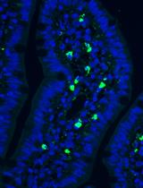
• Use of green fluorescent protein (GFP)-expressing donor tissue permits real-time visualization of graft location and growth.
• The protocol is readily adaptable for xenotransplantation and studies of immunological tolerance during fetal development.
Keywords: Fetal kidney transplantation (胎肾移植)Graphical overview
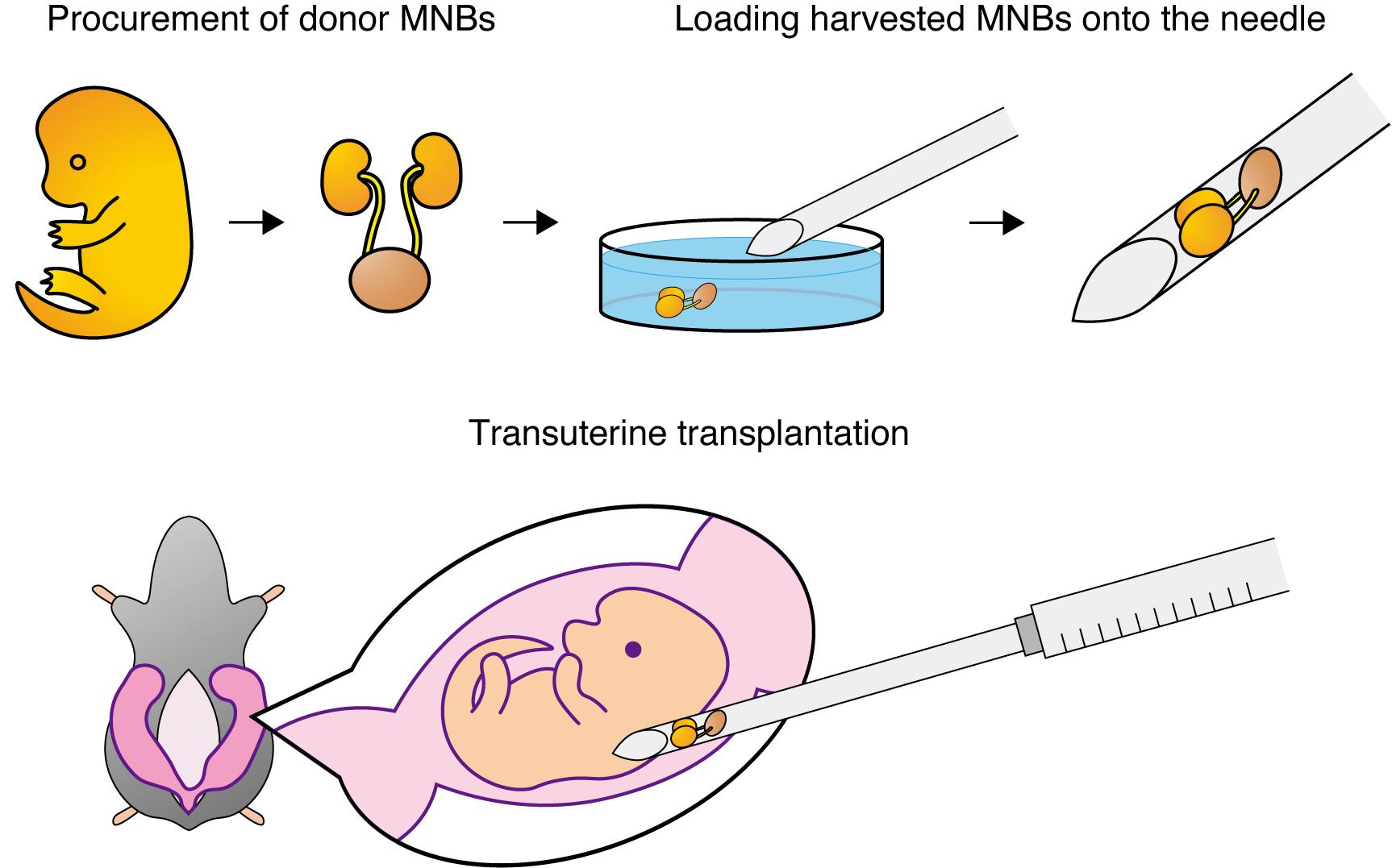
Fetal kidneys with attached bladders are first harvested from donor fetuses. Each fetal kidney–bladder unit is then loaded onto the bevel of a 15–16 G needle. After laparotomy in the pregnant rat, the uterus is exteriorized, and under a stereomicroscope, the needle is inserted transuterinely into the subcutaneous space of the recipient fetus. By expelling the contents of the needle, the donor fetal kidney is transplanted into the subcutaneous compartment of the fetus in utero. MNBs: metanephros–bladder units.
Background
The fetal environment offers a niche rich in growth factors and immunologically permissive conditions, fundamentally different from that of adult tissues [1,2]. Capitalizing on this favorable fetal environment, our protocol enables whole-organ transplantation into rat fetuses with longitudinal monitoring of graft maturation, establishing advanced research on developmental organogenesis, fetal immunology, and regenerative medicine.
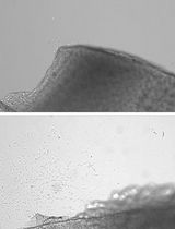
Previous in utero approaches have chiefly focused on microscale cell injections into the amniotic fluid, the peritoneal cavity, or the retroperitoneal space [3–6]. These methods accommodate fine-gauge needles, minimizing surgical trauma, but are limited to single-cell delivery and cannot support transplantation of intact organs. Whole-organ transplantation has remained challenging, as the use of larger-bore needles significantly increases fetal mortality. By introducing donor kidney–bladder units into the subcutaneous compartment, our protocol maintains fetal viability while providing a compliant space conducive to organ growth. This strategy overcomes the spatial limitation inherent to intraperitoneal and retroperitoneal delivery sites, enabling organ-scale transplantation during gestation. Furthermore, the transplanted donor kidneys become vascularized by recipient-derived vessels, allowing them to develop post-transplantation and form mature glomerular and tubular structures capable of producing urine.
Beyond modeling congenital kidney disease, this technique can be extended to evaluate xenogeneic grafts and investigate mechanisms of immune tolerance during gestation. It also offers potential for regenerative studies, as kidney organoids often exhibit limited maturation when implanted into adult hosts. Transplanting these organoids into fetal recipients may reveal developmental cues that promote more complete nephrogenesis. Therefore, this protocol not only fills a critical methodological gap in in utero transplantation but also opens new avenues for assessing organoid performance, immunological interventions, and organ-level developmental biology.
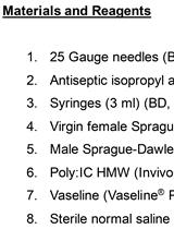
Materials and reagents
Biological materials
1. Sprague–Dawley (SD) pregnant rats (Sankyo Labo Service Corp., Japan)
2. SD-Tg [CAG-enhanced green fluorescent protein (EGFP)] pregnant rats (also called GFP-SD rats) (Sankyo Labo Service Corp., Japan)
Reagents
1. Isoflurane (Pfizer, catalog number: 2817774)
2. Pentobarbital sodium (Kyoritsu Seiyaku Corp.)
3. Ethanol, 70% (for disinfection)
4. Hank’s balanced salt solution (HBSS) (Thermo, catalog number: 14025134)
Laboratory supplies
1. Surgical scissors: standard pattern with serrations (FST, catalog number: 14007-14)
2. Ophthalmic scissors: fine iris scissors (FST, catalog number: 14094-11)
3. Microforceps: Dumont medical micro-blunted atraumatic tipped forceps (FST, catalog number: 11253-25)
4. Ring forceps (Natsume Seisakusho Co., catalog number: A-26)
5. Castroviejo needle holder (FST, catalog number: 12565-14)
6. 5-0 silk suture (Natsume Seisakusho Co., catalog number: CF1250B2NT)
7. 15–16 G needle (Saito Medical Instruments Inc.)
8. 10 cm sterile culture dish (AS ONE Co., catalog number: GD90-15)
9. Sterile gauze
10. 1 mL syringe (TERUMO, catalog number: SS-01T)
11. Warming pad
12. Polystyrene box (size L260 mm × H175 mm × W160 mm)
13. Ice
Equipment
1. Stereomicroscope (Leica Microsystems, model: M205FA)
2. Isoflurane anesthesia system (BioMedical Science, model: TK-40)
Procedure
文章信息
稿件历史记录
提交日期: Jul 31, 2025
接收日期: Dec 7, 2025
在线发布日期: Dec 29, 2025
出版日期: Jan 20, 2026
版权信息
© 2026 The Author(s); This is an open access article under the CC BY license (https://creativecommons.org/licenses/by/4.0/).
如何引用
Morimoto, K., Yamanaka, S. and Yokoo, T. (2026). Protocol for In Utero Fetal-to-Fetal Kidney Transplantation in Rats. Bio-protocol 16(2): e5565. DOI: 10.21769/BioProtoc.5565.
分类
发育生物学 > 器官形成
细胞生物学 > 细胞移植 > 同种异体移植
免疫学 > 动物模型 > 大鼠
您对这篇实验方法有问题吗?
在此处发布您的问题,我们将邀请本文作者来回答。同时,我们会将您的问题发布到Bio-protocol Exchange,以便寻求社区成员的帮助。