

Quantification of Protochlorophyllide (Pchlide) Content in Arabidopsis Seedlings Using a High-Performance Liquid Chromatography (HPLC) System
利用高效液相色谱法(HPLC)对拟南芥幼苗中原叶绿素酸酯(Pchlide)的含量进行定量分析
(*contributed equally to this work) 发布: 2026年01月05日第16卷第1期 DOI: 10.21769/BioProtoc.5553 浏览次数: 661
评审: Anonymous reviewer(s)
参见作者原研究论文
The authors used this protocol in:

Oct 2025
Advertisement


Abstract
The protochlorophyllide (Pchlide) level is a crucial indicator of plant fitness. Precise quantification of Pchlide content is necessary not only in studies of flu-related mutants that over-accumulate Pchlide in the dark but also for research on plants suffering from environmental stresses. Due to its low content and interference of chlorophylls, quantitative determination of Pchlide content is a challenge. Here, we describe an optimized protocol for Pchlide extraction from Arabidopsis thaliana seedlings and subsequent analysis using high-performance liquid chromatography (HPLC) coupled with fluorescence detection. Divinyl-Protochlorophyllide (DV-Pchlide, the major form of Pchlide in plants) quantification is achieved by interpolating fluorescence peak areas against an experimentally derived standard curve. This protocol provides a reliable workflow for Pchlide quantification, facilitating the deciphering of the underlying mechanism of plant environmental resilience.
Key features
• This method adopts acetone as a solvent for both Pchlide extraction and HPLC run.
• This protocol adopts a gradient HPLC system equipped with a fluorescence detector.
• This protocol applies an experimentally derived standard calibration curve using synthetic DV-Pchlide.
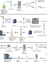
Keywords: DV-Pchlide (二乙烯基-原卟啉酸)Graphical overview

Background
Chlorophylls (Chls) are the most abundant organic pigment molecules on Earth. As components of photosynthesis machinery, Chls absorb light energy that is mostly used in photosynthesis. However, free Chl and most of its biosynthetic intermediates are particularly destructive once they become excited after light absorption [1]. These excited porphyrin molecules can interact with the surrounding ground-state oxygen molecule, leading to the highly reactive singlet oxygen (1O2) [2], which causes photooxidative damage and eventually programmed cell death (PCD) [3]. Thereby, the plant exerts strict control over tetrapyrrole biosynthesis [4,5]. In plants, Chl biosynthesis halts at the step of protochlorophyllide (Pchlide) generation due to the inhibitory effect of the FLU protein and resumes upon light illumination when Pchlide is photo-reduced to Chlide [6]. The Arabidopsis flu mutant over-accumulates Pchlide in the chloroplast in darkness and generates 1O2 when transferred to light due to the photosensitizing activity of the Pchlide molecules [7]. In the flu mutant, the level of generated 1O2 after dark-to-light shift is positively correlated with the amount of accumulated Pchlide, i.e., the duration of dark treatment [8]. However, when flu mutants are grown under continuous light, only a very low level of 1O2 is produced since Pchlide is immediately used for Chl biosynthesis, and the flu mutant grows exactly like the wild-type plants. These properties of the flu mutant make it an ideal tool for controlled generation of 1O2, and the exploration of this mutant has led to the identification of at least two chloroplastic 1O2-induced retrograde signaling pathways: the 1O2-EX1 pathway and 1O2-SAFE1 pathway [9–13].
Since Pchlide is a critical indicator of 1O2 level, precise quantification of its content is necessary not only for studies of flu-related mutants but also of plants suffering from environmental stresses [14–16]. In the etiolated flu seedlings, Pchlide accumulation can be directly visualized by its characteristic red fluorescence under blue light. However, Pchlide accumulation can hardly be detected in dark-incubated green leaves due to the interference of pigments, especially Chls, and is generally quantified using a fluorescence spectrophotometer [13,17] or high-performance liquid chromatography (HPLC) [11,18]. Compared with the former, HPLC-based Pchlide quantification is more accurate and reliable. Here, we provide a simple procedure for Pchlide extraction from Arabidopsis thaliana seedlings and a detailed workflow for Pchlide separation and detection using a fluorescence detector-equipped HPLC system, based on our recent publication [11]. Pchlide (DV-Pchlide) quantification is achieved based on fluorescence peak area and an experimentally derived standard curve.
Materials and reagents
Biological materials
1. 4-day-old etiolated Arabidopsis thaliana seedlings grown in darkness
2. 7-day-old Arabidopsis thaliana seedlings grown under continuous light and pretreated in darkness for 8 h
Reagents
1. Murashige & Skoog basal medium with vitamins (PhytoTech Labs, catalog number: M519); store at 2–8 °C
2. Agar (Merck KGaA, CAS number: 9008-12-0)
3. Acetone (HPLC grade) (SINOPHARM, CAS number: 67-64-1)
4. Water (HPLC grade)
5. Methanol (HPLC grade) (Thermo Fisher Scientific, CAS number: 67-56-1)
6. Acetic acid (Beijing Chemical Plant Co., Ltd., CAS number: 64-19-7)
7. Ammonia water (MODERN ORIENTAL FINE CHEMISTRY, CAS number: 1336-21-6)
8. Divinyl-protochlorophyllide (in solution) (HPLC grade) (ZZSTANDARD, CAS number: 18433-30-2); store at ≤-70 °C
9. KOH (Shanghai Macklin Biochemical Co., Ltd., CAS number: 1310-58-3)
10. Sucrose (Sigma, CAS number: 57-50-1)
11. MES (Sigma, CAS number: 145224-94-8)
Solutions
1. Half-strength Murashige and Skoog (1/2 MS) plant growth medium (see Recipes)
2. Pchlide extraction buffer (see Recipes)
Recipes
1. 1/2 MS plant growth medium
| Reagent | Final concentration | Quantity or volume |
|---|---|---|
| Murashige & Skoog basal medium with vitamins | n/a | 2.215 g |
| Sucrose | 1% (m/v) | 10 g |
| MES | 0.05% (m/v) | 0.5 g |
| ddH2O | n/a | Top to 1 L |
| Agar | 1% (m/v) | 10 g |
Dissolve all ingredients, except agar, in ddH2O in a beaker and adjust pH to 5.7–5.8 with KOH before agar is added. Autoclave for 15 min at 121 °C and store at room temperature.
2. Pchlide extraction buffer
| Reagent | Final concentration | Quantity or volume |
|---|---|---|
| Acetone | 90% (v/v) | 90 mL |
| Ammonia water | 0.1% (v/v) | 30 μL |
| Total | n/a | Top up to 100 mL with ddH2O |
Note: Store in darkness.
Laboratory supplies
1. 1.5 mL microcentrifuge tubes [Corning Life Sciences (Wujiang) Co., Ltd., catalog number: AXYMCT150C)
2. PES syringe filters, pore 0.22 μm (Tianjin JINTENG Experiment Equipment Co., Ltd., catalog number: JTSF025011)
3. Sterile 1 mL syringe (Shanghai Kindly Enterprise Development Group Co., Ltd. catalog number: 60017031)
4. HPLC 2 mL brown glass vials/caps (Shanghai Titan Scientific Co., Ltd., catalog number: 0204124-FXJYP-0016/02041969-FXJYP-0034)
5. HPLC 200 μL inserts [ANPEL Laboratory Technologies (Shanghai) Inc., catalog number: VDAP-4025PBS-631-100)
Equipment
1. Plant growth chamber [Xunon Instrument (Beijing) Co., Ltd, model: PT-G600)
2. Analytical balance [Shanghai Yueping Scientific Instrument (Suzhou) Manufacturing Co., Ltd., model: FA2204B)
3. Ball mill (DHS Life Science & Technology Co., Ltd., model: TL2010S)
4. Stainless steel grinding beads (Karryda Laboratory Solutions, model: YMZ-S3)
5. Refrigerated centrifuge (Eppendorf, model: 4025R)
6. Laboratory vacuum degassing unit (Sciencetool International Group Co., Ltd., model: T242/DV-9252)
7. Gradient HPLC system (Agilent, model: 1290 Infinity) with a fluorescence detector (Agilent, model: 1260 FLD)
8. Reversed-phase chromatography column (SinoPak, model: BEH T-C18, 5 μm, ID 4.6 mm × 250 mm)
Software and datasets
1. Origin (OriginLab, Version: 2021)
Procedure
文章信息
稿件历史记录
提交日期: Oct 21, 2025
接收日期: Nov 20, 2025
在线发布日期: Dec 3, 2025
出版日期: Jan 5, 2026
版权信息
© 2026 The Author(s); This is an open access article under the CC BY-NC license (https://creativecommons.org/licenses/by-nc/4.0/).
如何引用
Zhang, F., Zhang, L. and Wang, L. (2026). Quantification of Protochlorophyllide (Pchlide) Content in Arabidopsis Seedlings Using a High-Performance Liquid Chromatography (HPLC) System. Bio-protocol 16(1): e5553. DOI: 10.21769/BioProtoc.5553.
分类
植物科学 > 植物生物化学 > 其它化合物
生物化学 > 其它化合物 > 叶绿素
植物科学 > 植物生理学 > 非生物胁迫
您对这篇实验方法有问题吗?
在此处发布您的问题,我们将邀请本文作者来回答。同时,我们会将您的问题发布到Bio-protocol Exchange,以便寻求社区成员的帮助。