

Measuring Anti-aging Effects in Drosophila
果蝇中抗衰老效应的测量方法
发布: 2025年05月05日第15卷第9期 DOI: 10.21769/BioProtoc.5305 浏览次数: 2355
评审: Kai YuanAnonymous reviewer(s)
参见作者原研究论文
The authors used this protocol in:
Feb 2024
Advertisement


Abstract
One of the major factors contributing to aging and age-related diseases is the well-understood decline in the function of adult stem cells. Quantifying the degree of aging in adult stem cells is essential for advancing anti-aging mechanisms and developing anti-aging agents. However, no systematic approach to this exists. In this study, we developed a method to quantitatively assess the degree of aging in adult intestinal stem cells using a Drosophila midgut model and two aging markers. First, aging was induced in Drosophila with the desired genotype, and the anti-aging agent was administered 7 days before dissection. Then, the levels of two intestinal stem cell aging markers found in Drosophila (PH3 and γ-tubulin) were measured using immunohistochemistry. Finally, fluorescence microscopy was employed to count the number of aging markers and take images, which were analyzed using image analysis software. Using this approach, we quantitatively analyzed the effects of anti-aging agents on the aging of adult intestinal stem cells. This methodology is expected to significantly expedite the development of anti-aging agents and substantially reduce the research costs associated with aging-related studies.
Key features
• PH3 and γ-tubulin serve as reliable markers for quantitatively assessing aging in Drosophila intestinal stem cells.
• This method for discovering anti-aging agents involves processes such as aging induction, treatment with anti-aging agents, dissection, fixation, antibody staining, and analysis of the results.
• Vitamin D, similar to metformin and β-hydroxybutyrate, is an anti-aging agent.
• Quantitative analysis of adult stem cell aging will enable the rapid and accurate identification of anti-aging agents and efficacy validation.
Keywords: Drosophila (果蝇)Graphical overview
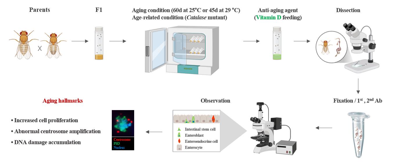
Experimental setup and procedure for measuring anti-aging effects
Background
Adult stem cells play a key role in tissue homeostasis and regeneration because of their ability to self-renew and generate differentiated cells. Age-related changes in these stem cells are closely associated with aging and age-related tissue diseases [1,2]. Many studies have aimed to identify changes in adult stem cells related to aging and develop anti-aging agents to slow the aging process [3–8]. Although studies have shown that various factors contribute to aging, there are limitations to quantifying these factors. Currently, there is no statistical method for quantifying adult stem cell aging.
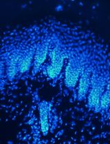
First, a suitable model system is required to quantitatively study adult stem cell aging. Drosophila, due to their short life span and genetically modifiable midgut function, are a widely utilized model organism for aging research, encompassing investigations of adult stem cells and their microenvironmental as well as changes associated with aging [2,8,9]. In the adult Drosophila midgut, intestinal stem cells (ISCs), marked by delta (Dl), are the only proliferation cells that give rise to two distinct differentiated progenies: enterocytes (ECs), which arise from enteroblasts (EBs) induced by strong Notch (N) signaling, and enteroendocrine (EE) cells, which are derived from EBs activated by weak N signaling [10]. These four types can be identified using specific markers such as Dl (ISCs), phospho-histone H3 (PH3, dividing ISCs), esg-GFP (ISCs and EBs), Su-GFP (EBs), Pdm and Myo-GFP (ECs), and Prospero (Pros) and Pros-GFP (EE cells) [7–8,10].
Second, utilizing appropriate markers is crucial for quantitatively assessing ISC aging. Using an anti-PH3 antibody, Choi et al. revealed age-related increases in Drosophila ISC division, which was corroborated by subsequent studies [3,11,12–16]. The anti-PH3 antibody specifically binds to phospho-histone H3 at serine 10, a modification that occurs during mitosis; therefore, it is widely used as a marker for identifying mitotic cells [14]. Using an anti-γH2AX antibody, Park et al. observed age-related increases in DNA damage accumulation in ISCs [17]. The anti-γH2AX antibody specifically recognizes and binds to phosphorylated histone H2AvD at serine 137, a modification analogous to the γH2AX phosphorylation in mammals [17]. Using an anti-γ-tubulin antibody, Park et al. revealed that age-related increases in abnormal centrosome amplification occur in ISCs and renal stem cells (RSCs) [14,15,18]. The anti-γ-tubulin antibody specifically recognizes and binds to γ-tubulin, a key protein in microtubule nucleation and organization in the division of eukaryotic cells [14,15].
Third, sufficient aging and age-related conditions are required to quantitatively study adult stem cell aging. It is well established that 60-day-old flies maintained at 25 °C and 45-day-old flies maintained at 29 °C serve as effective models for aging when compared to young flies [14–17]. In addition, the model of intrinsic oxidative stress Catalase heterozygous mutant flies is a widely used age-related model [14,18].
Fourth, appropriate anti-aging agents are essential for quantitatively assessing their effects on adult ISCs. Previous studies have reported that metformin, a commonly prescribed medication for the management of type 2 diabetes, and β-hydroxybutyrate, a type of ketone body produced during periods of fasting, prolonged exercise, or low-carbohydrate diets, exhibit anti-aging properties [15,19]. In recent studies, age-related declines in Vitamin D (VitD) synthesis and VitD receptor (VDR) expression have been associated with age-related diseases [16]. However, few studies have explored the role of VitD/VDR in adult stem cells, highlighting the need for further related investigations. In this study, by utilizing the age markers of Drosophila ISCs and age-related conditions, we aimed to provide a detailed and precise method for the quantitative measurement of adult stem cell aging. This method can be used for the quantitative analysis of anti-aging agent development and efficacy research, which will contribute to reducing research costs and shortening the research period spent in anti-aging fields in the future.
Materials and reagents
Biological materials
1. Oregon-R (Bloomington Drosophila Stock Center, BDSC, Bloomington, IN, USA, #5)
2. Catalase heterozygous mutant flies (Catn1 mutant) (BDSC, catalog number: 4014) [20]
3. UAS-hr96-RNAi lines (Vienna Drosophila RNAi Center, VDRC, Vienna, Austria, catalog number: 330288, catalog number: 10958)
4. Myo1A-Gal80ts flies, obtained from B.A. Edgar
5. Rat anti-GFP antibody (Nacalai Tesque Inc., Kyoto, Japan, catalog number: 04404-84)
6. Rabbit anti-phospho-histone H3 (PH3) (Millipore, Billerica, MA, USA, catalog number: 06-570)
7. Mouse anti-γ-tubulin (Sigma-Aldrich, St. Louis, MO, USA, catalog number: T6557-.2ML)
8. Goat anti-rat FITC antibody (Jackson ImmunoResearch, catalog number: 415-095-166)
9. Goat anti-rabbit Alexa Fluor® 647 antibody (Jackson ImmunoResearch, catalog number: 305-605-003)
10. Goat anti-mouse Cy3 antibody (Jackson ImmunoResearch, catalog number: 205-165-108)
Reagents
1. EM-grade paraformaldehyde 16% aqueous solution (Electron Microscopy Science, catalog number: 15710)
2. 10× phosphate-buffered saline (PBS) (Ambion, catalog number: AM9624)
3. Methanol (Sigma-Aldrich, CAS number: 67-56-1)
4. 10% Triton X-100 (Millipore, CAS number: 9036-19-5)
5. 4',6-diamidino-2-phenylindole (DAPI) (Molecular Probes, catalog number: D1306)
6. Vectashield (Vector Laboratories, catalog number: H-1000-10)
7. 1α,25-Dihydroxyvitamin D3, D1530 (VitD) (Sigma-Aldrich, catalog number: D1530)
8. Ultra-pure water (Welgene Inc., catalog number: ML 019-02)
9. Propionic acid (Junsei Chemical Co., CAS number 79-09-4)
10. Butyl 4-hydroxybenzoate (Junsei Chemical Co., Ltd., CAS number 94-26-8)
Solutions
1. Standard food (see Recipes)
2. Vitamin D food (VitD) (see Recipes)
3. Bokinin (see Recipes)
Recipes
1. Standard food
| Reagent | Final concentration | Quantity or Volume |
|---|---|---|
| Sucrose | 10% (w/v) | 100 g |
| Cornmeal | 7% (w/v) | 70 g |
| Yeast | 2% (w/v) | 20 g |
| Agar | 1% (w/v) | 10 g |
| Propionic acid | 0.5% (v/v) | 5 mL |
| Bokinin | 0.3% (v/v) | 3 mL |
| Water | 79.2% (v/v) | 1 L |
2. Vitamin D food (VitD)
| Reagent | Final concentration | Quantity or Volume |
|---|---|---|
| 10 mM Vitamin D | 100 nM (v/v) | 0.1 mL |
| Standard food | n/a | 9.9 mL |
| Total | n/a | 10 mL |
3. Bokinin
| Reagent | Final concentration | Quantity or Volume |
|---|---|---|
| Butyl 4-hydroxybenzoate | 10% (v/v) | 10 g |
| Ethanol | n/a | 70 mL |
| Water | n/a | 30 mL |
Laboratory supplies
1. Microtube (Axygen, model: MCT-175-X)
2. Microscopic slides (Paul Marienfeld GmbH & CoKG, model: 31593)
3. Microscope cover glasses (24 × 32 mm, Deckglaser, model: HSU-0101172)
4. Tip 20 μL (Axygen, model: TF-20-RSm TF-200-R-S, TF-1000-R-S)
5. Vonetex latex gloves (Hartalega Sdn. Bhd.)
Equipment
1. Microscope (Carl Zeiss Inc., model: Axioskop 2 Plus)
2. Aerobic incubator (Sanyo, model: MIR-254-PK)
3. Sigma Plot 14.5 (Systat Software Inc., San Jose, CA, USA)
4. Stereomicroscope with Axiocam 208 color camera (Zeiss, model: Stemi 508)
5. Fluorescent microscope with AxioCam MRm (Zeiss, model: Axioplan2)
6. Precision tweezer (Techni-Tool Tweezer Ni-Cr-Mo Superalloy, Type 5, Techni-Tool, Worcester, PA; catalog number: 33-04594-01)
7. Single orbital shaker (Daeil biotech., model: PM-6249)
Software and datasets
1. AxioVision Rel. 4.8 software (Carl Zeiss Inc.)
2. SigmaPlot 14.5 package (Systat, Software Inc., San Jose, California, USA)
Procedure
文章信息
稿件历史记录
提交日期: Feb 3, 2025
接收日期: Apr 7, 2025
在线发布日期: Apr 24, 2025
出版日期: May 5, 2025
版权信息
© 2025 The Author(s); This is an open access article under the CC BY license (https://creativecommons.org/licenses/by/4.0/).
如何引用
Na, H. and Park, J. (2025). Measuring Anti-aging Effects in Drosophila. Bio-protocol 15(9): e5305. DOI: 10.21769/BioProtoc.5305.
分类
发育生物学 > 细胞生长和命运决定 > 成熟衰老
细胞生物学 > 细胞染色 > 蛋白质
干细胞 > 成体干细胞 > 肠道干细胞
您对这篇实验方法有问题吗?
在此处发布您的问题,我们将邀请本文作者来回答。同时,我们会将您的问题发布到Bio-protocol Exchange,以便寻求社区成员的帮助。