

Cryo-SEM Investigation of Chlorella Using Filter Paper as Substrate
利用滤纸作为基质的冷冻扫描电镜研究绿球藻
发布: 2024年12月20日第14卷第24期 DOI: 10.21769/BioProtoc.5143 浏览次数: 1488
评审: Alba BlesaSimab KanwalAnonymous reviewer(s)
Advertisement


Abstract
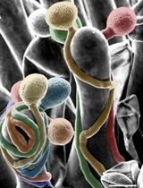
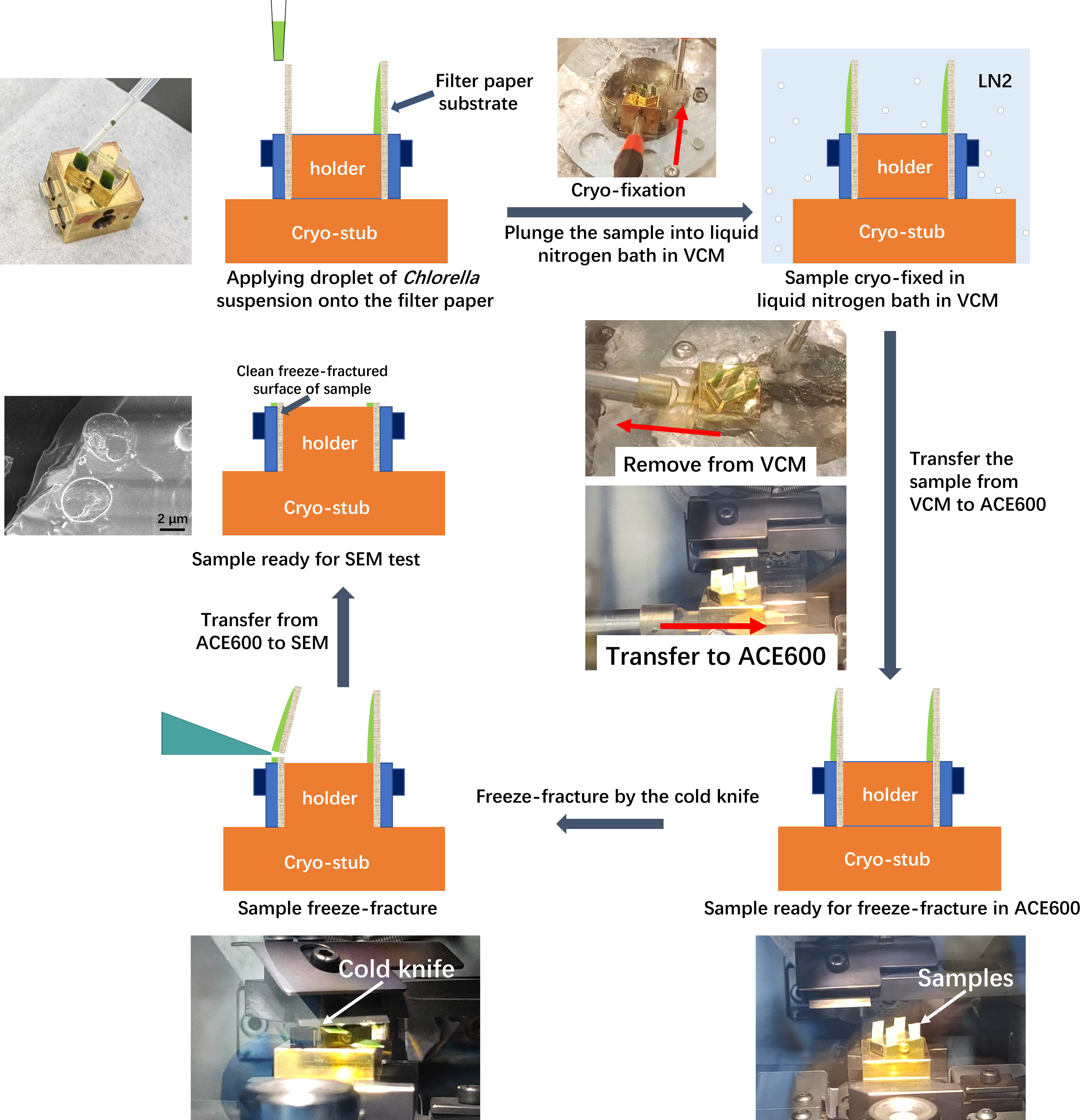
Cryo-electron microscopy (cryo-EM) is a powerful technique capable of investigating samples in a hydrated state, compared to conventional high-vacuum electron microscopy that requires samples to be completely dry. During the drying process, numerous features and details may be lost due to damage caused by dehydration. Cryo-EM circumvents these problems by cryo-fixing the samples, thereby retaining the intact and original features of hydrated samples. This protocol describes a step-by-step cryo-scanning electron microscopy (cryo-SEM) experimental procedure with Chlorella sorokiniana as the subject. By employing filter paper as the sample substrate, we propose a simple and reliable method for cryo-fixation and freeze-fracture of Chlorella sorokiniana in water suspension. The advantage of using filter paper as a substrate lies in its ability to support a thin film of sample, enabling a cold knife to make a cut effortlessly and produce a clean freeze-fractured surface for SEM investigation. By following the approach described in this protocol, both the internal structure and surface morphology of Chlorella sorokiniana can be easily resolved with high quality. This protocol is highly versatile and can be applied to samples dispersed in water or solvents, including cyanobacterial cells, algal cells, and any kind of sample that can be adsorbed onto filter paper.
Key features
• Introducing a reliable way for ideal freeze-fracture of a water-suspended sample using filter paper as substrate.
• Detailed step-by-step descriptions of the entire experiment, covering how to operate the instruments and including some practical experimental tips.
Keywords: Cryo-SEM (冷冻扫描电镜)Graphical overview
Background
Cryo-SEM is an irreplaceable approach for investigating biological samples in a hydrated state [1–5], and it has been proven to be a powerful tool for studying the microstructure and surface morphology of algae and cells [6–11]. There are four main procedures for sample treatment in cryo-SEM, including cryo-fixation, freeze-fracture, freeze-etching, and sputter coating [12,13]. Cryo-fixation is a technique for rapidly freezing a sample to preserve its structure in a near-native state [14,15]. Typically, a liquid nitrogen bath at atmospheric pressure, liquid nitrogen slush, liquid nitrogen under high pressure (for high-pressure freezing), and liquid ethane and propane are employed as cryogenic substances [14–17]. Freeze-fracture is a technique that fractures the cryo-fixed sample in order to reveal its internal structure, typically by a cold knife or a cryo-ultramicrotome [18]. Freeze-etching is the process of getting the fractured surface etched by sublimation of ice, which reveals fine details of the sample's structure. Sputter coating in cryo-state is similar to the conventional sputter coating, except that the sample is maintained in a cryogenic state.
There are some valuable protocols that have been published regarding plant and animal tissues as well as other biomaterials [1–6,9,13]. Nevertheless, only a limited number of protocols are available for cryo-SEM of algae. Research articles regarding cryo-SEM investigation of algae samples usually include a brief description of the cryo-SEM experiments but do not provide much information about the detailed and visualized experimental procedures. Particularly, information such as how the sample is prepared and how it is freeze-fractured is crucial for obtaining fine details of the internal structures.
The technique of freeze-fracture has been widely used in a broad range of samples, such as fluid samples, hydrated polymers and microgels, and plant tissues [18–20]. In the freeze-fracture procedure, a cold knife in a cryogenic state is commonly employed as the cutting tool: a blade with a sharp edge that is positioned above the sample. It sweeps across the top of the sample in parallel with the sample holder. When it hits the sample, preferably a prominent part or a tip, the sample is then freeze-fractured [1]. During this process, a certain type of sample holder or a specific way to hold the sample is employed. Due to the diverse characteristics of hydrated samples, such as water content, viscosity, strength, shape, or amount, various ways for sample holding are required. Consequently, more and more sample holders or approaches for holding samples are being developed. For example, Mo et al. developed a sample stage (holder) for the freeze-fracture of liquid, semi-liquid, and viscous samples, including chlorella [21]. Nonetheless, reliable protocols with easy and flexible sample-holding approaches that are compatible with various types of samples are still needed.
In this protocol, we propose an easy and reliable approach to hold and support the sample by employing a piece of filter paper as the substrate. The advantage of filter paper lies in its ability to support a thin film of sample, enabling a cold knife to make a cut and produce a clean freeze-fractured surface. This method is versatile and applicable to a broad range of samples, including cyanobacterial cells, algal cells, and any kind of sample that can be adsorbed onto a filter paper. Along with detailed, step-by-step operations for cryo-SEM tests, the authors believe that this protocol will be helpful for researchers to commence the design and operation of their cryo-SEM experiments.
Materials and reagents
Chlorella sorokiniana UTEX 1602 (Institute of Hydrobiology, Chinese Academy of Science, China, FACHB-275). In the following text, Chlorella sorokiniana UTEX 1602 is referred to as Chlorella sorokiniana
Filter paper (Hangzhou Special Paper Industry Co., Ltd, China, NEWSTAR) with a medium filtration speed. The thickness of the filter paper is 0.16 mm, and the pore size is approximately 7 μm. The diameter of a single piece of filter paper is 7 cm
Disposable transfer pipette (1 mL) (Jiangsu KangJian medical apparatus Co., Ltd, China, KANG JIAN, catalog number: KJ619)
Disposable sterile centrifuge tube (50 mL) (Hunan BKMAM Holding Co., Ltd, China, BKMAMLAB, catalog number: 20231206)
Liquid nitrogen (Dalian KeNa Technology Co., Ltd, China)
Equipment
Vacuum cryo transfer system (Leica, model: EM VCT500)
Field emission scanning electron microscopy (JEOL, model: JSM-7610 Plus)
Software and datasets
PC-SEM, v. 4.0.0.8 (Copyright 2006–2018 JEOL Ltd.)
Microsoft Office 2021 (Microsoft, https://www.office.com)
Jiangying Pro (v. 6.0.1, https://www.capcut.cn)
Procedure
文章信息
稿件历史记录
提交日期: Jul 24, 2024
接收日期: Oct 14, 2024
在线发布日期: Nov 4, 2024
出版日期: Dec 20, 2024
版权信息
© 2024 The Author(s); This is an open access article under the CC BY-NC license (https://creativecommons.org/licenses/by-nc/4.0/).
如何引用
Wan, P., Tao, M., Zhou, Y., Han, W., Wang, J. and Wang, J. (2024). Cryo-SEM Investigation of Chlorella Using Filter Paper as Substrate. Bio-protocol 14(24): e5143. DOI: 10.21769/BioProtoc.5143.
分类
微生物学 > 微生物细胞生物学 > 细胞成像
细胞生物学 > 细胞成像 > 电子显微镜
生物物理学 > 电子冷冻断层扫描
您对这篇实验方法有问题吗?
在此处发布您的问题,我们将邀请本文作者来回答。同时,我们会将您的问题发布到Bio-protocol Exchange,以便寻求社区成员的帮助。