

An Improved Focus-Forming Assay for Determination of the Dengue Virus Titer
登革病毒滴度测定改良聚焦形成法

发布: 2024年10月20日第14卷第20期 DOI: 10.21769/BioProtoc.5084 浏览次数: 2458
评审: Luis Alberto Sánchez VargasRan ChenDebashis Dutta
Advertisement


Abstract
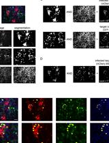
Dengue virus (DENV), a common and prevalent mosquito-borne endemic disease, is caused by four serotypes (DENV-1–4) and has spread rapidly on a global scale over the past decade. A crucial step in the development of antiviral therapeutics requires the utilization of in vitro cell-based techniques, such as plaque assays and focus-forming assays (FFA) for virus quantification. Vero cells have been widely used for FFA and plaque assay; however, there are instances when their efficacy and efficiency in the detection of certain clinical DENV isolates are low. Here, we showed that BHK-21 cells are more sensitive than Vero cells in the detection of all DENV-1–4 plaques and foci. In addition, we developed an improved FFA protocol for the quantification of all four DENV serotypes. Using a pan-flavivirus envelope (E) antibody, we reduce the possibility of false positives by defining a focus to consist of a minimum of eight infected cells. We outlined a protocol using the Operetta® high-content imaging system to automate the digital capture of these infected cells. A pipeline was also designed using the CellProfilerTM automated image analysis software to detect these foci. We then compare the results of the improved FFA with plaque assay. Notably, the improved FFA detected clear foci of the DENV-4 strain that does not form distinct plaques. We subsequently demonstrated the potential application of the improved FFA protocol in antiviral testing, utilizing a nucleoside inhibitor of DENV, NITD008 as a control. The protocol is amenable to a diverse array of applications, including high-throughput compound screening (HTS).
Key features
• An improved focus-forming assay that has the potential to quantify the DENV-4 strain, which was previously hard to plaque.
• Improvements have been made to reduce the possibility of false positives.
• Improved workflow using automated digital imaging process and counting of foci.
• Applicable to antiviral compounds screening and is amenable to high-throughput screening.
Keywords: DENV clinical isolates (登革病毒临床分离株)Background
Dengue represents a significant challenge to global public health systems, with over 40% of the global population at risk of infection. It is estimated that approximately 400 million people are infected with dengue virus (DENV) each year, of whom 100 million manifest clinical symptoms of dengue hemorrhagic fever with 25,000 deaths reported annually [1,2]. The symptoms include the abrupt onset of fever, which is followed by a multitude of other critical complications, including circulatory failure, hemorrhages in the skin and gastrointestinal tract, and shock. DENV comprises four antigenically distinct serotypes, exhibiting variations in both structural and nonstructural proteins. The four antigenically distinct serotypes of dengue virus are designated DENV-1, DENV-2, DENV-3, and DENV-4 [3,4]. The necessity for efficient, accurate, and robust assays for the quantification of infectious viruses, which contribute to the discovery of antiviral drugs, has thus increased. A variety of techniques have been explored and developed over the years, with plaque and/or focus-forming assays (FFA) still representing the gold standard for such assays. A variety of cell lines have been employed in these assays, with Vero (African green monkey kidney fibroblasts) cells being the most frequently utilized in plaque assay [5]. Furthermore, a number of optimization protocols for FFA have been documented for a range of viruses, including DENV [6,7]. However, this method typically necessitates a great number of steps, including the use of multiple distinct monoclonal antibodies for foci visualization; moreover, the method was not extended to DENV-3 and DENV-4 detection. In this work, we delineate the methodology for employing the FFA for the quantification of infectious virus particles using E protein detection in all serotypes and compare it with plaque assay quantification of infectious virus production. We demonstrate that the FFA approach was more sensitive than the plaque assay in detecting DENV-4, which exhibited poor plaque formation. In addition, all four DENV serotypes form clearer plaques and foci in BHK-21 cells than in Vero cells. Moreover, improvements have been made to the existing FFA. For example, we defined a focus to comprise a minimum of eight cells, thereby reducing the possibility of false positives. We also outlined a protocol using the Operetta® high-content imaging system and a pipeline using the CellProfilerTM software to automate foci imaging and foci counting. In addition, we demonstrate the immediate application of FFA as a tool for assessing the antiviral efficacy of compounds using a known adenosine nucleoside DENV inhibitor, NITD008 [9,10], as a control. Altogether, the improved FFA is a precise and reliable assay not only for quantifying clinical isolates but also for high-throughput screening of compounds for dengue drug discovery.
Materials and reagents
Biological materials
Representative Dengue virus strains from EDEN study [8]: DENV-1 clinical isolate (EDEN1: GenBank accession EU081230), DENV-2 clinical isolate (EDEN2: GenBank accession EU081177), DENV-3 clinical isolate (EDEN3: GenBank accession EU081190), DENV-4 clinical isolate (EDEN4: GenBank accession GQ398256)
Aedes albopictus C6/36 cell line (ATCC, CRL-1660TM)
Baby hamster kidney fibroblast, BHK-21 (ATCC, CCL-10TM)
African green monkey kidney fibroblast, Vero cell line (ATCC, CCL-81TM)
Hybridoma cells; 4G2 (ATCC, HB-112TM)
Reagents
RPMI1640 medium (Thermo Fisher Scientific, Gibco®, catalog number: 11875093)
DMEM (1×), high glucose, pyruvate medium (Thermo Fisher Scientific, Gibco®, catalog number: 11995-040)
Heat-inactivated fetal bovine serum (FBS) (Thermo Fisher Scientific, Gibco®, catalog number: 10082147)
200 mM L-glutamine (Thermo Fisher Scientific, Gibco®, catalog number: 25030081)
Penicillin and streptomycin (pen/strep) (Thermo Fisher Scientific, Gibco®, catalog number: 15140122)
1 M HEPES (Thermo Fisher Scientific, Gibco®, catalog number: 15630-080)
0.25% Trypsin-EDTA (Thermo Fisher Scientific, Gibco®, catalog number: 25200-056)
Protein-free hybridoma medium (PFHM-II medium) (Thermo Fisher Scientific, Gibco®, catalog number: 12040-077)
1× PBS (1st BASE, catalog number: BUF-2040-10X1L)
Ethanol (EtOH) molecular biology grade (Merck, Sigma-Aldrich, catalog number: 51976)
Hydrochloric acid (Merck, Sigma-Aldrich, catalog number: 258148)
RPMI1640 powder (Thermo Fisher Scientific, Gibco®, catalog number: 31800-022)
DMEM powder, high glucose (Thermo Fisher Scientific, Gibco®, catalog number: 12100046)
Sodium Bicarbonate (Thermo Fisher Scientific, catalog number: 25080094)
Tris (1st BASE, catalog number: BIO-1400)
Glycine (Merck, Sigma-Aldrich, catalog number: G7126)
Triton X-100 (Bio-Rad, catalog number: 161-0407)
Bovine serum albumin (BSA), heat shock isolation (Bio Basic, catalog number: AD0023)
Sodium azide (Merck, Sigma-Aldrich, catalog number: S8032)
DAPI (Merck, Sigma-Aldrich, catalog number: D9542)
37% Formaldehyde solution (Merck, Sigma-Aldrich, catalog number: F1635)
Crystal violet (Merck, Sigma-Aldrich, catalog number: C3886)
Alexa FluorTM 488 F(ab’) 2 fragment of goat anti-mouse IgG (H + L) (Thermo Fischer Scientific, Invitrogen, catalog number: A11017)
Alexa FluorTM 594 F(ab’) 2 fragment of goat anti-mouse IgG (H + L) (Thermo Fischer Scientific, catalog number: A-11020)
Alexa FluorTM 488 Phalloidin (Thermo Fisher Scientific, catalog number: A12379)
Solutions
0.8% methyl-cellulose medium supplemented with 2% FBS (see Recipes)
1% crystal violet (see Recipes)
0.1 M glycine (pH 2.7) (see Recipes)
1 M Tris- HCl (pH 9.0) (see Recipes)
3.7% formaldehyde (see Recipes)
10% formaldehyde (see Recipes)
Recipes
0.8% methyl-cellulose medium supplemented with 2% FBS
Add 8 g of methyl-cellulose powder into 500 mL of water and autoclave twice to dissolve the powder.
Prepare 500 mL of 2× RPMI1640 or DMEM media by dissolving RPMI1640 powder or DMEM powder in water followed by supplementing with 4% heat-inactivated FBS, 4 mM L-glutamine, 200 U/mL pen/strep, 0.075% sodium bicarbonate solution, and 50 mM HEPES.
After filtration of 2× RPMI1640 or DMEM media through a 0.2 μm membrane filter unit, mix well with 500 mL prepared methyl-cellulose.
Store at 4 °C.
1% crystal violet
Add 5 g of crystal violet to 100 mL of 100% EtOH and mix well to dissolve powder.
Add 400 mL of water.
Store at room temperature.
0.1 M glycine (pH 2.7)
Add 3.75 g of glycine into 500 mL of water.
Adjust the pH to 2.7 using 1 N HCl.
Store the solution at 4 °C
1 M Tris-HCl (pH 9.0)
Add 30.3 g of Tris into 250 mL of water.
Adjust the pH to 9.0 using 1 N HCl.
Store the solution at room temperature.
3.7% formaldehyde
Add 500 mL of 37% formaldehyde into 4,500 mL of Milli-Q water.
10% formaldehyde
Add 200 mL of 37% formaldehyde into 540 mL of Milli-Q water.
Laboratory supplies
NunclonTM MULTIDISH 24 (Thermo Fisher Scientific, catalog number: 142475)
NunclonTM MULTIDISH 48 (Thermo Fisher Scientific, catalog number: 150687)
PerkinElmer cell carrier ultra microplates, treated, black, 96-well with lid (Genomax Technologies, catalog number: 6055302)
Greiner Bio-One MASTERBLOCKTM 96 deep-well conical-bottom 2 mL storage plate (Fisher Scientific, catalog number: 07-000-873)
Cryotubes vials for freezing viruses (Thermo Fisher Scientific, catalog number: 368632)
175 cm2 angled-neck easy flasks (Nunc, catalog number: 159920)
50 mL centrifuge tubes (BD Falcon, catalog number: 357550)
Filter unit 0.45 µm (Merck, Sigma-Aldrich, Millex®-HP, catalog number: SLHPR33RS)
Filter unit 0.2 μm (Thermo Fisher Scientific, catalog number: 567-0020)
HiTrap protein G HP-5 mL (GE Healthcare, catalog number: 170-0405-01)
Snakeskin dialysis tubing 10 kDa (Thermo Fisher Scientific, catalog number: 68100)
15 mL centrifuge tubes (Merck, Sigma-Aldrich, catalog number: CLS430791)
Aluminum foil
Equipment
Incubator without CO2 atmosphere at 28 °C (Sanyo, model: MIR-262)
Humidified incubator with 5% CO2 atmosphere at 37 °C (Nuaire, model: NU-5710E)
VACUSAFE aspiration system (Integra Biosciences, model: 158310)
ProBlotTM 25 economy rocker single platform (Bio Laboratories, model: S2025-B-230V)
Operetta® high content imaging system (PerkinElmer, model: Operetta)
Cermax® Xenon fiber-optic light source (Excelitas Technologies, model: XL3000)
DELL computer swinging rotor centrifuge (for cells) (Thermo Electron)
-80 °C freezer (Thermo Fisher Scientific)
Visi-White transilluminator (Analytik Jena US, model: TW-26)
Autoclave (TOMY, model: SX-700)
AKTA purifierTM UPC 10 (GE Healthcare, catalog number: 28406268)
pH meter (Satorius, catalog number: PY-PW-A)
NanoDrop 2000 spectrophotometer (Thermo Fisher Scientific, catalog number: ND-2000)
Software and datasets
Harmony v4.8 (Operetta, 1/1/2013)
CellProfilerTM (Broad institute, 03/12/2022)
Prism v10.0 (GraphPad, 08/23/2023)
Procedure
文章信息
稿件历史记录
提交日期: May 6, 2024
接收日期: Aug 21, 2024
在线发布日期: Sep 29, 2024
出版日期: Oct 20, 2024
版权信息
© 2024 The Author(s); This is an open access article under the CC BY-NC license (https://creativecommons.org/licenses/by-nc/4.0/).
如何引用
Mahid, M. B. A., Bist, P., Sigmundsson, K., Mazlan, M. D. B. M., Watanabe, S., Choy, M. M., Vasudevan, S. G. and Chan, K. W. K. (2024). An Improved Focus-Forming Assay for Determination of the Dengue Virus Titer. Bio-protocol 14(20): e5084. DOI: 10.21769/BioProtoc.5084.
分类
微生物学 > 抗微生物试验 > 抗病毒试验
细胞生物学 > 细胞成像 > 荧光
药物发现 > 药物筛选 > 抗感染药物
您对这篇实验方法有问题吗?
在此处发布您的问题,我们将邀请本文作者来回答。同时,我们会将您的问题发布到Bio-protocol Exchange,以便寻求社区成员的帮助。