

Quantification of Proliferating and Mitotically Active Retinal Cells in Mice by Flow Cytometry
通过流式细胞术定量分析小鼠视网膜中增殖和有丝分裂活性细胞

发布: 2024年07月05日第14卷第13期 DOI: 10.21769/BioProtoc.5024 浏览次数: 2299
评审: Joel Jovanovic
Advertisement


Abstract
Adult mammals lack the ability to regenerate retinal neurons after injury. However, in previous studies from this lab, topical application of the selective alpha7 nicotinic acetylcholine receptor (nAChR) agonist, PNU-282987, has been associated with an increase in the number of retinal neurons in adult murine models both in the presence and absence of injury to the retina. Additionally, studies assaying mitotic markers have shown a substantial increase in the amount of mitotically active and proliferating cells with the topical application of the alpha7 nAChR agonist. However, these previous studies were performed using fluorescent immunolabeling and subsequent confocal microscopy, thus limiting the number of antibodies that can be multiplexed. As a result, we have developed a flow cytometry method that allows for the multiplexing and analysis of multiple external and internal markers in dissociated retinal cells. In this paper, a step-by-step protocol is described for the labeling of multiple retinal cell types such as retinal ganglion cells, rod photoreceptors, and Müller glia, concurrently with Müller glia–derived progenitor cells that arise after treatment with PNU-282987.
Key features
• Neurogenesis in the adult mammalian retina.
• Flow cytometry of retinal cells.
• PNU-282987-induced mitotic activity in the retina.
• Dissociation of the retina for flow cytometry analysis.
Keywords: Neurogenesis (神经发生)Graphical overview
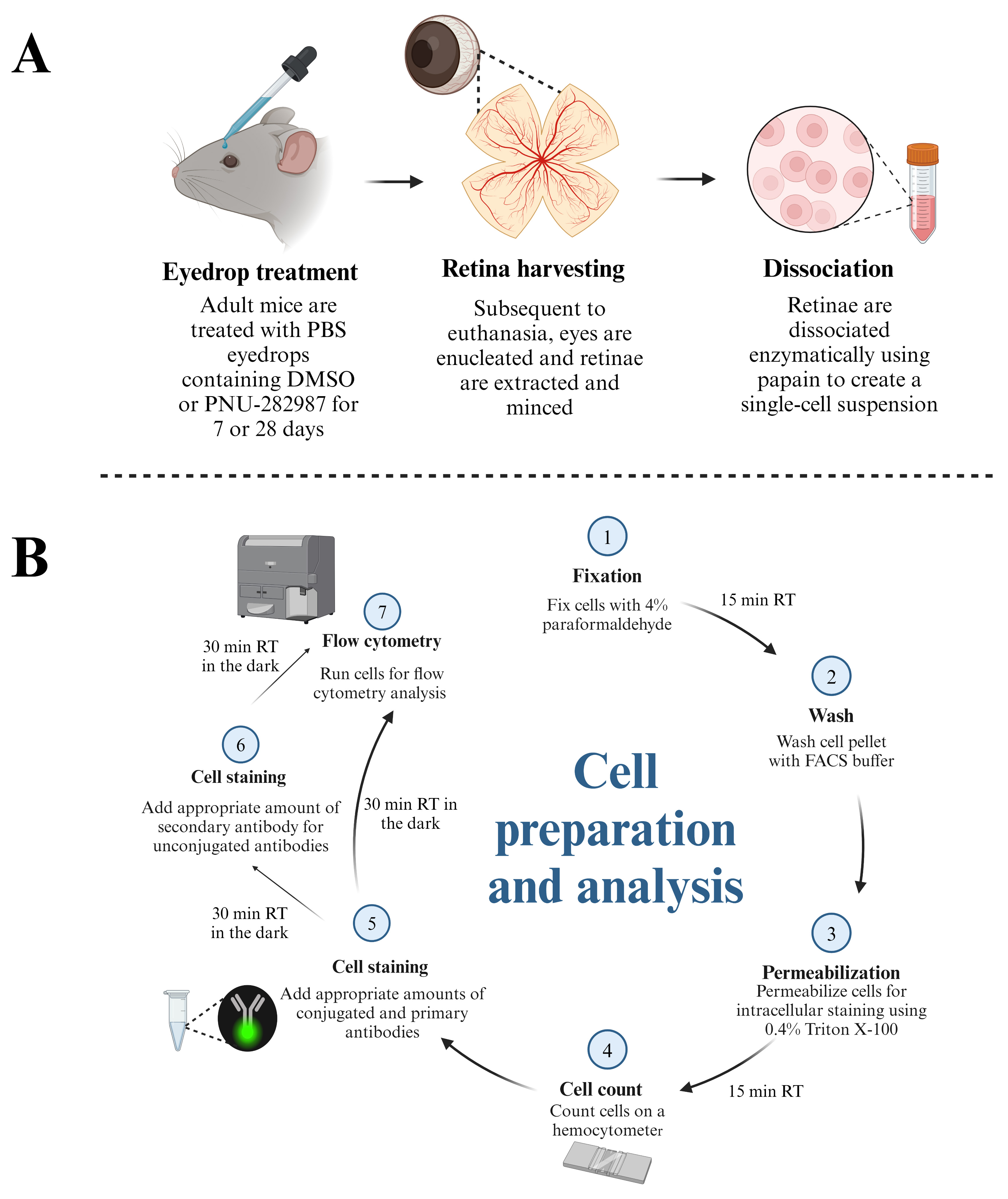
Schematic demonstrating the protocol for preparation of retinal cells for flow cytometry analysis. (A) Adult mice (3–6 months) are subjected to topical PBS eyedrop treatment containing DMSO (control groups) or PNU-282987 (experimental groups). Both eyedrop treatments contain 1 mg/mL of BrdU to label proliferating cells. After treatment, mice are euthanized, and retinae are harvested for dissociation using papain. (B) Dissociated retina cells are fixed and permeabilized before aliquots are taken for cell counts on a hemocytometer. After determining the number of cells present, conjugated antibodies and unconjugated primary antibodies are added at the appropriate dilutions. Fluorescent secondary antibodies are added for markers that are unconjugated. Cells are then subjected to flow cytometric analysis using a BD LSRFortessa.
Background
Adult mammals cannot typically regenerate retinal neurons after injury [1–3]. However, previous research from this lab using adult mice has shown that the selective alpha7 nicotinic acetylcholine receptor (nAChR) agonist, PNU-282987, can induce neurogenesis in adult mammals when applied as eyedrops in the presence or absence of any retinal injury [4–10]. PNU-282987 is believed to act on alpha7 nAChRs in the retinal pigment epithelium to release signaling molecules onto the end feet of Müller glia to introduce cell cycle re-entry. From there, Müller glia de-differentiate and form retinal progenitor cells that eventually develop into mature retinal neurons [6,9]. However, these studies were performed using fluorescent immunolabeling and subsequent confocal microscopy, thus restricting the number of antibodies that can be multiplexed and limiting the examination of cells undergoing mitosis and proliferation. As a result, we have developed a flow cytometry method that allows for the multiplexing and analysis of multiple external and internal markers in dissociated retinal cells. In this paper, a step-by-step protocol is described for the labeling of multiple retinal cell types such as retinal ganglion cells, rod photoreceptors, and Müller glia, concurrently with mitotically active and proliferating cells that arise after treatment with PNU-282987.
Materials and reagents
Papain dissociation system, which includes papain, DNase I, ovomucoid inhibitor, and EBSS (Worthington Biochemical Corporation, catalog number: LK003150)
Phosphate buffered saline (PBS) 10× concentrate (Sigma-Aldrich, catalog number: P5493)
PNU-282987 hydrate (N-[(3R)-1-azabicyclo[2.2.2]oct-3-yl]-4-chlorobenzamide hydrochloride) (Sigma-Aldrich, catalog number: P6499)
BrdU (5-Bromo-2’-deoxyuridine) (Sigma-Aldrich, catalog number: 19-160)
Fetal bovine serum (FBS) (Thermo Fisher Scientific, catalog number: A5670701)
Paraformaldehyde (PFA) (Sigma-Aldrich, catalog number: P6148)
Hydrochloric acid (HCl) (Sigma-Aldrich, catalog number: 258148)
Sodium hydroxide (NaOH) (Sigma-Aldrich, catalog number: S5881)
Triton X-100 (Sigma-Aldrich, catalog number: T8787)
Dimethyl sulfoxide (DMSO) (Sigma-Aldrich, catalog number: D8418)
BD HorizonTM brilliant stain buffer (BD Biosciences, catalog number: 563794)
Fc receptor binding inhibitor polyclonal antibody, eBioscienceTM (Thermo Fisher Scientific, catalog number: 14-9161-73)
Fluorescent antibodies and unconjugated blocking antibodies (see Table 1)
UltraComp eBeadsTM compensation beads (Invitrogen, catalog number: 01-2222-42)
Table 1. Antibody panel used in this protocol
Antibody Fluorophore Clone Host Company Catalog number Dilution (per 1 × 10⁶ cells) Rhodopsin Unconjugated RET-P1 Mouse Invitrogen MAS-11741 3 μL Zenon kit Alexa-405 - - Invitrogen Z25013 - Thy 1.2 APC-eFluor-780 53-2.1 Rat eBioscience 47-0902-82 1 μL Vimentin Biotinylated 280618 Rat R&D Systems BAM2105 2 μL Streptavidin BUV-395 - - BD Biosciences BD 564176 2 μL Ki67 BUV-737 20Raj1 Mouse eBioscience 367-5699-42 5 μL BrdU PerCP-eFluor-710 BU20A Mouse eBioscience 46-5071-42 1 μL
Solutions
DMSO/PNU-282987/BrdU eyedrop solution (see Recipes)
DMSO/BrdU eyedrop solution (see Recipes)
4% PFA (see Recipes)
FACS buffer with 4% FBS (see Recipes)
0.4% Triton X-100 (see Recipes)
0.1 M HCl (see Recipes)
Recipes
DMSO/PNU-282987/BrdU eyedrop solution
Caution: Appropriate PPE must be worn when handling chemicals.
Reconstitute 50 mg of PNU-282987 in 1.66 mL of DMSO to make a 100 mM stock that can be stored at 4 for several months. Dissolve 5 mg of BrdU in 5 mL of DMSO to make a stock solution of 100 mg/mL BrdU that can be stored at -20 for several months. To make PNU-282987/BrdU eye drop solution, mix 9.8 mL of 1× PBS, 100 μL of 100 mM PNU-282987, and 100 μL of BrdU to achieve a final solution of 1 mg/mL BrdU and 1 mM PNU-282987. Store at 4 for one month.
DMSO/BrdU eyedrop solution
Mix 9.8 mL of 1× PBS, 100 μL of DMSO, and 100 μL of BrdU to achieve a final solution of 1 mg/mL BrdU. Store at 4 for one month.
4% PFA
Caution: Preparation of PFA is hazardous. Avoid skin contact and inhalation. Use appropriate PPE and a chemical fume hood.
Mix 4 g of PFA and 100 mL of 1× PBS in a media storage bottle containing a stir bar. Add drops of 2 M NaOH while the solution is stirring until a pH of approximately 7.4 has been reached or the PFA has completely dissolved. Please note the solution may become more acidic as PFA dissolves and the addition of more NaOH may be necessary. Place the bottle on a hot plate heated to 85 for approximately 20 min or until there are no visible particles. Allow the solution to cool at 4 . Add 1 M HCl drops until the solution reaches a pH of 6.9. Store at 4 for one week.
FACS buffer with 4% FBS
Add 4 mL of FBS to 96 mL of 1× PBS. Store at 4 for one month.
0.4% Triton X-100
Add 4 mL of Triton X-100 to 96 mL of 1× PBS. Store at 4 for several months.
0.1 M HCl
Add 1.5 mL of 1 M HCl to 13.5 mL of MilliQ water for a final concentration of 0.1 M HCl. Store at room temperature for several months.
Laboratory supplies
10 mm Petri dish (VWR, catalog number: 10799-192)
90 mm Petri dish (Sigma-Aldrich, catalog number: P10903)
General lab supplies (pipettes, tips, tubes, etc.)
5 mL Falcon round-bottom Polystyrene test tubes with 70 μm cell strainer (Fisher, catalog number: 08-771-23)
Equipment
Dumont #7 curved forceps (Fine Science Tools, catalog number: 11274-20)
Dumont #5 forceps (Fine Science Tools, catalog number: 11251-10)
Spring scissors (Fine Science Tools, catalog number: 15000-00)
Scalpel blades #11 (Fine Science Tools, catalog number: 10011-00)
Scalpel handle (Fine Science Tools, catalog number: 10003-12)
Corning® LSETM digital water bath, 6 L, 120 V (Corning, catalog number: 6783)
FisherbrandTM analog hotplate stirrer (Fisher, catalog number: FB30786160)
pH indicator strips (Sigma-Aldrich, catalog number: 1095350001)
VWR® water jacketed CO incubator (VWR, catalog number: 10810-744)
Pipet-Aid (Corning, catalog number: 07-202-350)
Centrifuge (Thermo Fisher Scientific, model: Heraeus Multifuge X3)
Eppendorf benchtop centrifuge (for microcentrifuge tubes at room temperature)
Benchtop vortex
Bright-Line counting chamber, which includes slides (VWR, catalog number: 100503-092)
Note: Alternatively, an automated cell counter can be used for this protocol.
BD LSRFortessaTM cell analyzer (BD Biosciences)
Software and datasets
FlowJoTM software v10.10 (BD Life Sciences) was used for flow cytometry data analysis
Procedure
文章信息
稿件历史记录
提交日期: Feb 26, 2024
接收日期: May 23, 2024
在线发布日期: Jun 18, 2024
出版日期: Jul 5, 2024
版权信息
© 2024 The Author(s); This is an open access article under the CC BY license (https://creativecommons.org/licenses/by/4.0/).
如何引用
Vanzo-Sparks, H. K., Webster, S. E., Webster, M. K. and Linn, C. L. (2024). Quantification of Proliferating and Mitotically Active Retinal Cells in Mice by Flow Cytometry. Bio-protocol 14(13): e5024. DOI: 10.21769/BioProtoc.5024.
分类
神经科学 > 感觉和运动系统 > 视网膜
细胞生物学 > 基于细胞的分析方法 > 流式细胞术
您对这篇实验方法有问题吗?
在此处发布您的问题,我们将邀请本文作者来回答。同时,我们会将您的问题发布到Bio-protocol Exchange,以便寻求社区成员的帮助。