

Generation of Mouse Primitive Endoderm Stem Cells
小鼠原始内胚层干细胞的产生
发布: 2023年11月20日第13卷第22期 DOI: 10.21769/BioProtoc.4878 浏览次数: 2479
评审: Komuraiah MyakalaWei FanAbhijnya Kanugovi
参见作者原研究论文
The authors used this protocol in:
Feb 2022
Advertisement


Abstract
The blastocysts consist of dozens of cells of three distinct lineages: epiblast (Epi), trophoblast (TB), and primitive endoderm (PrE). All embryonic and extraembryonic tissues are derived from Epi, TB, and PrE. Stem cell lines representing preimplantation Epi and TB have been established and are known as embryonic stem cells (ESCs) and trophoblast stem cells (TSCs). Extraembryonic endoderm cells (XENCs) constitute a cell line that has been established from PrE. Although in vivo, PrE gives rise to visceral endoderm (VE), parietal endoderm (PE), and marginal zone endoderm (MZE); XENCs, on blastocyst injection into chimeras, primarily contribute to the distal region of PE. Here, we provide a comprehensive protocol for the establishment of fully potent primitive endoderm stem cell (PrESC) lines. PrESCs are established and maintained on mouse embryonic fibroblast (MEF) feeder cells in a serum-free medium supplemented with fibroblast growth factor 4 (FGF4), heparin, CHIR99021, and platelet-derived growth factor-AA (PDGF-AA). PrESCs co-express markers indicative of pluripotency and endoderm lineage commitment, exhibiting characteristics akin to those of PrE. On transplantation of PrESCs into blastocysts, they demonstrate a high efficiency in contributing to VE, PE, and MZE. PrESCs serve as a valuable model for studying PrE, sharing similarities in gene expression profiles and differentiation potential. PrESCs constitute a pivotal cornerstone for in vitro analysis of early developmental mechanisms and for studies of embryo reconstitution in vitro, particularly in conjunction with ESCs and TSCs.
Key features
• Establishment and maintenance of primitive endoderm stem cell (PrESCs) capable of recapitulating the developmental prowess inherent to PrE.
• Offering a source of PrE lineage for embryo-like organoid reconstitution studies.
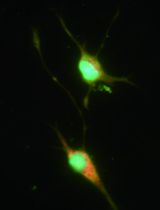
Graphical overview

Background
Epiblast (Epi), trophoblast (TB), and primitive endoderm (PrE) are differentiated from the zygote by the late blastocyst stage of preimplantation development. Epi lineage generates all embryonic tissues, extraembryonic mesodermal tissues (i.e., chorion, allantois, yolk sac mesoderm, amniotic mesoderm, and capillaries in the labyrinthine layer of the placenta), and amniotic ectoderm. TB lineage generates major parts of placenta and trophectoderm of parietal yolk sac (PYS). PrE lineage is vital for normal embryonic development and is the origin of the visceral endoderm (VE) layer of the visceral yolk sac (VYS), the parietal endoderm (PE) layer of the PYS, and the marginal zone endoderm (MZE) lining along the edge of the placenta/chorionic plate of the placental disk (Gardner, 1982; Igarashi et al., 2018). The extraembryonic endodermal tissues play a pivotal role in nourishing the embryo by facilitating nutrient provision at the maternal–fetal interface (Jollie, 1990; Lloyd, 1990), especially significant prior to the onset of placental circulation (~E10.5 in mice), anterior–posterior patterning (Brennan et al., 2001), and yolk sac hematopoiesis (Ueno and Weissman, 2010). In mice, the gestation period is 19–21 days, and the initial yolk sac is formed on the fourth day, so embryonic development is uniquely dependent on the yolk sac during the first quarter of development.
Stem cell lines representing preimplantation epiblasts and trophoblasts have been established and are known as embryonic stem cells (ESCs) (Evans and Kaufman, 1981; Nichols et al., 2009) and trophoblast stem cells (TSCs) (Tanaka et al., 1998; Ohinata and Tsukiyama, 2014), respectively. To gain further insight into how the PrE functions during pre- and early post-implantation development, establishment of stem cell lines that fully retain PrE developmental potential has long been awaited. Extraembryonic endoderm cells (XENCs) turned out to be derivatives of the PrE but could only contribute to the distal region of the PE in chimeras (Kunath et al., 2005). Efforts to improve XEN cell culture conditions enabled the generation of XEN-P (extraembryonic endoderm precursor) cells in rats (Debeb et al., 2009; Galat et al., 2009) and pXEN (primitive XEN) cells in mice (Zhong et al., 2018), which express pluripotent makers and endoderm markers simultaneously. XEN-P cells have a slightly improved ability to contribute to cells other than in the distal PE at gastrula stages in chimeras. Further improvement of XENCs was performed by treating them with Nodal or Cripto, a ligand and coreceptor in the nodal signaling, respectively, but the impact of this treatment was again limited (Julio et al., 2011). Therefore, a robust protocol to capture early PrE-derived cells that can generate all derivatives of the PrE in the chimeric conceptus awaits discovery.
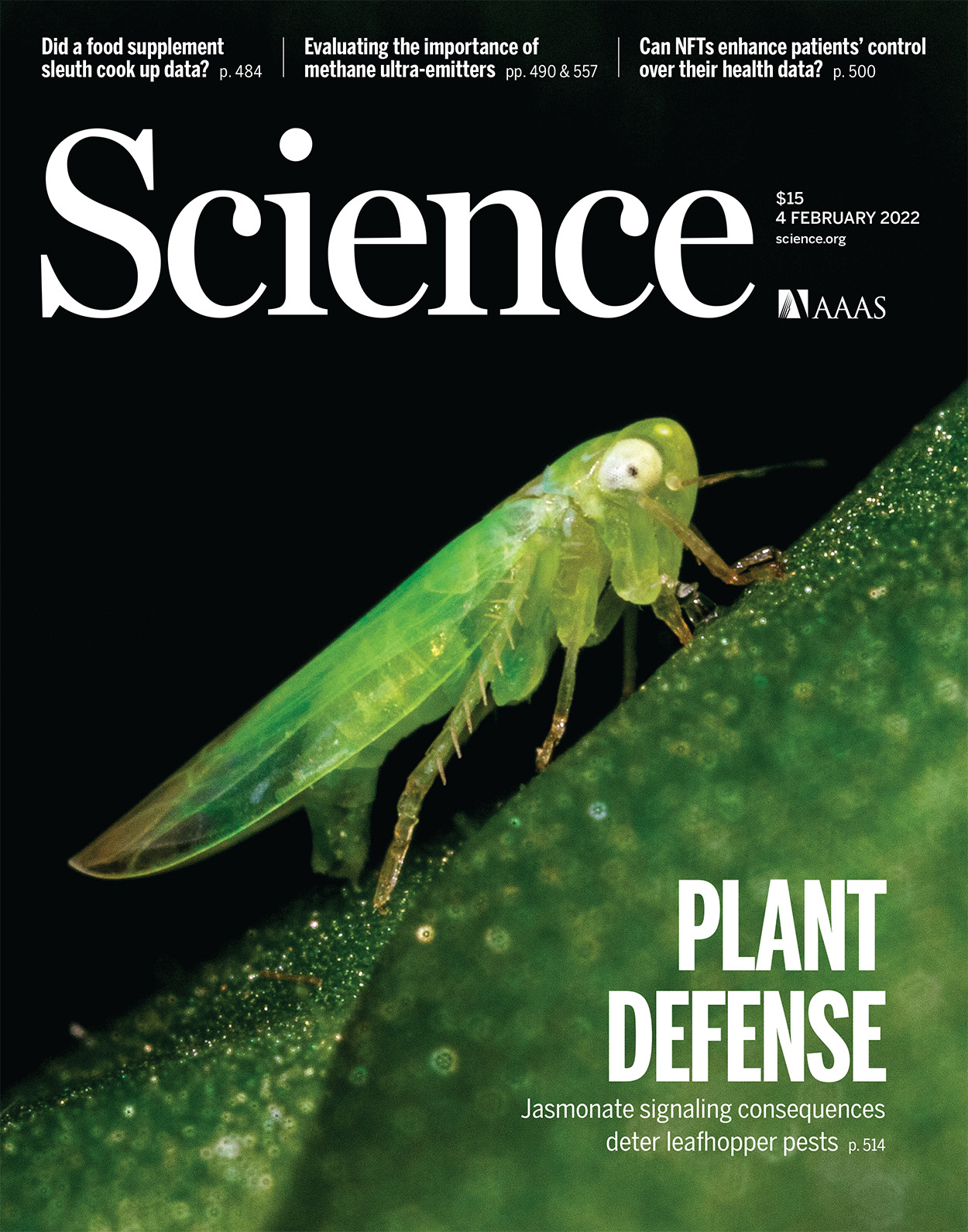
Recently, we succeeded in establishing primitive endoderm stem cells (PrESCs) that can fully recapitulate the developmental potential of PrE in mice. PrESCs are established and maintained on MEF feeder cells in a serum-free medium supplemented with FGF4, heparin, CHIR99021, and PDGF-AA. PrESCs co-express markers indicative of pluripotency (i.e., OCT4 and E-Cadherin) and endoderm lineage commitment (i.e., GATA4, GATA6, and SOX17), exhibiting characteristics akin to those of PrE. On transplantation of PrESCs into blastocysts, they demonstrate a high efficiency in contributing to all PrE derivatives, VE, PE, and MZE (Ohinata et al., 2022). In this protocol, we describe the detailed procedure for efficient generation of PrESC lines in mice. PrESCs therefore represent not only a regenerative resource to complement the PrE, but also an intervention tool to elucidate the mechanisms of PrE specification and subsequent pre- and post-implantation development. Importantly, PrESCs can be the final component of stem cell line assemblage required to generate development-competent blastocysts in vitro, in collaboration with ESCs and TSCs. PrESCs will contribute to the opening of a new research field linking pre- and early post-implantation development.
Materials and reagents
Biological materials
Pregnant mouse, 3.5 days post coitum (dpc) (8–20 weeks old) (e.g., CLEA Japan, CD-1). We have confirmed this protocol in CD-1, BALB/c, and B6PWKF1 genetic backgrounds
Mouse embryonic fibroblasts (MEFs) (ATCC, SCRC-1045TM). MEFs can also be derived from DR4 mice [Jackson, Dnmt1tm3Jae Hprt1b-m3 Tg (pPWL512hyg) 1Ems/J (DR4)] that are dissected 14.5 dpc; cells are passaged five times before inactivation by mitomycin C treatment. We routinely use DR4 MEFs, but the widely used CD-1 MEFs can also be used
Human fibroblast growth factor 4 (FGF4) (Sigma, catalog number: F8424)
Human platelet-derived growth factor-AA (PDGF-AA) (R&D, catalog number: 221-AA)
Rabbit anti-OCT4 antibody (MBL, catalog number: PM048)
Goat anti-SOX17 antibody, stock solution: 10 mg/mL in PBS (R&D, catalog number: AF1924)
Donkey anti-rabbit IgG antibody Alexa 488 conjugated (Invitrogen, catalog number: A21206)
Donkey anti-goat IgG antibody Alexa 568 conjugated (Invitrogen, catalog number: A11057)
Reagents
Mitomycin C (MMC) (Sigma, catalog number: M4287)
Fetal bovine serum (FBS) (biosera, catalog number: FB-1380/500)
Dulbecco’s modified Eagle’s medium (DMEM) (Sigma, catalog number: D5796)
β-mercaptoethanol (100×) (Millipore, catalog number: ES-007-E)
Penicillin-streptomycin (100×) (Gibco, catalog number: 15140-122)
Gelatin from porcine skin (Sigma, catalog number: G1890)
TrypLETM Select Enzyme (1×), no phenol red (Gibco, catalog number: 12563-029)
StemFit® AK02N (or StemFit® Basic02, StemFit® Basic03) (Ajinomoto)
Heparin (Sigma, catalog number: H3149)
CHIR99021, GSK3 inhibitor (REPROCELL, catalog number: 04-0004)
CELLBANKER® 1 plus (ZENOGEN PHARMA)
Ethanol (e.g., Nacalai Tesque, catalog number: 14712-34)
Paraformaldehyde (PFA) (TAAB, catalog number: P001)
Tween 20 (Chem Cruz, catalog number: sc-29113)
Normal donkey serum (Sigma, catalog number: D9663)
Hoechst 33342 10 mg/mL solution (Invitrogen, catalog number: H3570)
Solutions
MEF medium (see Recipes)
AK02N medium (see Recipes)
PrESC medium (see Recipes)
100× Mitomycin C stock solution (see Recipes)
0.2% Gelatin (see Recipes)
70% Ethanol (see Recipes)
PBST (see Recipes)
10 N NaOH (see Recipes)
4% PFA/PBST (see Recipes)
Recipes
MEF medium
Reagent Final concentration Quantity DMEM 500 mL FBS 10% 55 mL β-mercaptoethanol 1× 5.6 mL Penicillin-streptomycin 1× 5.6 mL Total n/a 566.2 mL AK02N medium
Reagent Quantity Liquid A 400 mL Liquid B 100 mL Penicillin-streptomycin 5 mL Total 505 mL PrESC medium
Reagent Final concentration Quantity AK02N medium (liquid A+B) 50 mL CHIR99021 10 μM 50 μL (10 mM stock solution) Human FGF4 50 ng/mL 50 μL (50 μg/mL stock solution) Heparin 10 μg/mL 50 μL (50 mg/mL stock solution) Human PDGF-AA 25 ng/mL 50 μL (25 μg/mL stock solution) Total 50 mL 100× Mitomycin C stock solution
Reagent Final concentration Quantity Mitomycin C 0.5 mg/mL 5 mg ddH2O n/a 10 mL Total n/a 10 mL 0.2% Gelatin
Reagent Final concentration Quantity Gelatin 0.2% 1 g ddH2O n/a 500 mL Total n/a 500 mL Sterilize 0.2% gelatin solution by autoclaving.
70% Ethanol
Reagent Final concentration Quantity Ethanol 70% 350 mL ddH2O n/a 150 mL Total n/a 500 mL PBST
Reagent Final concentration Quantity Tween 20 0.2% 1 mL PBS n/a 500 mL Total n/a 501 mL 10 N NaOH
Reagent Final concentration Quantity NaOH 0.4 g/mL 40 g ddH2O n/a 100 mL Total n/a 100 mL NaOH liberates thermal energy upon dissolution. It is prudent to refrain from dissolving it in its entirety in a single instance. Instead, endeavor to dissolve it incrementally in multiple aliquots before culminating in a final volume of 100 mL. Employ safety spectacles to shield your eyes.
4% PFA/PBST
Reagent Final concentration Quantity PFA 4% 2 g PBST n/a 50 mL 10 N NAOH n/a 10 μL Total n/a 50 mL Incubate at 65 °C and mix several times until the PFA melts.
Equipment
Conical tube (15 mL) (Corning, catalog number: 352196)
Conical tube (50 mL) (Corning, catalog number: 352098)
Tissue culture plate (24 well) (Corning, catalog number: 351147)
Tissue culture plate (12 well) (Corning, catalog number: 351143)
Tissue culture plate (6 well) (Corning, catalog number: 351146)
Culture dish (35 mm) (Thermo Scientific, catalog number: 150460)
Culture dish (150 mm) (Corning, catalog number: 353025)
Petri dish (60 mm) (Corning, catalog number: 351007)
Glass bottom culture dish (35 mm) (IWAKI, catalog number: 3910-035)
Cryotube vial (Thermo Scientific, catalog number: 377224)
P2 micro-pipette (e.g., Gilson, catalog number: F144054M)
P20 micro-pipette (e.g., Gilson, catalog number: F144056M)
P1000 micro-pipette (e.g., Gilson, catalog number: 144059M)
Dissection microscope (e.g., Evident, model: SZX7)
Inverted microscope (e.g., Evident, model: IX71)
Water bath (e.g., TAITEC, model: SJ-07N)
Centrifuge (e.g., TOMY, model: LCX-100)
Hemocytometer (e.g., Biomedical Science, model: BMS-OCC01)
CO2 incubator (e.g., Panasonic, model: MCO-170AICV-PJVH)
Scissors (e.g., Natsume, model: NAPOX B-68T)
Forceps (e.g., Inox-Med, L5, catalog number: 11253-27)
Syringe (5 mL) (e.g., Terumo, model: SS-05LZ)
21-gauge needle (e.g., Terumo, model: NN-2138R)
Freezing container (e.g., Nihon Freezer, model: BICELL)
Ultra-low temperature freezer (-80 °C) (e.g., Panasonic, model: MDF-DU502VHS1-PJ)
Ultra-low temperature freezer (-150 °C) (e.g., Panasonic, model: MDF-1156ATA-PJ)
Confocal microscope (e.g., Evident, model: FV1200)
Procedure
文章信息
版权信息
© 2023 The Author(s); This is an open access article under the CC BY-NC license (https://creativecommons.org/licenses/by-nc/4.0/).
如何引用
Readers should cite both the Bio-protocol article and the original research article where this protocol was used:
- Ohinata, Y., Saraya, A. and Koseki, H. (2023). Generation of Mouse Primitive Endoderm Stem Cells. Bio-protocol 13(22): e4878. DOI: 10.21769/BioProtoc.4878.
- Ohinata, Y., Endo, T. A., Sugishita, H., Watanabe, T., Iizuka, Y., Kawamoto, Y., Saraya, A., Kumon, M., Koseki, Y., Kondo, T., et al. (2022). Establishment of mouse stem cells that can recapitulate the developmental potential of primitive endoderm. Science 375(6580): 574–578.
分类
干细胞 > 胚胎干细胞 > 维持和分化
干细胞 > 生殖细胞 > 体外培养
细胞生物学 > 细胞分离和培养 > 细胞分离
您对这篇实验方法有问题吗?
在此处发布您的问题,我们将邀请本文作者来回答。同时,我们会将您的问题发布到Bio-protocol Exchange,以便寻求社区成员的帮助。