

Mitochondrial Replication Assay (MIRA) for Efficient in situ Quantification of Nascent mtDNA and Protein Interactions with Nascent mtDNA (mitoSIRF)
线粒体复制测定(MIRA)可有效原位定量新生线粒体DNA和蛋白质与新生线粒体DNA(mitoSIRF)的相互作用

(*contributed equally to this work) 发布: 2023年05月20日第13卷第10期 DOI: 10.21769/BioProtoc.4680 浏览次数: 2257
评审: Gal HaimovichDavid PaulVaibhav B. ShahAnonymous reviewer(s)
参见作者原研究论文
The authors used this protocol in:

Dec 2021
Advertisement


Abstract
Mitochondria play decisive roles in bioenergetics and intracellular communication. These organelles contain a circular mitochondrial DNA (mtDNA) genome that is duplicated within one to two hours by a mitochondrial replisome, independently from the nuclear replisome. mtDNA stability is regulated in part at the level of mtDNA replication. Consequently, mutations in mitochondrial replisome components result in mtDNA instability and are associated with diverse disease phenotypes, including premature aging, aberrant cellular energetics, and developmental defects. The mechanisms ensuring mtDNA replication stability are not completely understood. Thus, there remains a need to develop tools to specifically and quantifiably examine mtDNA replication. To date, methods for labeling mtDNA have relied on prolonged exposures of 5′-bromo-2′-deoxyuridine (BrdU) or 5′-ethynyl-2′-deoxyuridine (EdU). However, labeling with these nucleoside analogs for a sufficiently short time in order to monitor nascent mtDNA replication, such as under two hours, does not produce signals suited for efficient or accurate quantitative analysis. The assay system described here, termed Mitochondrial Replication Assay (MIRA), utilizes proximity ligation assay (PLA) combined with EdU-coupled Click-IT chemistry to address this limitation, thereby enabling sensitive and quantitative analysis of nascent in situ mtDNA replication with single-cell resolution. This method can be further paired with conventional immunofluorescence (IF) for multi-parameter cell analysis. By enabling monitoring nascent mtDNA prior to the complete replication of the entire mtDNA genome, this new assay system allowed the discovery of a new mitochondrial stability pathway, mtDNA fork protection. Moreover, a modification in primary antibodies application allows the adaptation of our previously described in situ protein Interactions with nascent DNA Replication Forks (SIRF) for the detection of proteins of interest to nascent mtDNA replication forks on a single molecule level (mitoSIRF).
Graphical overview

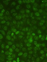
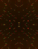
Schematic overview of Mitochondrial Replication Assay (MIRA). 5′-ethynyl-2′-deoxyuridine (EdU; green) incorporated in DNA is tagged with biotin (blue) using Click-IT chemistry. Subsequent proximity ligation assay (PLA, pink circles) using antibodies against biotin allows the fluorescent tagging of the nascent EdU and amplification of the signal sufficient for visualization by standard immunofluorescence. PLA signals outside the nucleus denote mitochondrial DNA (mtDNA) signals. Ab, antibody. In in situ protein interactions with nascent DNA replication forks (mitoSIRF), one of the primary antibodies is directed against a protein of interest, while the other detects nascent biotinylated EdU, thus enabling in situ protein interactions with nascent mtDNA.
Background
Mitochondria, the organelles best known for their orchestration of cellular bioenergetics, operate semi-autonomously within eukaryotic cells and are key signaling hubs for intracellular communication (Friedman and Nunnari, 2014). Each mitochondrion harbors multiple copies of its ~16 kb circular genome, which encodes essential subunits of the electron transport chain as well as a set of transfer and ribosomal RNAs (Falkenberg et al., 2007). Mitochondrial DNA (mtDNA) is replicated separately from nuclear DNA (nDNA): the mitochondrial genome is replicated by its organelle-specific replisome, comprised of DNA polymerase POLγ and other nuclear-encoded proteins (Holt and Reyes, 2012; Gustafsson et al., 2016; Bailey and Doherty, 2017). mtDNA replication can occur at any cell cycle stage. Because of its proximity to events during oxidative phosphorylation, mtDNA is constantly exposed to reactive oxygen species, rendering it highly susceptible to oxidative DNA damage (Kang and Hamasaki, 2002). Failure to repair damage of the mitochondrial genome additionally results in instability of mtDNA and mitochondrial dysfunction, which is associated with premature aging, tumorigenesis, and neurological disorders (Trifunovic et al., 2004; Chatterjee et al., 2006; Ishikawa et al., 2008; Park and Larsson, 2011). Apart from mtDNA repair pathways, mtDNA stability is also regulated at the level of mtDNA replication with the exonuclease activity of POLγ polymerase and MGME1 (mitochondrial genome maintenance exonuclease 1) degrading damaged mtDNA and generating new mtDNA (Torregrosa-Munumer et al., 2019).
To date, detailed mechanisms of mtDNA replication linking to stability are incompletely understood. A comprehensive understanding of these mechanisms is imperative to allow the development of effective and specific agents targeting disease outcomes. For this purpose, efficient tools to specifically monitor and quantifiably measure nascent mtDNA replication are needed. mtDNA is replicated primarily by POLγ within one to two hours, yet methods for labeling mtDNA have historically relied on prolonged exposures (up to 24 h) of 5′-bromo-2′-deoxyuridine (BrdU) or 5′-ethynyl-2′-deoxyuridine (EdU) (Clayton, 1982; Korhonen et al., 2004). Incorporation of these nucleoside analogs for labeling times sufficiently short to monitor nascent mtDNA replication before completion of the entire mtDNA genome, e.g. less than two hours, does not produce signals suited for efficient quantitative analysis (Luzwick et al., 2021). The assay system described here, termed Mitochondrial replication assay (MIRA), utilizes proximity ligation assay (PLA) as a means of signal amplification of the mtDNA-incorporated nucleoside analogs to overcome this limitation. Specifically, mtDNA is labeled with the thymidine analog EdU, which is then biotinylated using Click-IT chemistry (Moses and Moorhouse, 2007). The PLA technology was developed by Soderberg et al. (2008) for single-molecule protein–protein interaction studies, utilizing antibodies with oligonucleotide conjugates that can be ligated into a circular DNA molecule when two antibodies are in close proximity to each other (<40 nm). Once ligated, the DNA circle can be amplified by rolling circle DNA polymerase. After annealing of fluorescent DNA probes that are complementary to the DNA circle, this procedure results in a highly amplified fluorescent signal, readily detectable by conventional immunofluorescence (IF) microscopy. In MIRA, PLA is adapted to signal-amplify the nascent mtDNA signals by utilizing complementary antibodies against biotin (detecting biotinylated EdU). As with conventional EdU and BrdU labeling, MIRA also detects and visualizes nDNA. Nonetheless, only cytoplasmic MIRA signals are considered for analysis, which are present irrespective of the cell cycle state and do not form without EdU or in mtDNA-depleted Rho zero cells (Luzwick et al., 2021). With amplification, the nascent mtDNA can be readily detected and quantified with a 1 h EdU pulse, which is less time than needed to replicate the entire genome. This assays system enables the monitorization of ongoing mtDNA replication prior to completion of mtDNA genome replication. It therefore differs from most common mtDNA label schemes, which use prolonged nucleoside analog incorporation for extended time over multiple hours. Since the mtDNA is replicated within one to two hours, prolonged labeling will predominantly result in post-replicative signals. Moreover, as an adaptation from our single-cell assay for in situ protein Interactions with nascent DNA Replication Forks (SIRF) protocol (Roy et al., 2018; Roy and Schlacher, 2019), MIRA can be combined with a primary antibody against a protein of interest to detect protein mtDNA interactions (mitoSIRF). The MIRA and mitoSIRF assay systems allowed the discovery of a new mitochondrial stability pathway, mtDNA fork protection (Luzwick et al., 2021). Specifically, BRCA and FANC tumor-suppressor proteins protect nascent mtDNA from instability during ongoing mtDNA replication; this would not have been detectable by previous assay systems that visualize post-replicative events. The instability is caused by MRE11-dependent nuclease degradation, which results in cGAS-mediated inflammatory signaling (Luzwick et al., 2021).
The MIRA method requires a minimal number of cells (~100–1,000), preserves the single-cell resolution as seen with IF, which provides valuable information regarding cell morphology, and can be readily and accurately quantified. The MIRA assay can furthermore be combined with conventional IF for multi-parameter biomarker analysis, including cell cycle stage or cell identity. If combined with other biomarkers, primary and secondary antibody staining for conventional IF is performed following the Click-IT and PLA reaction to avoid interference of PLA secondary antibodies from cross-reacting with IF primary antibodies.
Materials and Reagents
8-well chamber microscope slides (Thermo Scientific, Nunc, catalog number: 177402)
Plastic coverslips (Electron Microscopy Sciences, catalog number: 72261-50)
Glass coverslips (Fisher Scientific, catalog number: 12-548-5M)
Kimwipes (Fisher Scientific, catalog number: NC9855580)
Aluminum foil (Fisher Scientific, catalog number: 01-213-105)
Paper towel (Envision, catalog number: 23304)
0.22 μm bottle top filter (Corning, catalog number: 430758)
Rabbit anti-biotin antibody (Cell Signaling, catalog number: 5597S)
Mouse anti-biotin antibody (Sigma-Aldrich, catalog number: B7653)
5′-ethylene-2′-deoxyuridine (EdU) (Invitrogen, catalog number: A10044)
Paraformaldehyde 32% solution, EM grade (PFA) (EMS, catalog number: 15714)
Triton X-100 (Sigma-Aldrich, catalog number: T8787)
Biotin azide (Invitrogen, catalog number: B10184)
Alexa Fluor 488 azide (Invitrogen, catalog number: A10266)
Copper sulfate solution (Fluka Analytical, catalog number: 35185)
Sodium ascorbate (Sigma-Aldrich, catalog number: 11140)
Phosphate buffered saline (PBS) (Sigma-Aldrich, catalog number: P4417)
Duolink® mouse plus PLA probe (Sigma-Aldrich, catalog number: DUO92001-100RXN)
Duolink® rabbit minus PLA probe (Sigma-Aldrich, catalog number: DUO92005-100RXN)
Duolink® blocking solution (Sigma-Aldrich, catalog number: DUO82007-8ML)
Duolink® antibody diluent (Sigma-Aldrich, catalog number: DUO82008-8ML)
Duolink® PLA detection reagent red (Sigma-Aldrich, catalog number: DUO92008-100RXN)
Duolink® in situ wash buffers (Sigma-Aldrich, catalog number: DUO82049-20L)
4’,6-diamidino-2-phenylindole (DAPI) (Life Technologies, catalog number: 62248)
Prolong Gold antifade reagent (Invitrogen, catalog number: P36934)
Cell-specific culturing media
Dideoxycytidine (ddC, 20 μM) (Sigma-Aldrich, catalog number: D5782)
MitoPQ (10 μM) (Abcam, catalog number: 1821370-28-8)
mtOX (Wisnovsky et al., 2016)
Sodium chloride (NaCl) (Fisher Scientific, catalog number: 7647-14-5)
Tween 20 (Sigma-Aldrich, catalog number: P7949)
Hydrochloric acid (HCl) (Fisher Scientific, catalog number: 7647-01-0)
Trizma hydrochloride (Tris-HCl) (Sigma-Aldrich, catalog number: T5941)
Tris base (Fisher Scientific, catalog number: BP152-1)
Dimethyl sulfoxide (DMSO) (Santa Cruz Biotechnology, catalog number: sc-358801)
Fixation solution (see Recipes)
Permeabilization solution (see Recipes)
Wash buffer A (see Recipes)
Wash buffer B (see Recipes)
EdU stock solution (see Recipes)
Biotin-azide stock solution (see Recipes)
Alexa488-azide stock solution (see Recipes)
Equipment
Autoflow IR water jacketed CO2 incubator (NUAIRE, model: NU-4750)
Nikon eclipse Ti-U inverted microscope (Nikon, model: Ti-U)
Colin jar (Thermo ScientificTM E94)
Fine curved forceps (Fine Science Tools, Dumont #7, catalog number: 11271-30)
Slide box (VWR, catalog number: 82003-414)
Vortexer (VWR analog vortex mixer, catalog number: 10153-838)
4 °C refrigerator (BSI, model: SCGP21OW1AREF)
-20 °C freezer (BSI, model: ABT-2020MB)
Software
NIS-elements (Nikon, https://www.nikoninstruments.com/Products/Software)
ImageJ (ImageJ, https://imagej.net/Welcome)
Microsoft Excel (Microsoft, https://products.office.com/en-us/excel)
GraphPad Prism (GraphPad, https://www.graphpad.com/scientific-software/prism/)
Procedure
文章信息
版权信息
© 2023 The Author(s); This is an open access article under the CC BY license (https://creativecommons.org/licenses/by/4.0/).
如何引用
Readers should cite both the Bio-protocol article and the original research article where this protocol was used:
- Lozen, M., Chen, Y., Boisvert, R. A. and Schlacher, K. (2023). Mitochondrial Replication Assay (MIRA) for Efficient in situ Quantification of Nascent mtDNA and Protein Interactions with Nascent mtDNA (mitoSIRF). Bio-protocol 13(10): e4680. DOI: 10.21769/BioProtoc.4680.
- Luzwick, J. W., Dombi, E., Boisvert, R. A., Roy, S., Park, S., Kunnimalaiyaan, S., Goffart, S., Schindler, D. and Schlacher, K. (2021). MRE11-dependent instability in mitochondrial DNA fork protection activates a cGAS immune signaling pathway. Sci Adv 7(51): eabf9441.
分类
癌症生物学 > 基因组不稳定性及突变 > 细胞生物学试验
分子生物学 > DNA > DNA 检测
您对这篇实验方法有问题吗?
在此处发布您的问题,我们将邀请本文作者来回答。同时,我们会将您的问题发布到Bio-protocol Exchange,以便寻求社区成员的帮助。