

THRIFTY—A High-throughput Single Muscle Fiber Typing Method Based on Immunofluorescence Detection
THRIFTY-一种基于免疫荧光检测的高通量单肌纤维分型方法
(*contributed equally to this work) 发布: 2023年05月20日第13卷第10期 DOI: 10.21769/BioProtoc.4678 浏览次数: 1638
评审: Alessandro DidonnaMarco Pagliusi Jr.Xin Xu
参见作者原研究论文
The authors used this protocol in:

Oct 2022
Advertisement


Abstract
Skeletal muscle consists of a mixture of fiber types with different functional and metabolic characteristics. The relative composition of these muscle fiber types has implications for muscle performance, whole-body metabolism, and health. However, analyses of muscle samples in a fiber type–dependent manner are very time consuming. Therefore, these are often neglected in favor of more time-efficient analyses on mixed muscle samples. Methods such as western blot and myosin heavy chain separation by SDS-PAGE have previously been utilized to fiber type–isolated muscle fibers. More recently, the introduction of the dot blot method significantly increased the speed of fiber typing. However, despite recent advancements, none of the current methodologies are feasible for large-scale investigations because of their time requirements. Here, we present the protocol for a new method, which we have named THRIFTY (high-THRoughput Immunofluorescence Fiber TYping), that enables rapid fiber type identification using antibodies towards the different myosin heavy chain (MyHC) isoforms of fast and slow twitch muscle fibers. First, a short segment (<1 mm) is cut off from isolated muscle fibers and mounted on a customized gridded microscope slide holding up to 200 fiber segments. Second, the fiber segments attached to the microscope slide are stained with MyHC-specific antibodies and then visualized using a fluorescence microscope. Lastly, the remaining pieces of the fibers can either be collected individually or pooled together with fibers of the same type for subsequent analyses. The THRIFTY protocol is approximately three times as fast as the dot blot method, which enables not only time-sensitive assays to be performed but also increases the feasibility to conduct large-scale investigations into fiber type specific physiology.
Graphical Overview

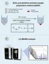
Graphical overview of the THRIFTY workflow. Cut off a small segment (0.5 mm) of an individually dissected muscle fiber and mount it onto the customized microscope slide containing a printed grid system. Using a Hamilton syringe, fixate the fiber segment by applying a small droplet of distilled water on the segment and let it fully dry (1A). The remaining large segment of the fiber should be placed in the corresponding square on a black A4 paper (1B). Once the microscope slide has been fully mounted with fiber segments, submerge the slide in a polypropylene slide mailer (illustrated as a Coplin jar in the figure) containing acetone to permeabilize the fiber segments. Thereafter, incubate the slide with primary antibodies targeting MyHC-I and MyHC-II. Following washes in PBS solution, incubate the slides with fluorescently labeled secondary antibodies, wash again, and mount with a cover glass and antifade reagent (2). Identification of fiber type can be performed using a digital fluorescence microscope (3), whereafter the remaining pieces of the fiber segments (large) are pooled together according to their fiber type or individually collected for experiments on single fibers (4). Image modified from Horwath et al. (2022).
Background
Skeletal muscle consists of a mixture of muscle fiber types, unique in their contractile and metabolic properties, as well as in their proteomic profile (Murgia et al., 2021). At a whole-muscle level, the relative abundance of the fiber types determines whether the muscle is primarily adapted to low-intensity repetitive activities or to short bursts of high-intensity contractions. The relative abundance of fiber types has also been linked to a number of physiological outcomes at a whole-body level, such as obesity and weight loss (Tanner et al., 2002), glucose-stimulated insulin secretion (Blackwood et al., 2022), and risk of cardiovascular disease (Karjalainen et al., 2006). In addition, many reports have demonstrated that skeletal muscle responds to acute exercise stimuli in a fiber type–dependent manner (Koopman et al., 2006; Tannerstedt et al., 2009; Kristensen et al., 2015). Similar observations have been reported for muscle adaptations in response to long-term training regimens, such as muscle fiber hypertrophy, improved glucose transport, and increased mitochondrial content (Daugaard et al., 2000; Verdijk et al., 2009; Skelly et al., 2021). Understanding the molecular intricacies of the different fiber types of skeletal muscle is therefore important for both disease prevention and physical performance.
Fiber type–specific analyses of isolated muscle fibers are typically associated with a tedious and time-consuming workflow, including isolating and fiber typing of hundreds to thousands of individual fibers prior to the experimental analysis. Even though the process of fiber type identification was recently improved by the introduction of the dot blot protocol (Christiansen et al., 2019), this method is still limited by the time required to fiber type large quantities of muscle fibers. The dot blot protocol also requires the entire muscle fiber to be denatured, thus limiting the number of analytical applications that can be performed on the same sample lysate. Therefore, we set out to develop the THRIFTY (high-THRoughput Immunofluorescence Fiber TYping) method to further facilitate the workflow associated with fiber type identification of single muscle fibers. The THRIFTY protocol presented herein will drastically reduce the time consumption of fiber type identification, thus allowing researchers to more easily capture a representative portion of the total muscle fiber pool. Our method also allows for experimental techniques in which a large sample mass is required to obtain proper measurement resolution, e.g., analyses of muscle protein synthesis using stable isotope tracers.
Materials and Reagents
Microscope slides (VWR, catalog number: 631-1554)
Cover slips 24 × 50 mm (VWR, catalog number: 631-1574)
Test tubes, soda glass 40 × 8 mm (VWR, catalog number: 212-0011)
Versilic peroxide-cured silicone stoppers (Lab Pure, catalog number: 263006-50)
CryoPure tubes 1.2 mL (Sarstedt, catalog number: 72.377)
Silica gel, SiO2 (VWR, catalog number: 83001.260)
Milli-Q ultrapure water
Acetone (C3H6O) (Fisher Scientific, catalog number: 1000220500)
Phosphate buffered saline (PBS) tablets (Fisher Scientific, catalog number: 10388739)
TritonTM X-100 (Sigma-Aldrich, catalog number: T8787)
Normal goat serum (NGS) 10% (Thermo Fisher Scientific, catalog number: 50062Z), store at 4 °C
Mouse anti-myosin heavy chain isoform I (DSHB, catalog number: BA-F8-c), store at 4 °C
Mouse anti-myosin heavy chain isoform II (DSHB, catalog number: SC-71-c), store at 4 °C
Alexa Fluor goat anti-mouse IgG2b 488 (Thermo Fisher Scientific, catalog number: A-21141), store at 4 °C in the dark
Alexa Fluor goat anti-mouse IgG1 647 (Thermo Fisher Scientific, catalog number: A-21240), store at 4 °C and in the dark
ProlongTM Gold antifade mountant (Thermo Scientific, catalog number: P36934)
Triton 10% stock solution (see Recipes)
Phosphate buffered saline (PBS) solution (see Recipes)
Primary antibody solution (see Recipes)
Secondary antibody solution (see Recipes)
Equipment
Freeze-dryer system (Heto FD 1.0 cooling unit, Edwards VacuumTM nXDS6i Vacuum Pump and Vacuubrand VAP5 Vacuum Gauge)
Climate controlled room (<40% humidity)
VisiScope® Stereo microscope (VWR, catalog number: 630-3073)
Dumont forceps (FST, catalog number: 11252-00)
Needles (sharpened sewing needles glued to a wooden handle)
Scalpel (Swamm-Morton, carbon steel scalpel, catalog number: 11)
HamiltonTM syringe 10 μL (Fisher Scientific, catalog number: 203560)
Black A4 paper (Common office supply)
Set of micropipettes (Tacta®, mechanical pipettes 0.5–1,000 μL, Sartorius)
Pipette tips (Safetyspace®, filtered pipette tips 0.5–1,000 μL, Sartorius)
Vortex-Genie® (VWR, catalog number: 444-5900)
Magnetic stirrer (Heidolph Instruments, catalog number: 503-02000-00)
Polypropylene slide mailer (Histolab, catalog number: 05309)
Celena S fluorescence microscope (Logos Biosystems)
Celena S LED filter cube (EYFP Ex500/20, Em535/30; Logos Biosystems, catalog number: I10106)
Celena S LED filter cube (Cy5 Long Pass Ex620/60, Em665 lp; Logos Biosystems, catalog number: I10112)
Pen and paper
Software
Celena S Digital Imaging Software (Logos Biosystem)
Procedure
文章信息
版权信息
© 2023 The Author(s); This is an open access article under the CC BY-NC license (https://creativecommons.org/licenses/by-nc/4.0/).
如何引用
Edman, S., Horwath, O. and Apró, W. (2023). THRIFTY—A High-throughput Single Muscle Fiber Typing Method Based on Immunofluorescence Detection. Bio-protocol 13(10): e4678. DOI: 10.21769/BioProtoc.4678.
分类
分子生物学 > 蛋白质 > 表达
细胞生物学 > 组织分析 > 组织成像
您对这篇实验方法有问题吗?
在此处发布您的问题,我们将邀请本文作者来回答。同时,我们会将您的问题发布到Bio-protocol Exchange,以便寻求社区成员的帮助。