

Fluorescence Assays for Real-Time Tracking of Cell Surface Protein Internalization and Endosomal Sorting in Axons of Primary Mouse Hippocampal Neurons
荧光法实时跟踪原代小鼠海马神经元轴突中细胞表面蛋白内化和内体分选
发布: 2023年04月05日第13卷第7期 DOI: 10.21769/BioProtoc.4651 浏览次数: 2442
评审: Xi FengElma FriasAnonymous reviewer(s)
参见作者原研究论文
The authors used this protocol in:

Dec 2021
Advertisement


Abstract
The trafficking and sorting of proteins through the secretory-endolysosomal system is critical for the proper functioning of neurons. Defects in steps of these pathways are associated with neuronal toxicity in various neurodegenerative disorders. The prion protein (PrP) is a glycosylphosphatidylinositol (GPI)-anchored protein that follows the secretory pathway before reaching the cell surface. Following endocytosis from the cell surface, PrP sorts into endosomes and lysosomes for further recycling and degradation, respectively. A few detailed protocols using drug treatments and fluorescent dyes have previously allowed the tracking of PrP trafficking routes in real time in non-neuronal cells. Here, we present a protocol optimized for primary neurons that aims to monitor and/or manipulate the trafficking and sorting of PrP particles at several steps during their secretory-endolysosomal itineraries, including (a) ER export, (b) endocytosis, (c) lysosomal degradation, and (d) accumulation in axonal endolysosomes. These primary neuron live assays allow for the robust quantitation of accumulation and/or degradation of PrP or of other membrane-associated proteins that transition from the ER to the Golgi via the cell surface.
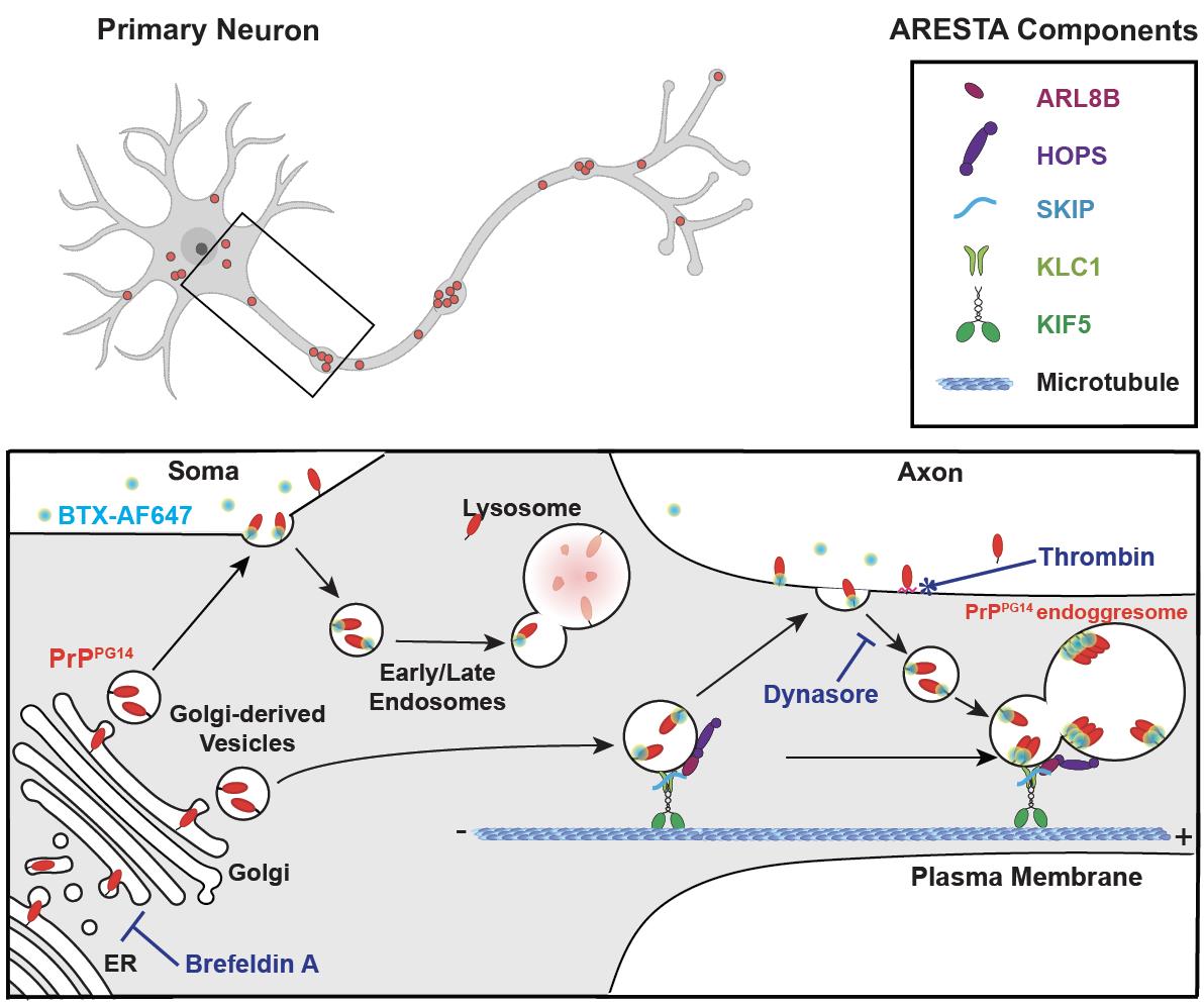
Graphical abstract
Background
Both wild-type (PrPWT) and mutant prion proteins (PrPmut) exit the ER and Golgi and follow secretory and endolysosomal trafficking pathways en route to somatodendritic and axonal cell surfaces, where PrP attaches via its glycosylphosphatidylinositol (GPI) anchor prior to internalization (Satpute-Krishnan et al., 2014; Zavodszky and Hegde, 2019; Chassefeyre et al., 2021). From the cell surface, PrPWT is endocytosed and sorted into endosomes or lysosomes for recycling or degradation, respectively, while PrPmut accumulates inside non-acidic/non-degradative enlarged endolysosomes where it can form aggregates called endoggresomes (Chassefeyre et al., 2021). PrPmut endoggresome formation in axons occurs via an axonal rapid endosomal sorting and transport-dependent aggregation (ARESTA) pathway (Chassefeyre et al., 2021). In ARESTA, vesicles containing luminal PrPmut exit from the Golgi after association with a protein complex composed of the small endolysosomal guanosine triphosphatase (GTPase) ARL8b. ARL8b recruits its effectors SifA and kinesin-interacting protein (SKIP), the kinesin-1 (KIF5C) molecular motor and associated kinesin light chain 1 (KLC1) subunit, and the vacuolar protein sorting 41 (VPS41), a member of the multiunit homotypic fusion and protein sorting (HOPS) complex (Hofmann and Munro, 2006; Khatter et al., 2015), which associate to endosomes carrying PrPmut and promote their entry into the axon and their homotypic fusion to form endoggresomes (Graphical abstract). For aggregation to occur in axons, endosomes carrying PrPmut homotypically fuse within axons after direct trafficking from the Golgi, or after undergoing rapid bouts of exocytosis and endocytosis throughout the axonal surface prior to fusion via the HOPS fusion complex. The rapid and very transient residence at the cell surface of PrPmut compared to PrPWT is key for the formation of endoggresomes vs. their degradation in axons. To understand the biology of misfolded PrPmut endoggresome formation vs. the normal trafficking of PrPWT, it is therefore critical to follow the transit of PrP particles throughout these subcellular itineraries.
Previous studies using drug treatments and fluorescent assays could monitor the trafficking routes of misfolded PrP in non-neuronal cells grown in culture (Satpute-Krishnan et al., 2014; Zavodszky and Hegde, 2019). However, as neurons are primary targets in prion diseases, it is important to follow the subcellular sorting of misfolded PrP in neurons, including in highly polarized compartments such as axons, which are primarily affected in disease. Here, we present live assays to image and quantitate the formation of PrPmut endoggresomes along their subcellular transit inside vesicles at the single-particle level using standard fluorescence microscopy, in either the soma or in axons of primary neurons, in the presence or absence of compounds that can modulate ER export and internalization capacity. These assays also monitor PrPmut accumulation in endolysosomal compartments. These approaches allowed the identification of trafficking and sorting pathways of PrPmut endoggresome biogenesis and would allow the monitoring of the trafficking of various other membrane proteins, including of the amyloid precursor protein (APP) or amyloid beta peptides implicated in the pathophysiology of Alzheimer’s disease, using cultured primary neurons as a model system.
Materials and Reagents
60 × 15 mm NuncTM Cell Culture/Petri Dishes (Thermo Fisher, catalog number: 150288)
150 × 21 mm NuncTM Cell Culture/Petri Dishes (Thermo Fisher, catalog number: 168381)
15 mL Falcon tube (Thermo Fisher, catalog number: 12565268)
#1.5 thickness 12 mm coverslip (Neuvitro, catalog number: NC0319857)
Note: Wash 3× acetone, 3× ethanol, and rinse 3× with sterile water; store in sterile water.
Cell culture plate, size 24-wells, sterile, flat bottom, tissue-culture treated (Sigma, catalog number: SIAL0524)
Syringe filter Millex-GP, 33 mm, PES, 0.22 µm, gamma-sterile, hydrophilic, FDA, CE, 50 pc/PAK (Millipore, catalog number: MPSLGPM33RS)
Eppendorf® Safe-Lock microcentrifuge tubes, 1.5 mL (Sigma, catalog number: T9661)
45 units of papain (Worthington, catalog number: PAPL LS003119)
Hanks' Balanced Salt Solution (HBSS), calcium, magnesium (Gibco, catalog number: 24020-117), store at 4 °C
LipofectamineTM 2000 transfection reagent (Invitrogen, catalog number: 11668030), store at 4 °C
Brefeldin A solution (1,000×) (BioLegend, catalog number: 420601), 5 mg/mL, store at 4 °C
Bungarotoxin (BTX), Alexa Fluor® 647 (AF647) conjugate (Thermo Fisher, catalog number: B35450)
Resuspend in H2O at a concentration of 1 mg/mL
Add sodium azide (Sigma, catalog number: 199931) at 2 mM
Store small aliquots at -20 °C. Avoid freezing and thawing. Protect from light
Tubocurarine hydrochloride pentahydrate (Sigma, catalog number: T2379-100MG)
Resuspend in H2O at 10 mM stock concentration
Store aliquots at 4 °C
Dynasore hydrate (Sigma, catalog number: D7693-5MG)
Resuspend in DMSO to make 80 mM stock solution, store at -20 °C.
LysoTrackerTM Green DND-26 (Invitrogen, catalog number: L7526), 1 mM, store at -20 °C
Magic Red Cathepsin-B Assay kit (ImmunoChemistry Technologies, catalog number: 937), store at 4 °C
Resuspend in 200 µL DMSO to the vial to make 250× stock, store at 4 °C.
Before each use, dilute the 250× DMSO stock in sterile H2O at 1:10 to make 25× sub-stock. Use immediately.
Thrombin from rat plasma (Sigma, catalog number: T5772-100UN), lyophilized powder
Resuspend in 1 mL to make a stock of 100 U/ml (20×), store at -20 °C.
Dimethyl sulfoxide (DMSO) (Sigma, catalog number: D4540), ≥99.5% (GC), suitable for cell culture
Phosphate-buffered saline (PBS), pH 7.2 (Gibco, catalog number: 20012027), suitable for cell culture
Water, cell culture grade (Corning, catalog number: 15363651)
Borate buffer (5 mL) (see Recipes)
Boric acid (Fisher, catalog number: A73-500)
Borax (Sigma, catalog number: B-9876)
1 mL of 0.05% DNaseI (see Recipes)
DNaseI (Boehringer, catalog number: 10104159001, 100 mg from bovine pancreas, grade II)
MgSO4 (Fisher, catalog number: BP213-1)
HBSS (Gibco, catalog number: 24020-117) (without Pen-Strep)
Poly-L-lysine stock solution (see Recipes)
5 mg bottle of Poly-L-lysine hydrobromide, molecular weight = 300,000, lyophilized powder, gamma-irradiated, BioXtra, suitable for cell culture (Sigma, catalog number: P5899-5MG)
Dissection media (see Recipes)
HBSS (Gibco, catalog number: 24020117)
D-glucose (Sigma, catalog number: G6152-100g or G5767-500g)
HEPES (Sigma, catalog number: H4034-100g or H-7523-50g)
Pen-Strep (Gibco, catalog number: 15140148 for 20 mL)
Brain digestion enzyme (Mixture A) (see Recipes)
PBS (Gibco, catalog number: 21600069, or DPBS, catalog number: 14190-144)
DL-cysteine HCl (Sigma, catalog number: C9768)
BSA (Sigma, catalog number: A7906-100G)
D-glucose (Sigma, catalog number: G6152)
Plating media (see Recipes)
DMEM High Glucose (1×) with phenol red + L-Glut w/o Sod. Pyr. (Gibco, catalog number: 11965092)
FBS (Gibco, catalog number: 26050-070)
Growth media (see Recipes)
Neurobasal A (NBA) medium with phenol red (Gibco, catalog number: 10888022)
B-27 Supplement (50×) (Gibco, catalog number: 17504044)
GlutaMAX-I 100× (Gibco, catalog number: 35050061)
Equipment
Surgical scissors (sharp/blunt) 4–6” (Fine Science Tools, catalog number: 14028-10)
Dumont forceps Super Fine #7 (Fine Science Tools, catalog number: 11271-30)
For brain dissections: stereo microscope equipped with dual top and bottom illumination, and with 6× and 12× magnification eyepieces/objectives.
5 mL NuncTM serological pipettes (Thermo Scientific, catalog number: 170355N)
10 mL NuncTM serological pipettes (Thermo Scientific, catalog number: 170356N)
25 mL NuncTM serological pipettes (Thermo Scientific, catalog number: 170357N)
Nikon Ti-E Perfect Focus inverted fluorescence microscope, with a total internal reflection fluorescence (TIRF) setup, equipped with 100×/1.49NA objectives and Andor iXon + DU897 EM Camera
CO2 incubator for live imaging on the microscope
35 mm Dish | No. 1.5 Coverslip | 14 mm glass diameter | uncoated (MatTek, catalog number: P35G-1.5-14-C)
Biosafety cabinet Class II
SciSpin MINI microfuge BLUE, with rotor for 8 × 1.5/2.2 mL, (7,000 rpm/2,680 × g) (SciQuip, catalog numbers: SS-6050 and SS-6058)
Software
ImageJ (Fiji) software (free download from https://imagej.net/software/fiji/)
Microsoft Excel
Procedure
文章信息
版权信息
© 2023 The Author(s); This is an open access article under the CC BY-NC license (https://creativecommons.org/licenses/by-nc/4.0/).
如何引用
Readers should cite both the Bio-protocol article and the original research article where this protocol was used:
- Chaiamarit, T., Wu, Y., Verhelle, A. and Encalada, S. E. (2023). Fluorescence Assays for Real-Time Tracking of Cell Surface Protein Internalization and Endosomal Sorting in Axons of Primary Mouse Hippocampal Neurons. Bio-protocol 13(7): e4651. DOI: 10.21769/BioProtoc.4651.
- Chassefeyre, R., Chaiamarit, T., Verhelle, A., Novak, S. W., Andrade, L. R., Leitao, A. D. G., Manor, U. and Encalada, S. E. (2021). Endosomal sorting drives the formation of axonal prion protein endoggresomes. Sci Adv 7(52): eabg3693.
分类
神经科学 > 细胞机理
细胞生物学 > 细胞成像
您对这篇实验方法有问题吗?
在此处发布您的问题,我们将邀请本文作者来回答。同时,我们会将您的问题发布到Bio-protocol Exchange,以便寻求社区成员的帮助。