

In situ Microinflammation Detection Using Gold Nanoclusters and a Tissue-clearing Method
利用金纳米团簇和组织清除方法进行原位微炎症检测
(*contributed equally to this work) 发布: 2023年04月05日第13卷第7期 DOI: 10.21769/BioProtoc.4644 浏览次数: 2710
评审: Meenal SinhaJulie WeidnerMarieta RusevaAnonymous reviewer(s)
参见作者原研究论文
The authors used this protocol in:
Feb 2012
Advertisement


Abstract
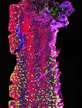
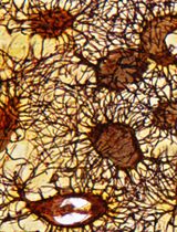
Microinflammation enhances the permeability of specific blood vessel sites through an elevation of local inflammatory mediators, such as interleukin (IL)-6 and tumor necrosis factor (TNF)-α. By a two-dimensional immunohistochemistry analysis of tissue sections from mice with experimental autoimmune encephalomyelitis (EAE), an animal model for multiple sclerosis (MS), we previously showed that pathogenic immune cells, including CD4+ T cells, specifically accumulate and cause microinflammation at the dorsal vessels of the fifth lumbar cord (L5), resulting in the onset of disease. However, usual pathological analyses by using immunohistochemistry on sections are not effective at identifying the microinflammation sites in organs. Here, we developed a new three-dimensional visualization method of microinflammation using luminescent gold nanoclusters (AuNCs) and the clear, unobstructed brain/body imaging cocktails and computational analysis (CUBIC) tissue-clearing method. Our protocol is based on the detection of leaked AuNCs from the blood vessels due to an enhanced vascular permeability caused by the microinflammation. When we injected ultrasmall coordinated Au13 nanoclusters intravenously (i.v.) to EAE mice, and then subjected the spinal cords to tissue clearing, we detected Au signals leaked from the blood vessels at L5 by light sheet microscopy, which enabled the visualization of complex tissue structures at the whole organ level, consistent with our previous report that microinflammation occurs specifically at this site. Our method will be useful to specify and track the stepwise development of microinflammation in whole organs that is triggered by the recruitment of pathogenic immune cells at specific blood vessels in various inflammatory diseases.
Keywords: Experimental autoimmune encephalomyelitis (EAE) (实验性自身免疫性脑脊髓炎(EAE))Background
Multiple sclerosis (MS) is a chronic inflammatory disease of the central nervous system (CNS), mediated by myelin-specific autoreactive CD4+ T cells (International Multiple Sclerosis Genetics et al., 2011). Active experimental autoimmune encephalomyelitis (aEAE) is a well-established animal model of MS that is induced by immunizing mice with myelin oligodendrocyte protein–derived peptide (MOG) and complete Freund's adjuvant (CFA) together with pertussis toxin (PTx). However, where and how pathogenic CD4+ T cells enter the CNS from peripheral blood and cause the disease had been unclear, since it was believed that the CNS is protected from immune cells by the blood–brain barrier (Liu et al., 2012). Using a passive transfer model of EAE, in which pathogenic CD4+ T cells including autoreactive CD4+ T cells from the secondary lymphoid tissues of aEAE mice are transferred to naïve mice, we demonstrated that sensory-sympathetic crosstalk, triggered by gravity stimulation via the soleus muscle, leads to the accumulation of pathogenic immune cells at the dorsal vessels of the fifth lumbar cord (L5) locally, which in turn causes microinflammation, a chronic inflammation that enhances the permeability of specific blood vessel sites by elevated local inflammatory mediators, such as IL-6 and TNF-α (Arima et al., 2012). Importantly, this permeability enables autoreactive CD4+ T cells to enter the CNS. We named this specific neuro-immune interaction the gateway reflex and have since demonstrated it can be activated by six types of environmental stimuli: gravity, electricity, pain, stress, light, and inflammation (Arima et al., 2012, 2015 and 2017; Hasebe et al., 2022; Murakami et al., 2019; Stofkova et al., 2019).
Coordinated gold nanoclusters (AuNCs) have an atomically precise molecular structure, ultrasmall size, facile surface modification, and characteristic optical properties (Jin et al., 2016). Some AuNCs have been reported to exhibit unique photoluminescence emission with long lifetimes and large Stokes shift from the visible to near-infrared wavelengths (Chen et al., 2015; Yu et al., 2019), as well as high compatibility and photostability. Thus, they are good candidates as luminophores for long-term imaging, high-sensitivity detection, and target-specific treatment (Luo and Liu, 2022; Palmal and Jana, 2014; Zhang et al., 2022). The usefulness of AuNCs as fluorescent probes has already been shown in imaging, detection, and therapy. Phosphine-coordinated AuNCs represent a luminescent gold cluster family (Konishi et al., 2018; Sugiuchi et al., 2017). Among them, diphosphine-coordinated Au13 nanoclusters with an icosahedral gold core motif are interesting, because of their easy accessibility, robustness, and moderately intense photoluminescence in the near-infrared region (Shichibu and Konishi, 2010; Sugiuchi et al., 2015).
The development of tissue-clearing reagents and protocols, efficient fluorescent labeling, and rapid volumetric imaging by light sheet microscopy has enabled new tissue-clearing methods for the 3D imaging of intact tissues and even entire organisms (Hama et al., 2015; Ueda et al., 2020). Despite tissue-clearing approaches enabling high tissue transparency within a few days using organic chemicals (e.g., benzyl alcohol and benzyl benzoate), concerns about the quenching of fluorescent proteins and safety have remained. In response, Tainaka et al. (2014) developed the hydrophilic unobstructed brain/body imaging cocktails and computational analysis (CUBIC) tissue-clearing method, which efficiently removes lipids, pigments, and calcium phosphate to retain the fluorescent signals and enable whole-body imaging with single-cell resolution in mice. Combining light sheet microscopy and CUBIC can visualize rare cells and complex structures in various tissues at the whole organ level.
Chronic microinflammation is a hallmark of many inflammatory diseases, including autoimmune, neurodegenerative, and metabolic syndrome–driven diseases. In such inflammatory diseases with stochastic and proliferative processes, single-cell events ultimately affect the health status of the entire organism. Therefore, a detection system that enables quick identification of the development and progression of microinflammation is desired. However, conventional protocols for identifying inflammation sites are mainly based on 2D histology using inflammatory mediator-specific reporter mice or antibodies, which makes it difficult to accurately identify the sites depending on the tissue sections.
In this protocol, we show a rapid 3D-imaging method to detect microinflammation in aEAE mice using Au13 nanoclusters and CUBIC. Au13 signals leaked out from the blood vessels around the L5, indicating enhanced vascular permeability caused by microinflammation, supporting our previous findings of a vascular gate formed for pathogenic immune cells to enter the CNS in this region (Arima et al., 2012). Thus, our method is useful for studying the stepwise development of microinflammation at the whole organ level in various inflammatory diseases and will also provide future perspectives of Au13 nanocluster-based imaging techniques to study biological events, which are induced by the enhancement of vascular permeability at specific vessel sites.
Materials and Reagents
C57BL/6NCrSlc mice (6–8-week-old male; Japan SLC)
50 mL polypropylene conical tube (Corning, Falcon®, catalog number: 352070)
15 mL polypropylene conical tube (Corning, Falcon®, catalog number: 352096)
1 mL syringe for tuberculin slip tip (TERUMO, catalog number: SS-01T)
Injection needle 25G × 1 (TERUMO, catalog number: NN-2525R)
Insulin syringe (with 27G × 1/2 needle) (TERUMO, catalog number: SS-10M2713)
Normal winged needle for vein D type (25G × 5/8 needle) (TERUMO, catalog number: SV-25DLK)
Three-way stopcock (TERUMO, catalog number: TS-TL1K)
30 mL syringe lock type 30 (TERUMO, catalog number: SS-30LZ)
5 mL Costar Stripette (Corning, catalog number: 4487)
10 mL Costar Stripette (Corning, catalog number: 4488)
25 mL Costar Stripette (Corning, catalog number: 4489)
MOG35-55 (MEVGWYRSPFSRVVHLYRNGK, Sigma-Aldrich)
Incomplete Freunds’ adjuvant (IFA) (Sigma-Aldrich, catalog number: F5506)
Mycobacterium tuberculosis H37RA (DIFCO, catalog number: 231141)
PTx from Bordetella pertussis (Sigma-Aldrich, catalog number: P7208-50UG)
Saline (Otsuka Pharmaceutical, catalog number: K6E85)
Isoflurane (Pfizer)
Sodium chloride (NaCl) (Sigma-Aldrich, catalog number: S9625)
Potassium chloride (KCl) (Kanto Chemical, catalog number: 32326-00)
Disodium hydrogen phosphate (Na2HPO4) (Nacalai Tesque, catalog number: 31801-05)
Potassium dihydrogen phosphate (KH2PO4) (Kishida Chemical, catalog number: 000-63955)
2-[4-(2-Hydroxyethyl)-1-piperazinyl]ethanesulfonic acid (HEPES) (Sigma-Aldrich, catalog number: H4034)
Sodium hydroxide (NaOH) (FUJIFILM Wako Chemicals, catalog number: 198-13765)
4% paraformaldehyde phosphate buffer solution (PFA) (Nacalai Tesque, catalog number: 09154-85)
Triton X-100 (Sigma-Aldrich, catalog number: T8787)
Sodium azide (FUJIFILM Wako Chemicals, catalog number: 197-11091)
Casein from bovine milk (Sigma-Aldrich, catalog number: C3400)
1,2-Hexanediol (Tokyo Chemical Industry, catalog number: H0688)
1-Methylimidazole (Tokyo Chemical Industry, catalog number: M0508)
2,3-Dimethyl-1-phenyl-5-pyrazolone (Tokyo Chemical Industry, catalog number: D1876)
Nicotinamide (Tokyo Chemical Industry, catalog number: N0078)
Silicone oil (Shin-Etsu Silicone, catalog number: HIVAC-F4)
Mineral oil (Sigma-Aldrich, catalog number: M8410-1L)
Anti-actin, α-smooth muscle (α-SMA)–FITC antibody, mouse monoclonal (clone: 1A4) (Sigma-Aldrich, catalog number: F3777)
Purified rat anti-mouse CD31 (clone: MEC 13.3) (BD Biosciences, catalog number: 550274)
Goat anti-rat IgG (H+L) cross-absorbed secondary antibody, Alexa FluorTM 555 (Thermo Fisher Scientific, catalog number: A-21434)
Dimethyl sulfoxide (DMSO) for spectrochemical analysis (FUJIFILM Wako Chemicals, catalog number: 045-28335)
Sodium borohydride (NaBH4) (Tokyo Chemical Industry, catalog number: S0480)
Ethanol (Kanto Chemical, catalog number: 14033-00)
[1,2-Bis(diphenylphosphino)ethane]dichlorodigold(I) (Au2(dppe)Cl2) (Sigma-Aldrich, catalog number: 717363)
Dichloromethane (Kanto Chemical, catalog number: 10158-81)
Hydrochloric acid (FUJIFILM Wako Chemicals, catalog number: 080-01061)
Acetone (Kanto Chemical, catalog number: 01026-81)
Methanol (Kanto Chemical, catalog number: 25183-81)
[Au13(dppe)5Cl2]Cl3 (Au13) [synthesized according to the literature (Shichibu and Konishi, 2010), see Recipes]
1 M HEPES buffer, pH 7.2 (see Recipes)
Phosphate buffered saline (PBS) (see Recipes)
Delipidation/decoloration buffer (see Recipes)
Staining buffer (see Recipes)
CUBIC-R (see Recipes)
Observation oil (see Recipes)
Equipment
Micro-dissecting scissors (Hammacher, catalog number: HSB014-11)
Micro scissors 105 mm straight sword (AS ONE, catalog number: YS-7105)
Angled serrated tip forceps (Hammacher, catalog number: HSC187-11)
KFI tweezers GG (Kowa Forceps Industry, catalog number: K-3 GG)
PIPETMAN P (Gilson, model: P20, catalog number: F123600)
PIPETMAN P (Gilson, model: P200, catalog number: F123601)
PIPETMAN P (Gilson, model: P1000, catalog number: F123602)
Pipet-aid (FALCON/catalog number: 357590)
Light sheet microscopy (LaVision BioTec/UltraMicroscope II)
Shake-LR (TAITEC, catalog number: 0054809-000)
Wave-SI (TAITEC, catalog number: 0054334-000)
Double shaker NR-30 (TAITEC, catalog number: 0000205-000)
Neo shaker NS-LR (AS ONE, catalog number: 2-7827-01)
Stereomicroscope (OLYMPUS, model: SZ61)
LED illuminator stand (OLYMPUS, model: SZ2-ILST)
Refractive index measurement equipment (ATAGO, catalog number: PAL-RI)
Software
Imaris version 9.0.0 (Bitplane, https://imaris.oxinst.com)
Procedure
文章信息
版权信息
© 2023 The Author(s); This is an open access article under the CC BY-NC license (https://creativecommons.org/licenses/by-nc/4.0/).
如何引用
Naim, F., Hasebe, R., Hojyo, S., Shichibu, Y., Ishii, A., Tanaka, Y., Tainaka, K., Kubota, S. I., Konishi, K. and Murakami, M. (2023). In situ Microinflammation Detection Using Gold Nanoclusters and a Tissue-clearing Method. Bio-protocol 13(7): e4644. DOI: 10.21769/BioProtoc.4644.
分类
免疫学 > 炎症性疾病 > 多发性硬化
神经科学 > 神经系统疾病 > 动物模型
细胞生物学 > 组织分析 > 组织成像
您对这篇实验方法有问题吗?
在此处发布您的问题,我们将邀请本文作者来回答。同时,我们会将您的问题发布到Bio-protocol Exchange,以便寻求社区成员的帮助。