

Determining Bone-forming Ability and Frequency of Skeletal Stem Cells by Kidney Capsule Transplantation and Limiting Dilution Assay
肾囊移植和有限稀释法测定骨骼干细胞的成骨能力和频率
发布: 2023年03月20日第13卷第6期 DOI: 10.21769/BioProtoc.4639 浏览次数: 1735
评审: Olga KopachAnonymous reviewer(s)
参见作者原研究论文
The authors used this protocol in:
Mar 2021
Advertisement


Abstract
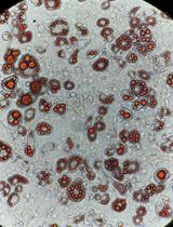
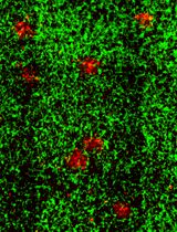
Adult stem cells not only maintain tissue homeostasis but are also critical for tissue regeneration during injury. Skeletal stem cells are multipotent stem cells that can even generate bones and cartilage upon transplantation to an ectopic site. This tissue generation process requires essential stem cell characteristics including self-renewal, engraftment, proliferation, and differentiation in the microenvironment. Our research team has successfully characterized and isolated skeletal stem cells (SSCs) from the cranial suture called suture stem cells (SuSCs), which are responsible for craniofacial bone development, homeostasis, and injury-induced repair. To assess their stemness features, we have demonstrated the use of kidney capsule transplantation for an in vivo clonal expansion study. The results show bone formation at a single-cell level, thus permitting a faithful assessment of stem cell numbers at the ectopic site. The sensitivity in assessing stem cell presence permits using kidney capsule transplantation to determine stem cell frequency by limiting dilution assay. Here, we described detailed protocols for kidney capsule transplantation and limiting dilution assay. These methods are extremely valuable both for the evaluation of skeletogenic ability and the determination of stem cell frequency.
Background
Conventional methods have shown the skeletogenic abilities of mesenchymal stromal cells (MSCs) isolated from bone marrow and other tissues in a Petri dish. However, in vivo transplantation studies indicate that the majority of MSCs lack engraftment, survival, and differentiation abilities (Caplan and Correa, 2011; Zeitouni et al., 2012). Only a small portion of MSCs are genuine skeletal stem cells (SSCs) (Robey et al., 2014; Sacchetti et al., 2007). In addition, in vitro experiments cannot examine certain features that are essential for stem cell stemness, thus lacking critical characteristics to fit into the rigorous criteria for the modern definition of SSCs. Animal models of ectopic bone formation have clear advantages over orthotopic transplantation because of the environments lacking cytokine interference and interactions with endogenous cell types, e.g., bone-forming cells. Three ectopic locations— subcutaneous, intramuscular, and kidney capsule—have been mainly used for transplantation studies (Scott et al., 2012). Subcutaneous implantation is the simplest method, but pertinent concerns arise because of several caveats related to the paucity of bone formation (Yang et al., 1996). Intramuscular implantation implies close contact with native skeletal muscle progenitors, raising concerns about the cellular origin of the ectopic bone that could be converted from host cells by osteogenic stimulus or traumatic injury (Takaoka et al., 1988; Yu et al., 2008). However, no endogenous cell interferes with the analysis of the transplant, which can be easily identified through kidney capsule transplantation. Although technically more demanding, the environment provides the most nutrient-rich resource for robust bone formation from significantly fewer cell numbers (Maruyama et al., 2021). Using kidney capsule transplantation, we have demonstrated ectopic bone generation at a single-cell level (Maruyama et al., 2016). This method also enables the determination of stem cells’ switch from an osteogenic to a chondrogenic fate for cartilage generation (Maruyama et al., 2016; Maruyama et al., 2022a). Furthermore, because kidney capsule transplantation permits in vivo clonal expansion analysis, it is sensitive enough to determine stem cell frequency by limiting dilution analysis (Maruyama et al., 2016; Maruyama et al., 2021). These analyses utilizing kidney capsule transplantation are powerful tools with clear advantages to advance skeletal stem cell research.
Materials and Reagents
50 mL conical polypropylene centrifuge tube (Fisher Scientific, catalog number: 12-565-271)
1.5 mL microtube (Fisher Scientific, catalog number: 05-408-129)
Insulin syringes with Ultra-FineTM needle (BD Biosciences, catalog number: 08290-3284-38)
Artificial tears ointment (Rugby Laboratories, catalog number: 0536-6550-91)
Cotton swab, puritan 6” sterile (Puritan, catalog number: 25-806 1WC)
Povidone-Iodine cotton swab stick, individual packet 10% strength (Medicine, catalog number: MDS093901)
Alcohol pads (PDI/Professional Disposables, catalog number: B60307)
Nonidet P-40 substitute (Sigma-Aldrich, catalog number: 98379)
Capillary tube (Drummond Scientific, catalog number: 1-000-800)
POLYSYN violet braided coated polyglycolic acid absorbable suture (catalog number: G391NV, TruStitch)
Latex gloves (VWR, catalog number: 56617-180), 5 years shelf life at room temperature
SCID mouse (NOD.CB17-Prkdcscid/NCrCrl, Charles River) older than two months of age
Dulbecco’s phosphate-buffered saline without calcium & magnesium (Gibco, catalog number: 10010-023), two years shelf life at room temperature
Dulbecco’s phosphate-buffered saline with calcium & magnesium (Gibco, catalog number: 14040-133), two years shelf life at room temperature
Matrigel (BD Biosciences, catalog number: 354234), two years of shelf life at -20 °C
Ketamine HCl (10 mg/mL) (COVETRUS, catalog number: 071069), two years shelf life at room temperature
Xylazine (1 mg/mL) (COVETRUS, catalog number: 061035), two years shelf life at room temperature
NYLON blue monofilament suture (TruStitch, catalog number: A662NV)
Ethiqa XR (Fidelis Pharmaceuticals LLC, catalog number: 86084-100-30), one year shelf life at room temperature
Paraformaldehyde (J.T. Baker, catalog number: S898-0), seven years of shelf life at room temperature
Silver nitrate (Sigma-Aldrich, catalog number: 10220-100G), >5 years shelf life at room temperature
4% PFA (see Recipes)
2% Nonidet P-40 (see Recipes)
0.02% NP40, 2% PFA (see Recipes)
5% sodium thiosulfate (see Recipes)
Anesthesia (see Recipes)
Equipment
Centrifuge (Thermo Scientific, Sorvall ST 16R Centrifuge)
Incubator (Thermo Forma, Series II Water Jacketed CO2 Incubator)
Procedure/Dissection Board (Aims Lab Products, GPM2)
Fiber-Optic Illuminator with dual goosenecks (Nikon)
Scissors (Iris scissors, Fine Science Tools)
Forceps (Dumont #5SF, Ring, and Graefe forceps, Fine Science Tools)
Needle holder: Halsted-Mosquito Hemostats (Fine Science Tools, catalog number: 91308-12)
Reflex Wound Clips (VWR, catalog number: 203-1000101326-476)
Stapler: Reflex Wound Clips (VWR, catalog number: 203-1000101326-476)
Electrocautery: Cautery Loop Tip High-Temperature 2200 °F (Bovie Medical Corporation)
Heat pad, microwavable (K&H Pet Products, catalog number: 69553)
Electric shaver (OSTER, catalog number: 078996-101-001)
Nikon stereoscope (NIKON, catalog number: SMZ1500)
Spot imaging system (Insight CMOS Digital Camera, SPOT)
Procedure
文章信息
版权信息
© 2023 The Author(s); This is an open access article under the CC BY-NC license (https://creativecommons.org/licenses/by-nc/4.0/).
如何引用
Readers should cite both the Bio-protocol article and the original research article where this protocol was used:
- Uchida, H., Maruyama, T. and Hsu, W. (2023). Determining Bone-forming Ability and Frequency of Skeletal Stem Cells by Kidney Capsule Transplantation and Limiting Dilution Assay. Bio-protocol 13(6): e4639. DOI: 10.21769/BioProtoc.4639.
- Maruyama, T., Hasegawa, D., Valenta, T., Haigh, J., Bouchard, M., Basler, K. and Hsu, W. (2022a). GATA3 mediates nonclassical beta-catenin signaling in skeletal cell fate determination and ectopic chondrogenesis. Sci Adv 8(48): eadd6172.
分类
干细胞 > 成体干细胞 > 细胞移植
发育生物学 > 形态建成 > 器官形成
细胞生物学 > 细胞移植 > 异种移植
您对这篇实验方法有问题吗?
在此处发布您的问题,我们将邀请本文作者来回答。同时,我们会将您的问题发布到Bio-protocol Exchange,以便寻求社区成员的帮助。