- EN - English
- CN - 中文
Establishment of an in vitro Differentiation and Dedifferentiation System of Rat Schwann Cells
大鼠雪旺细胞体外分化和去分化体系的建立
发布: 2023年03月05日第13卷第5期 DOI: 10.21769/BioProtoc.4631 浏览次数: 1811
评审: Gal HaimovichPhilipp WörsdörferAnonymous reviewer(s)
Abstract
In the peripheral nervous system, Schwann cells are the primary type of glia. This protocol describes an in vitro differentiation and dedifferentiation system for rat Schwann cells. These cultures and systems can be used to investigate the morphological and biochemical effects of pharmacological intervention or lentivirus-mediated gene transfer on the process of Schwann cell differentiation or dedifferentiation.
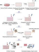
Graphical abstract

Background
Schwann cells are the primary glial cells of the peripheral nervous system. During axonal sorting and myelination in the peripheral nerves, Schwann cells originate from neural crest cells that differentiate into mature phenotypes (Cristobal and Lee, 2022). Moreover, Schwann cells’ incredible plasticity is one of the most important characteristics following nerve damage or demyelination (Nocera and Jacob, 2020). Schwann cells undergo dedifferentiation after injury and redifferentiate to promote nerve regeneration and complete functional recovery (Jessen and Mirsky, 2019). Therefore, it is important to study Schwann cells’ differentiation and dedifferentiation status to understand their role in nerve development and injury. As reported previously, dibutyryl adenosine 3',5'-cyclic monophosphate (dbcAMP) induces Schwann cells to acquire a differentiated phenotype (Yamauchi et al., 2011). After stimulation with 1 mM dbcAMP, Schwann cells from rats change from bipolar or tripolar to flat within 24 h. By contrast, mouse Schwann cells retain their bipolar or tripolar morphology after dbcAMP treatment (Arthur-Farraj et al., 2011). And a previous study described in vitro assays of scalable differentiation and dedifferentiation of Schwann cell in the absence of neurons (Monje PV., 2018). Based on previous studies, we established a detailed and simplified system for differentiation and dedifferentiation of Schwann cells. In order to clearly distinguish morphological changes, we performed the differentiation and dedifferentiation assay using rat Schwann cells. In brief, the first step was to obtain Schwann cells from the spinal nerves of rats based on a previous study (Wen et al., 2017). During the second step, differentiation and dedifferentiation assays were conducted. Schwann cells were purified and passaged, and experiments were conducted on the third passage. Finally, Schwann cell status was determined by morphological examination, western blotting analysis, and immunofluorescence detection.
Materials and reagents
Cell culture dish (3.5 and 6 cm) (JET BIOFILT, catalog numbers: CD000035, TCD-010-060)
50 mL centrifuge tubes (Corning, catalog number: 430828)
15 mL centrifuge tubes (Corning, catalog number: 430790)
1.5 mL tube (JET BIOFILT, catalog number: CFT000015)
Polyvinylidene fluoride membrane (PVDF) (Bio-Rad, catalog number: 1620177)
Cell glass coverslips (diameter: 12 mm, thickness: 0.13–0.17 mm) (Fisherbrand, catalog number: FIS12-545-80)
Neonatal Sprague-Dawley (SD) rat [postnatal 1–2 days (P1–P2)]
Distilled water
75% ethanol
Phosphate buffered saline (PBS) (Gibco, catalog number: 10010023)
Poly-L-lysine hydrobromide (PLL) (Sigma-Aldrich, catalog number: P1274)
0.25% trypsin-EDTA (Gibco, catalog number: 25200072)
Fetal bovine serum (FBS) (Corning, catalog number: 35-076-CV)
DMEM/F12 (Gibco, catalog number: 11330057)
Cytosine arabinoside (Ara-C) (Sigma-Aldrich, catalog number: C1768)
Recombinant human heregulin β-1 (PeproTech, catalog number: 100-03)
Forskolin (Sigma-Aldrich, catalog number: F6886)
4% paraformaldehyde (PFA) (Biosharp, catalog number: BL539A)
Dimethyl sulfoxide (DMSO), suitable for cell culture (Beyotime, catalog number: ST038)
Dibutyryl adenosine 3',5'-cyclic monophosphate (dbcAMP) (Sigma-Aldrich, catalog number: D0627)
Triton X-100 (Sigma-Aldrich, catalog number: V900502)
Tween-20 (Sigma-Aldrich, catalog number: P1379)
Phalloidin (Abcam, catalog number: ab176759)
RIPA lysis buffer (FUDE Biological Technology, catalog number: FD009)
Rabbit monoclonal (EP1039Y) anti-p75 (Abcam, catalog number: ab52987)
Rabbit anti-Krox20 (Novus Biologicals, catalog number: 13491-1-AP)
Mouse monoclonal anti-c-Jun (BD Biosciences, catalog number: 610326)
Mouse monoclonal (9-9-3) anti-Sox2 (Abcam, catalog number: ab79351)
Alexa Fluor® 488 goat anti-mouse IgG (H+L) (Thermo Fisher Scientific, catalog number: A11001)
Alexa Fluor® 568 goat anti-rabbit IgG (H+L) (Thermo Fisher Scientific, catalog number: A11011)
4’,6-diamidino-2’-phenylindole (DAPI) (Sigma-Aldrich, catalog number: D8417)
Penicillin–streptomycin (Gibco, catalog number: 15140-122)
Omni-ECLTM Light Chemiluminescence kit (EpiZyme, catalog number: SQ201)
10% dodecyl sulfate sodium salt-polyacrylamide gel (EpiZyme, catalog number: PG112)
5% non-fat milk (Solarbio, catalog number: D8340)
Tris-buffered saline (Sigma-Aldrich, catalog number: 93318)
Horseradish peroxidase (HRP)-conjugated secondary antibody (Invitrogen, catalog numbers: 31430, 31460)
1,000× Ara-C (10 mM in distilled water, see Recipes)
1,000× dbcAMP (1,000 mM in DMSO, see Recipes)
30 mM forskolin stock solution (see Recipes)
Complete growth medium of rat Schwann cells (see Recipes)
10% FBS (see Recipes)
3% FBS (see Recipes)
1% FBS (see Recipes)
1 mM dbcAMP (see Recipes)
0.1% Triton X-100 (see Recipes)
5% gelatin (see Recipes)
PBST (see Recipes)
Blocking buffer (see Recipes)
Equipment
Pipettes (Thermo Fisher Scientific, 10, 200, and 1,000 μL)
CO2 incubator (Thermo Fisher Scientific, model: Heracell 150i)
Surgical scissors and forceps (Shenzhen RWD, catalog numbers: S14014 and F12029-09)
Spring scissors (Shenzhen RWD, catalog number: S11001)
Superfine forceps (Shenzhen RWD, catalog number: F13002)
Stereomicroscope (Jiangnan Novel, model: SZ6060)
Centrifuge (Eppendorf, model: Micro21)
Phase contrast microscope (Zeiss, model: Primovert)
Fluorescence microscope (Zeiss, model: Axio Imager A2)
Software
ImageJ (Version 1.8.0, https://imagej.en.softonic.com/)
Procedure
文章信息
版权信息
© 2023 The Author(s); This is an open access article under the CC BY-NC license (https://creativecommons.org/licenses/by-nc/4.0/).
如何引用
Ying, Z. (2023). Establishment of an in vitro Differentiation and Dedifferentiation System of Rat Schwann Cells. Bio-protocol 13(5): e4631. DOI: 10.21769/BioProtoc.4631.
分类
神经科学 > 周围神经系统 > 施万细胞
细胞生物学 > 细胞分离和培养 > 细胞分化
细胞生物学 > 细胞成像 > 固定细胞成像
您对这篇实验方法有问题吗?
在此处发布您的问题,我们将邀请本文作者来回答。同时,我们会将您的问题发布到Bio-protocol Exchange,以便寻求社区成员的帮助。
Share
Bluesky
X
Copy link


