

Far-western Blotting Detection of the Binding of Insulin Receptor Substrate to the Insulin Receptor
Far-western blotting 检测胰岛素受体底物与胰岛素受体的结合
发布: 2023年02月20日第13卷第4期 DOI: 10.21769/BioProtoc.4619 浏览次数: 2530
评审: Suresh KumarRitu GuptaAnonymous reviewer(s)
参见作者原研究论文
The authors used this protocol in:
Mar 2022
Advertisement


Abstract
Far-western blotting, derived from the western blot, has been used to detect interactions between proteins in vitro, such as receptor–ligand interactions. The insulin signaling pathway plays a critical role in the regulation of both metabolism and cell growth. The binding of the insulin receptor substrate (IRS) to the insulin receptor is essential for the propagation of downstream signaling after the activation of the insulin receptor by insulin. Here, we describe a step-by-step far-western blotting protocol for determining the binding of IRS to the insulin receptor.
Keywords: Signal transduction ( 信号转导)Background
Insulin, secreted by the pancreatic β-cells, is the most powerful anabolic hormone known to regulate the metabolism of glucose, lipids, and amino acid metabolism through the activation of the insulin signaling pathway. Insulin binding to the insulin receptor (IR), a tetrameric complex consisting of two extracellular α-subunits and two transmembrane β-subunits, leads to a conformational change, the activation of tyrosine kinase activity in the β-subunits, and the transphosphorylation of β-subunits at Y972 (Sweet et al., 1987; Cheatham and Kahn, 1995; Yip and Ottensmeyer, 2003). The phosphorylation of the β-subunit at Y972 generates a NPXpY motif, which the downstream mediator insulin receptor substrate (IRS) subsequently recognizes and binds to in the insulin receptor b (IRβ), resulting in the activation of PI3K-AKT signaling (Machado-Neto et al., 2018; White et al., 1988). Therefore, the binding of the IRS to the IRβ subunit plays a critical role in the activation of insulin signaling (Peng and He, 2018).
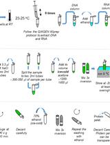
Far-western blotting is an effective technique to assess protein–protein interactions, including receptor–ligand interactions (Kaido et al., 2007; Wu et al., 2007), in in vitro assays. In a far-western blotting analysis, one protein is first separated in an SDS-PAGE gel and transferred to a membrane, followed by the binding of a non-antibody secondary protein. Then, a specific antibody against the secondary protein will be employed to determine its binding to the first protein that is being transferred onto the membrane (Figure 1). This method can be used in a regular laboratory, without the need of expensive equipment, to determine the interaction of proteins in other methods, such as the surface plasmon resonance system. We introduce a far-western blotting analysis that has been successfully used to determine the interaction of IRβ to its downstream mediator, the IRS, and to assess the importance of post-translational acetylation of IRS in affecting its binding to IRβ, as well as the activation of the insulin signaling pathway in our studies (Cao et al., 2017; Peng et al., 2022).

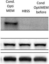
Figure 1. Procedure of far-western blotting to examine the binding of IRS to IRβ. A. Sequential steps of the procedure. B. Diagram depicts the detection of the binding with antibody. C. Purified IRS1 and IRS2 proteins were subjected to SDS-PAGE and stained with colloidal blue. D. Similar quantities of IRS2 and acetylated IRS2 by acetyltransferase P300 protein were employed in an SDS-PAGE and transferred onto a membrane; after renaturation, membranes were incubated with IRβ, followed by incubation with anti-IRβ antibody.
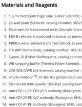
Materials and Reagents
1.5 mL Eppendorf tubes (Eppendorf, catalog number: 022363204)
Immuno-Blot PVDF membrane (Bio-Rad, catalog number: 1620177)
Tris-HCl (pH 6.8) (Sigma-Aldrich, catalog number: T5941)
Sodium dodecyl sulfate (Sigma-Aldrich, catalog number: L3771)
Glycerol (Sigma-Aldrich, catalog number: G5516)
β-mercaptoethanol (Sigma-Aldrich, catalog number: M3148)
EDTA (Sigma-Aldrich, catalog number: E9884)
Bromophenol blue (Sigma-Aldrich, catalog number: B8026)
NuPAGETM LDS sample buffer (4×) (Thermo Fisher Scientific, catalog number: NP0007)
NuPAGETM 3%–8%, NovexTM tris-acetate 1.5 mm mini protein gel, 10 well (Thermo Fisher Scientific, catalog number: EA0378BOX)
NuPAGETM tris-acetate SDS running buffer (20×) (Thermo Fisher Scientific, catalog number: LA0041)
IRS proteins were prepared as described previously (Cao et al., 2017; Peng et al., 2022). To determine the effect of IRS acetylation on its binding to IRb, IRS1 and IRS2 proteins were acetylated by acetyltransferase P300 protein. In the acetylation assay, 2 μg of IRS1 or IRS2 were added to the reaction containing 50 mM Tris-HCl (pH 8.0), 5% glycerol, 0.1 mM EDTA, 50 mM KCl, 1 mM dithiothreitol (DTT), 1 mM PMSF, 10 mM sodium butyrate, 0.2 μg of acetyl-CoA, and 0.2 μg of P300 (Active Motif). Samples were incubated at 30 °C for 1 h. In another acetylation assay set, acetyl-CoA was not added but it served as a positive control.
Recombinant human IRβ protein (Creative Biomart, catalog number: INSR-5093H)
Anti-IRβ (Cell Signaling Technology, catalog number: 3020); dilution: 1:500
Protein ladder (Thermo Fisher Scientific, catalog number: 26634)
Tris (Sigma-Aldrich, catalog number: T1503)
Glycine (Sigma-Aldrich, catalog number: G8898)
Methanol (Sigma-Aldrich, catalog number: 34860)
NaCl (Sigma-Aldrich, catalog number: 9888)
Tween-20 (Sigma-Aldrich, catalog number: P1379)
Skim milk powder (Bio-Rad, catalog number: 1706404XTU)
DTT (Sigma-Aldrich, catalog number: D0632)
KH2PO4 (Sigma-Aldrich, catalog number: P5655)
Na2HPO4 (Sigma-Aldrich, catalog number: S9763)
Guanidine-HCl (Sigma-Aldrich, catalog number: G3272)
ECL kit (PierceTM ECL western blotting substrate) (Thermo Fisher Scientific, catalog number: 32106)
Loading buffer (see Recipes)
Wet transfer buffer (see Recipes)
Denaturing and renaturing buffers (see Table 1) (see Recipes)
Protein-binding buffer (see Recipes)
PBST buffer (see Recipes)
Equipment
Mini gel tank (Thermo Fisher Scientific, catalog number: A25977)
PowerPacTM basic power supply (Bio-Rad, catalog number: 1645050)
ChemiDoc XRS+ gel imaging system (Bio-Rad)
Mini trans-blot electrophoretic transfer cell (Bio-Rad, catalog number: 1703930)
Procedure
文章信息
版权信息
© 2023 The Author(s); This is an open access article under the CC BY-NC license (https://creativecommons.org/licenses/by-nc/4.0/).
如何引用
Readers should cite both the Bio-protocol article and the original research article where this protocol was used:
- Peng, J., Ramatchandirin, B., Pearah, A. and He, L. (2023). Far-western Blotting Detection of the Binding of Insulin Receptor Substrate to the Insulin Receptor. Bio-protocol 13(4): e4619. DOI: 10.21769/BioProtoc.4619.
- Peng, J., Ramatchandirin, B., Wang, Y., Pearah, A., Namachivayam, K., Wolf, R. M., Steele, K., MohanKumar, K., Yu, L., Guo, S., White, M. F., Maheshwari, A. and He, L. (2022). The P300 acetyltransferase inhibitor C646 promotes membrane translocation of insulin receptor protein substrate and interaction with the insulin receptor. J Biol Chem 298(3): 101621.
分类
生物化学 > 蛋白质 > 免疫检测 > 免疫印迹法(WB )
细胞生物学 > 细胞信号传导 > 胞内信号传导
您对这篇实验方法有问题吗?
在此处发布您的问题,我们将邀请本文作者来回答。同时,我们会将您的问题发布到Bio-protocol Exchange,以便寻求社区成员的帮助。