

Staining and Scanning Protocol for Micro-Computed Tomography to Observe the Morphology of Soft Tissues in Ambrosia Beetles
用于观察食菌小蠹软组织形态的微型计算机断层扫描染色方法
发布: 2023年01月05日第13卷第1期 DOI: 10.21769/BioProtoc.4584 浏览次数: 1366
评审: Khyati Hitesh ShahSailendra SinghJegan Sekar
参见作者原研究论文
The authors used this protocol in:

Sep 2020
Advertisement


Abstract
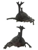
Advances in imaging technology offer new opportunities in developmental biology. To observe the development of internal structures, microtome cross-sectioning followed by H&E staining on glass slides is a common procedure; however, this technique can be destructive, and artifacts can be introduced during the process. In this protocol, we describe a less invasive procedure with which we can stain insect samples and obtain reconstructed three-dimensional images using micro-computed tomography, or micro-CT (µCT). Specifically, we utilize the fungus-farming ambrosia beetle species Euwallacea validus to observe the morphology of mycangia, a critical internal organ with which beetles transport fungal symbionts. Not only this protocol is ideal to observe mycangia, our staining/scanning procedure can also be applied to observe other delicate tissues and small organs in arthropods.
Graphical abstract

Background
The study of developmental biology often requires visualization across life stages as it concerns the process through which certain organs or tissues form. Microscopy has been the core means to observe the morphology of organs under development. To observe external features, stereo microscopy is adequate to visualize structures across developmental stages; however, internal structures are often difficult to observe and measure without destructive sampling. Employing micro-computed tomography (µCT) visualization protocols allows for the observation of structures throughout their development, in hard- and soft-bodied life stages alike (Spahr et al., 2020). The success of staining and scanning protocols can allow for detailed internal visualization and quantification of structures after genetic functional analyses, where the use of a non-destructive visualization technique is integral. Here, we describe stepwise methods for µCT scanning in the fungus-farming ambrosia Euwallacea validus .
Ambrosia beetle symbiosis is an emerging, global topic in forest pathology and entomology due to many species being invasive pests that vector phytopathogenic fungi across native and introduced ranges. Notably, Raffaelea lauricola , the causal agent of laurel wilt disease, is vectored by the redbay ambrosia beetle Xyleborus glabratus (Fraedrich et al., 2008). Other phytopathogenic fungi, such as Fusarium species carried by Euwallacea ambrosia beetles, cause significant agricultural and economic damages to avocado, citrus, and tea (Freeman et al., 2013; O'Donnell et al., 2015). Understanding the developmental process of tissues in beetles or fungi may lead to the development of effective means to control these pests.
The beetles rely on novel pits or pouches called mycangia, in which they house and transmit fungal propagules between tree hosts. The location of these structures varies between beetle species. External mycangia, such as the pronotal pouches of the bark beetle Dendroctonus frontalis and related species, are commonly observed by electron scanning microscopy; ease of study has allowed for detailed structural descriptions (Yuceer et al., 2011). However, the internal mycangia, such as the preoral mycangia found in Euwallacea , are more difficult to observe and describe. Published studies of internal mycangia have employed microtome cross-sectioning, laser ablation tomography (LAT) scanning, and micro-computed tomography (Li et al., 2015, 2018). The destructive nature of microtome cross-sectioning can require many samples to produce a viable result, especially when using small arthropod samples. By employing scanning techniques, the artifacts caused by slicing and staining of cross-sections can be reduced, which allows us to achieve detailed results. This technique can assist other studies in observing and describing soft tissues in arthropods and can be combined with other techniques (such as RNA interference, allometry, etc.) to strengthen resources for small, internal, or otherwise difficult to measure structures.
Materials and Reagents
Eppendorf 1.5 mL microcentrifuge tubes (Sigma-Aldrich, catalog number: T6649)
200 μL pipette tips (Sigma-Aldrich, Corning, catalog number: CS4860)
Paint brush (any fine brush)
Scissors
100% ethanol (200 proof) (Sigma-Aldrich, catalog number: E7023)
Deionized/sterile water (Sigma-Aldrich, catalog number: 8483331000)
5% Lugol’s solution (Fisher Scientific, Spectrum Chemical, catalog number: 18-611-063)
Dental wax ortho tray strips (Kerr, Benco, catalog number: 1021-752)
Insect samples
70% ethanol (see Recipes)
Equipment
Bruker Skyscan 1272 micro-computed tomography scanner
Computer that can run the software (see Note 1 below)
Software
Skyscan Micro-CT scanning software (version 1.1.10, Bruker; https://www.bruker.com/en/products-and-solutions/microscopes/3d-x-ray-microscopes/skyscan-1272.html)
NRecon Reconstruction (Bruker, 3D.SUITE software includes Dataviewer, CTAn, CTVol, CTVox) (Bruker, https://www.bruker.com/en/products-and-solutions/preclinical-imaging/micro-ct/3d-suite-software.html)
Procedure
文章信息
版权信息
© 2023 The Authors; exclusive licensee Bio-protocol LLC.
如何引用
Spahr, E. J., McLaughlin, S. L., Tichinel, A. M., Kasson, M. T. and Kijimoto, T. (2023). Staining and Scanning Protocol for Micro-Computed Tomography to Observe the Morphology of Soft Tissues in Ambrosia Beetles. Bio-protocol 13(1): e4584. DOI: 10.21769/BioProtoc.4584.
分类
分子生物学 > RNA > RNA 干扰
发育生物学 > 形态建成
您对这篇实验方法有问题吗?
在此处发布您的问题,我们将邀请本文作者来回答。同时,我们会将您的问题发布到Bio-protocol Exchange,以便寻求社区成员的帮助。