

Monitoring Mitochondrial Protein Import Using Mitochondrial Targeting Sequence (MTS)-eGFP
使用线粒体靶向序列 (MTS)-eGFP 监测线粒体蛋白导入
(*contributed equally to this work) 发布: 2022年12月20日第12卷第24期 DOI: 10.21769/BioProtoc.4578 浏览次数: 3598
评审: Julie WeidnerOlli MatilainenAlberto RissoneAnonymous reviewer(s)
参见作者原研究论文
The authors used this protocol in:

Jan 2022
Advertisement


Abstract
Mitochondria are cellular organelles essential for the function and survival of eukaryotic cells. Nearly all mitochondrial proteins are nuclear-encoded and require mitochondrial import upon their synthesis in the cytosol. Various approaches have been described to study mitochondrial protein import, such as monitoring the entry of radiolabeled proteins into purified mitochondria or quantifying newly synthesized proteins within mitochondria by proteomics. Here, we provide a detailed protocol for a commonly used and straightforward assay that quantitatively examines mitochondrial protein import by monitoring the co-localization of mitochondrially targeted enhanced green fluorescent protein (eGFP) with the mitochondrial fluorescence dye MitoTracker TM Deep Red FM by live cell imaging. We describe the preparation and use of a stable mammalian cell line inducibly expressing a mitochondrial targeting sequence (MTS)-eGFP, followed by quantitative image analysis using an open-source ImageJ-based plugin. This inducible expression system avoids the need for transient transfection while enabling titration of MTS-eGFP expression and thereby avoiding protein folding stress. Overall, the assay provides a simple and robust approach to assess mitochondrial import capacity of cells in various disease-related settings.
Graphical abstract

Background
Mitochondria are crucial for cellular survival. Almost all mitochondrial proteins are encoded in the nuclear genome and proper mitochondrial function and biogenesis rely on mitochondrial protein import (Dimogkioka et al., 2021). Multiple mitochondrial protein import machineries exist; each mediates protein translocation into a distinct mitochondrial sub-compartment (Schmidt et al., 2010). Stress conditions, such as the loss of mitochondrial membrane potential, are known to disturb mitochondrial protein import, and defective import can lead to a series of pathologies, such as neurodegenerative and cardiovascular diseases and cancer (Geissler et al., 2000; Palmer et al., 2021).
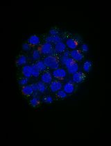
To investigate mitochondrial protein import, several methods have been established. Radiolabeling of individual mitochondrial proteins followed by incubation with purified mitochondria allows following mitochondrial import of the labeled protein (Murschall et al., 2021; Poveda-Huertes et al., 2021). Besides the technical (e.g., purification of fully active mitochondria) and bureaucratic requirements of performing radioactive lab work, this in vitro approach lacks cellular context and thus does not account for potential contribution of cytosolic factors on protein import. Recently developed proteomics methods allow monitoring the import of newly synthesized proteins into mitochondria on the global level (Schafer et al., 2022). This method is carried out within the cellular context and allows monitoring hundreds of mitochondrial proteins; however, it is a complex proteomics method that requires a mass spectrometry infrastructure not available in most laboratories. Here, we describe a fluorescence microscopy–based approach that allows monitoring mitochondrial import of the previously established mitochondrial targeting sequence (MTS)–enhanced green fluorescent protein (eGFP) reporter protein by live cell imaging (Chen et al., 2003). The reporter consists of eGFP that is selectively targeted to the mitochondrial matrix by means of a N-terminal MTS, which consists of the first 69 amino acids of subunit 9 of the F0-ATPase. Transcription of the reporter gene can be induced with the Tet-On system by treating cells with doxycycline, which allows for a controlled and titratable MTS-eGFP expression. Defective import of the reporter is assessed by its mislocalization to the cytosol, which is monitored by co-staining of mitochondria with MitoTrackerTM Deep Red FM. Subsequent co-localization analysis with the ImageJ-implemented Coloc2 plugin allows assessing mitochondrial protein import defects in a quantitative manner.
Materials and Reagents
Cultivating cell lines
10 cm tissue culture dish (Sarstedt, catalog number: 83.3902)
6-well cell culture plate (Sarstedt, catalog number: 83.3920.005)
Cell counting slides (dual-chamber) (Bio-Rad, catalog number: 1450015)
0.45 µm filters (Whatman, catalog number: 10462100)
HeLa FlpIn TRex cell line (Invitrogen, catalog number: R71407)
Note: This protocol was established for HeLa FlpIn TRex cell lines. It can be transferred to other cell lines that are suitable for microscopy.
HEK 293T cells (human embryonic kidney 293T) (ATCC, catalog number: CRL-3216)
RPMI 1640 medium (+L-Glutamine) (Thermo Fisher Scientific, catalog number: 21875-034)
DMEM medium (+L-Glutamine) (Thermo Fisher Scientific, catalog number 41966029)
Fetal bovine serum (FBS) (Thermo Fisher Scientific, catalog number: 10270-106)
Trypsin-EDTA (0.25%), phenol red (Thermo Fisher Scientific, catalog number: 25200056)
Dulbecco's phosphate buffered saline (PBS) (Thermo Fisher Scientific, catalog number: 14190-169)
Trypan blue solution (Sigma-Aldrich, catalog number: T8154)
Stable and inducible MTS-eGFP cell line generation
15 mL tube (Greiner, catalog number: 188271-N)
Lipofectamine 2000 transfection reagent (Thermo Fisher Scientific, catalog number: 11668027)
Puromycin (InvivoGen, catalog number: ant-pr-1)
Opti-MEM I (Thermo Fisher Scientific, catalog number: 31985-047)
Polybrene (Sigma-Aldrich, catalog number: TR-1003-G)
pHDM-VSV-G (Addgene, catalog number: 164440); encodes VSV-G protein
pHDM-Hgpm2 (Addgene, catalog number: 164441); encodes codon-optimized HIV gag-pol
pHDM-tatIB (Addgene, catalog number: 164442); encodes HIV-1 Tat accessory protein
pRC-CMV-revIB (Addgene, catalog number: 164443); encodes HIV-1 Rev accessory protein
pLD-puro-MTS-EGFP (Addgene, catalog number: 190270); contains tetracycline/doxycycline-inducible MTS-eGFP and lentiviral packaging signal
MTS-eGFP mitochondrial import assay
Cell culture microplate, 96-well, black (Greiner, catalog number: 655090)
MitoTrackerTM Deep Red FM (Thermo Fisher Scientific, catalog number: M22426)
Doxycycline (0.25 µg/mL stock in RNAse-free H2O) (Sigma-Aldrich, catalog number: D9891-10G)
Carbonyl cyanide 3-chlorophenylhydrazone (CCCP) (Sigma-Aldrich, catalog number: C2759)
Note: CCCP is toxic if swallowed. Avoid skin contact and wash hands thoroughly after handling.
Dimethyl sulfoxide for cell culture (DMSO) (VWR, catalog number: A3672.0100)
Gamitrinib-triphenylphosphonium (GTPP) (custom synthesized)
Equipment
-80 °C freezer (Ewald, catalog number: V86-520.1)
TC20 automated cell counter (Bio-Rad, catalog number: 1450102)
CQ-1 confocal quantitative (Yokogawa) or other live cell microscope with 60× magnification (488 nm, 640 nm laser)
Emission filters 525/50 nm for eGFP (CQ-1, Yokogawa)
Emission filters 685/40 nm for MitoTrackerTM Deep Red FM (CQ-1, Yokogawa)
Incubator for mammalian cell culture (Thermo Fisher Scientific, catalog number: 16416639)
Software
ImageJ (NIH, version: 1.53e)
Procedure
文章信息
版权信息
© 2022 The Authors; exclusive licensee Bio-protocol LLC.
如何引用
Michaelis, J. B., Bozkurt, S., Schäfer, J. A. and Münch, C. (2022). Monitoring Mitochondrial Protein Import Using Mitochondrial Targeting Sequence (MTS)-eGFP. Bio-protocol 12(24): e4578. DOI: 10.21769/BioProtoc.4578.
分类
细胞生物学 > 基于细胞的分析方法
细胞生物学 > 细胞成像 > 荧光
您对这篇实验方法有问题吗?
在此处发布您的问题,我们将邀请本文作者来回答。同时,我们会将您的问题发布到Bio-protocol Exchange,以便寻求社区成员的帮助。