

A Novel Imaging Technique for The On-site Assessment of Renal Biopsy Specimens
一种用于肾活检标本现场评估的新型成像技术
(*contributed equally to this work) 发布: 2022年09月20日第12卷第18期 DOI: 10.21769/BioProtoc.4517 浏览次数: 1761
评审: Alessandro DidonnaAnonymous reviewer(s)
参见作者原研究论文
The authors used this protocol in:

Jul 2020
Advertisement


Abstract
When performing renal biopsy, it is necessary to identify the cortex, where glomeruli are exclusively distributed, to ensure the quality of the specimen for histological diagnosis. However, conventional methods using a stereomicroscope or magnifying lens often fail to clarify the quality of the specimen. We have established a fluorescent-based imaging technique for the on-site assessment of renal biopsy specimens. The fluorescent images can be easily obtained by adding an optical filter to the microscope and with a short incubation of an activatable fluorescent probe. This novel imaging technique can be applied to renal biopsy specimens for distinguishing the renal cortex.
Keywords: Activatable fluorescent probe (可激活荧光探针)Background
Renal biopsy is one of the most important procedures for the assessment of kidney diseases. Upon performing renal biopsy, it is necessary to obtain a sufficient number of glomeruli, which are exclusively distributed in the renal cortex. A stereomicroscope or magnifying lens are usually used for this purpose; however, it is often difficult to clearly identify the cortex. Therefore, novel methods for the on-site assessment of renal biopsy specimens need to be established.
Gamma-glutamyl hydroxymethyl rhodamine green (gGlu-HMRG) is a recently developed activatable fluorescent probe (Urano et al., 2011). This probe is characterized by immediate fluorescence emission upon enzymatic catalysis by gamma-glutamyl transpeptidase (GGTP). gGlu-HMRG was originally developed for the detection of several types of cancer that express high levels of GGTP (Mitsunaga et al., 2013), and has not been applied to renal biopsy specimens.
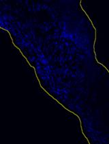
Our recent work has focused on investigating the feasibility of gGlu-HMRG for the on-site assessment of renal biopsy specimens (Iyama et al., 2020). Renal cortex, in which most of the glomeruli are contained, showed rapid induction of fluorescence upon the incubation of gGlu-HMRG and could be clearly distinguished from renal medulla. We herein present a protocol for the induction of fluorescence by gGlu-HMRG with some modifications for better clarity of the images.
Materials and Reagents
Pipettes (M&S Instruments, catalog numbers: F144059M, F144058, and F144055M)
Pipette tips (Violamo, catalog numbers: V-1000, V-200, and V-10)
Eppendorf centrifuge tubes, 1.5 mL
6 cm dishes (AS ONE, catalog number: 2-8590-02)
Phosphate buffer saline (PBS) (FUJIFILM, catalog number: 166-23555)
Dimethyl sulfoxide (FUJIFILM, catalog number: 041-29351)
Normal saline (Otsuka Pharmaceutical Factory, catalog number: 035081517)
ProteoGREENTM-gGlu (GORYO Chemical, catalog number: GC801). Dissolve in dimethyl sulfoxide at 1 mM and store at -20 °C before use (unnecessary to filter)
Equipment
Stereomicroscope (BioTools, catalog number: BS-3048BT)
Fluorescent unit (BioTools, catalog number: BT-ExSM)
Band pass filter (FUJIFILM, catalog number: BPB-45). This filter should be set at the light source of the fluorescent unit
Sharp cut filter (FUJIFILM, catalog number: SC-52). This filter should be set at the lens side of the fluorescent unit (Figure 1)

Figure 1. Setup of the fluorescent unit for the imaging. Band pass filter and sharp cut filter should be set as illustrated.
Procedure
文章信息
版权信息
© 2022 The Authors; exclusive licensee Bio-protocol LLC.
如何引用
Takata, T., Isomoto, H., Iyama, T. and Yamada, K. (2022). A Novel Imaging Technique for The On-site Assessment of Renal Biopsy Specimens. Bio-protocol 12(18): e4517. DOI: 10.21769/BioProtoc.4517.
分类
生物物理学 > 生物工程 > 医用生物材料
分子生物学 > 蛋白质 > 活性
生物化学 > 其它化合物 > 肽
您对这篇实验方法有问题吗?
在此处发布您的问题,我们将邀请本文作者来回答。同时,我们会将您的问题发布到Bio-protocol Exchange,以便寻求社区成员的帮助。