

Von Willebrand Factor Multimer Analysis by Low Resolution SDS-Agarose Gel Electrophoresis
通过低分辨率 SDS-琼脂糖凝胶电泳进行血友病因子多聚体分析
发布: 2022年08月20日第12卷第16期 DOI: 10.21769/BioProtoc.4495 浏览次数: 3380
评审: Anonymous reviewer(s)
参见作者原研究论文
The authors used this protocol in:

May 2021
Advertisement


Abstract
Von Willebrand factor (VWF) is a complex glycoprotein found in plasma, composed of disulfide-bond-linked multimers with apparent molecular weights between 500 kDa and 20,000 kDa. After release of VWF from storage granules, it is cleaved in flowing blood by the specific metalloproteinase ADAMTS13, resulting in a highly characteristic cleavage pattern and structure. As the structure of VWF multimers determines diagnosis of von Willebrand disease, which has different sub-types with different multimer- and cleavage patterns, VWF analysis is performed using low-resolution horizontal SDS-agarose gel electrophoresis. However, almost every laboratory uses a different protocol, and all experimental details are rarely, if at all, described. Therefore, the results from similar methods may be substantially different. Here, we present a detailed description of a validated VWF multimer method that we have developed. It has been successfully used for over more than 20 years in quality control of recombinant and plasma-derived VWF drug products, and in preclinical and clinical studies with VWF drug candidates. As most of the published methods, it enables visualization of VWF multimers separated by electrophoresis by immunostaining with a polyclonal anti-human VWF antibody followed by a secondary antibody coupled to alkaline phosphatase. VWF appears as a series of regularly spaced bands on the low and middle molecular weight range of the gel, with an unresolved zone in the high molecular weight (HMW) range, where ultra-large multimers are found. An example is shown below. This low-resolution agarose gel electrophoresis allows the determination of the number of VWF multimers with high reproducibility.
Graphical abstract:

Example of electrophoretic analysis of multimer structure of four batches of a recombinant VWF drug substance.
Background
Von Willebrand factor (VWF) is the largest soluble plasma protein, with essential functions in blood coagulation, control of bleeding, and angiogenesis. It is a complex glycoprotein with a mature subunit of 2,050 amino acids (Turecek et al., 2017). Approximately 19% of its molecular size are N- and O-linked carbohydrates, some of which resembling important blood group antigens. VWF is synthesized in endothelial cells and megakaryocytes. Intracellular polymerization starts with the formation of disulfide bridged dimers, which subsequently assemble into multimeric structures via additional disulfide bonds, creating multimers with apparent molecular masses between 500 kDa and 20,000 kDa. VWF is released into the circulation from its storage granules as ultra-large VWF with ultra-high molecular weight (UHMW). These UHMW multimers are potent mediators of platelet adhesion to other platelets and to subendothelial structures exposed upon vessel damage. However, under normal circumstances, UHMW VWF multimers are rapidly cleaved by the plasma protease ADAMTS13 (A disintegrin-like and metalloprotease with thrombospondin type 1 repeats) to smaller sized multimers with lower hemostatic and, thus, lower thrombogenic potential. ADAMTS13 specifically cleaves VWF at the bond between Tyr1605 and Met1606, generating 140-kDa N-terminal and 176-kDa C-terminal fragments, thus, regulating multimer size. The number of VWF multimers is generally seen as an indicator of functionality because more binding sites (e.g., for collagen and platelets) are exposed from larger multimers when the molecules become elongated under shear stress in the vasculature (Favaloro et al., 2021).
The main function of VWF is to mediate platelet binding and aggregation at sites of vascular injury. This process is known as primary hemostasis. VWF is critical for the formation of the primary hemostatic platelet plug by serving as glue between platelets through binding to platelet receptors glycoprotein (GP) Ib-IX-V and αIIbβ3 integrin, in promoting primary platelet adhesion and aggregation following vessel injury, and promoting binding of platelet aggregates to subendothelial matrix proteins such as collagens, which become exposed to flowing blood when lesions occur in vessels (Bryckaert et al., 2015). Shear forces in blood vessels promote folding and unfolding of VWF, essentially regulating its binding properties to receptors, which are further controlled by limited proteolytic cleavage by ADAMTS13. Furthermore, VWF serves as chaperone protein for coagulation factor VIII (FVIII) through tight binding, maintaining a highly controlled equilibrium between free and bound FVIII, which is also dependent on multimer size. Additionally, VWF determines the circulatory half-life of FVIII. All these functions of VWF depend on multimer size, and the degree of VWF multimerization correlates with the ability to promote platelet aggregation, as an essential step in blood coagulation.
Defects in VWF and ADAMTS13 are involved in hemostatic and thrombotic disorders. Deficiencies in VWF result in von Willebrand disease (VWD). VWD is the most common inherited bleeding disorder in the world, with an estimated prevalence of symptomatic disease of approximately 1 in 1000 individuals. VWD is a heterogeneous disease with different genetic subtypes, called type 1, type 2 (with subtypes 2A, 2B, 2M, and 2N), and type 3 (Peyvandi et al., 2019). In addition to genetic analysis, differential diagnosis of VWD and its subtypes requires determination of VWF activity by different functional assays and determination of the multimer pattern. Phenotypic von VWD classification largely relies on analysis of multimeric distributions of VWF and evaluation of its structure. Examples of typical multimer patterns, as seen in VWF subtype patients, can be found in textbooks and review articles (e.g., Schneppenheim and Budde, 2011). Although VWF multimer analysis is labor intensive, non-standardized, and limited to specialized laboratories, efforts have been made recently to establish ranges for VWF multimer distribution (Vangenechten and Gadisseur, 2020).
Inherited or immune mediated deficiencies of ADAMTS13 impair VWF cleavage, resulting in an increased portion of ultra-large multimers, which can cause thrombosis in the microvasculature. This disease is called thrombotic thrombocytopenic purpura (TTP), and its differential diagnosis requires VWF multimer analysis. Most recently, an impairment of the ratio of VWF and ADAMTS13 was identified as the main determinant of severity of COVID-19, with highly elevated levels of VWF and reduced ADAMTS13 resulting in thrombosis of the microvasculature, particularly in the lungs (Turecek et al., 2021; Seth et al., 2022).
Since the development of the first electrophoretic methods to separate and visualize multimers of VWF, the principle of these methods has not changed (Ruggeri and Zimmerman, 1981). Despite some modest improvements, the quality of test results largely depends on the quality and concentration of the agarose gel and the equipment used. Thus, the most demanding diagnostic test for VWD is still the VWF multimer analysis, an investigation that challenges even the most seasoned laboratory technologist (Lillicrap, 2013). Therefore, it is a limitation that methods are often not fully described with all the experimental details required to make the analytical system robust and reproducible. While multimer analysis of VWF is now performed for more than 40 years (Meyer et al., 1980), only a few publications describe the experimental methods, and even these do not give precise indications on how to perform the analysis. Here, we describe a validated method for electrophoretic separation of VWF multimers in low resolution SDS-agarose gels, with subsequent visualization by immunostaining (Aihara et al., 1986), allowing densitometric analysis. The method equally qualifies for analysis of VWF in blood from humans and other species, diagnosis of VWD subtypes, and characterization of VWF preparations derived from human or animal blood, produced by expression in cell culture systems, or by vectors used in gene therapy (Turecek et al., 2010). This method has been successfully used in quality control of recombinant and plasma-derived VWF drug products, and in preclinical and clinical studies with VWF drug candidates (Turecek et al., 1997).
Materials and Reagents
Material
Flatbed electrophoresis apparatus and Multiphor II electrophoresis unit (with ceramic cooling plate), GE Healthcare, IL, USA (or equivalent)
Power Supply
Cryo thermostat, Modell MultiTemp III, (Cytiva, Uppsala, Sweden)
Hairdryer
Horizontal shaker
pH-meter
Vortex mixer
Water bath (up to 90 °C)
Pipettes and tips
Plastic test tubes 12 × 55 mm
Glass and plastic laboratory trays, approximately 14 × 26 × 6 cm
Glass plates, float glass, 11.5 × 23.0 × 0.2 cm (or equivalent)
Clamps for electrophoresis plates
Rubber seals for gel plates, U-shaped cut, approximately 1.5 mm thick
Rubber seals rectangular cut, 10 mm long, 4 mm wide, 1.5 mm thick
Disposable syringes, cannulas, and locking cone
GelbondFilm for agarose gel 124 × 258 mm (product: 80112932; Cytiva, Uppsala, Sweden)
Chromatography paper 1CHR (Whatman WHA1001917; Sigma, Vienna, Austria) and 3CHR (Whatman WHA3003917; Sigma, Vienna, Austria)
Paper towels/kitchen roll
Laboratory balances
Reagents
Purified water
Agarose SEAKEM HGT (prod. 50040; Lonza, Rockland, USA)
Agarose Biozym Plaque Agarose (prod. 840101; Biozym Scientific GmbH, Oldendorf, Germany)
Tris(hydroxymethyl-aminomethane)
Sodium dodecyl sulfate pellets (SDS)
Ethylen-diamin-tetraacetic acid-di-sodium salt EDTA
NaCl
Glycine
Bromophenol-blue
Triton X100
Na2HPO4·2H2O
NaH2PO4·H2O
Antibody rabbit anti-human-VWF (product A0082 DAKO, Agilent Technologies, Glostrup, Denmark)
Secondary antibody goat-anti-rabbit-IgG-ALP-conjugated. (product 111-055-003; Jackson ImmunoResearch Laboratories, Inc., West Grove, PA, USA)
AP Conjugate Substrate Kit (product 1706432, BioRad Laboratories, Hercules, CA, USA)
Human normal plasma pool (e.g., Precision Biologics, Rockville, USA)
0.9% NaCl
Separation gel buffer (see Recipes)
Stacking gel buffer (see Recipes)
Sample buffer (see Recipes)
Running buffer for electrophoresis apparatus (see Recipes)
Bromophenol blue solution (see Recipes)
PBS-Buffer for immuno-staining (in gel staining) (see Recipes)
Washing buffer/antibody dilution buffer (see Recipes)
Equipment
Densitometer GS-900 (BioRad Laboratories, Hercules, CA, USA)
Software Image Lab (BioRad Laboratories, Hercules, CA, USA) and/or Image Quant TL (Cytiva, Uppsala, Sweden)
Multiphor II Electrophoresis-System with cooling unit (Figure 1).
Microwave oven
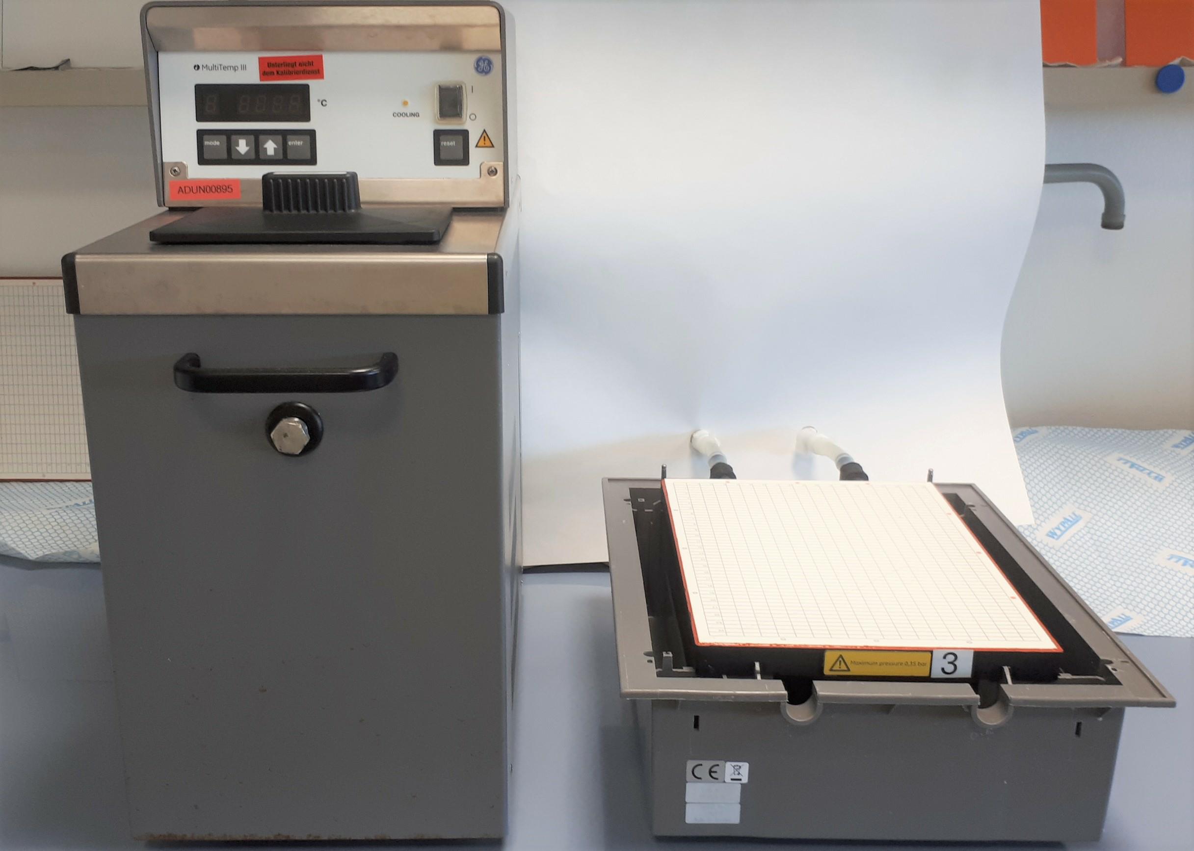
Figure 1. Electrophoresis system: Cooling unit (left) and flatbed apparatus (right)
Procedure
文章信息
版权信息
© 2022 The Authors; exclusive licensee Bio-protocol LLC.
如何引用
Gritsch, H., Stimpfl, M. and Turecek, P. L. (2022). Von Willebrand Factor Multimer Analysis by Low Resolution SDS-Agarose Gel Electrophoresis. Bio-protocol 12(16): e4495. DOI: 10.21769/BioProtoc.4495.
分类
医学 > 心血管疾病
生物化学 > 蛋白质 > 电泳
您对这篇实验方法有问题吗?
在此处发布您的问题,我们将邀请本文作者来回答。同时,我们会将您的问题发布到Bio-protocol Exchange,以便寻求社区成员的帮助。