

Evaluation of Urine Proteins by Capillary Electrophoresis
毛细管电泳法评价尿蛋白
(*contributed equally to this work) 发布: 2022年08月05日第12卷第15期 DOI: 10.21769/BioProtoc.4466 浏览次数: 2199
评审: Manjula MummadisettiSaptashati BiswasAnonymous reviewer(s)
参见作者原研究论文
The authors used this protocol in:

2021
Advertisement


Abstract
Capillary electrophoresis (CE) is a laboratory method usually used to separate proteins in body fluids such as serum, cerebrospinal fluid, or urine. Separation of proteins in urine can have clinical applications for evaluating samples from healthy dogs and dogs with proteinuria in a qualitative way, which would not be possible with gel electrophoresis. Other advantages of CE over gel electrophoresis in serum include the reduced separation time (2 min vs. 20 min in a gel), reduction of waste harmful to humans and the environment, and ability to obtain a curve without the need for additional staining. This protocol is divided into four steps. Firstly, urine needs to be prepared prior to dialysis. Secondly, urine needs to undergo dialysis to eliminate compounds that could interfere with separation, and to concentrate the urine. The third step is CE using specific equipment. The last step is to separate the fractions of the phoretograms obtained in the previous step. This method is mostly an automatized process, easily reproducible, and that can be performed in any laboratory, as a part of the diagnostic or follow-up of patients with renal disease.
Graphical abstract:
Background
Serum protein analysis by capillary electrophoresis (CE) is a well-established laboratory method used for the diagnosis and follow up of infectious, inflammatory, immune-mediated, and neoplastic conditions in human and veterinary medicine (Jenkins, 2009; Giordano and Paltrinieri, 2010). CE is one of the most frequent techniques used to separate serum proteins, as it is a simple, adaptable, and quick technique, and does not require a large sample amount. The CE laboratory technique is based on the separation of charged molecules by their electrophoretic mobility in an alkaline buffer at a specific pH. The separation occurs according to the electrolyte pH and electroosmotic flow, yielding different electrophoretic fractions (Osatinsky, 2007). In serum, the phoretogram is commonly divided into five different fractions, from low to high molecular weight and charge: albumin, alpha1 globulin, alpha2 globulin, beta globulin, and gamma globulin (Gay-Bellile et al., 2003; Tappin et al., 2011).
Recently, the analysis of proteins in urine by CE has proven to be a suitable method in human medicine to detect the presence of characteristic electrophoretic patterns in renal and extrarenal disorders, such as myelomas (Jenkins, 1997; Theodorescu et al., 2005; Mischak et al., 2010). Although quantitative proteinuria can only be assessed by calculation of the urine protein/creatinine ratio (UPC), electrophoretic techniques can be used as a qualitative method to assess the loss of proteins through the urine, as different patterns can be identified (Lees et al., 2005). CE associated with mass spectrophotometry techniques is also useful to identify peptide biomarkers associated with chronic kidney disease (Pelander et al., 2019). The use of urine to identify such abnormalities represents a great advantage over the use of blood, because urine can be collected in quantity, does not require trained staff for its collection, and urinary proteins remain stable for at least 3 days at 4ºC or 3 months at -20°C (Théron et al., 2017). They can provide information about kidney functionality earlier than blood biomarkers, such as SDMA or Cystatin C (Yalçin and Çetin, 2004; Pelander et al., 2019).
Comparison with other electrophoretic techniques that evaluate proteinuria could be challenging because the principle of migration is different. In sodium dodecyl sulfate agarose/polyacrylamide gel (SDS-AGE or SDS-PAGE) electrophoretic techniques, particle migration is only based on their molecular weight. Initially, it might be easier to identify proteins that migrate in the different phoretogram fractions (Yalçin and Çetin, 2004; Zini et al., 2004; Giori et al., 2011; Lavoue et al., 2015; Chacar et al., 2017; Hokamp et al., 2017). Nevertheless, the result of the technique presented in this manuscript is a profile, where abnormalities associated with protein excretions can be easily detected when compared against curves from healthy dogs and any other mammals.
The aim of this study is to establish a standardized protocol to prepare urine adequately, to evaluate the proteins in dog urine by CE on an instrument normally used for serum samples. It can be a useful tool to assess pathological proteinuria in dogs, alongside quantitative methods such as UPC.
Materials and Reagents
Ultrafiltration column 4 mL Vivaspin Turbo 4 10000 MWCO (Sartorius, Vivaspin Turbo 4, catalog number: VS04T02)
Eppendorfs 1.5 mL (Lambda, Eppendorf, catalog number: 1003/G)
Polystyrene tube for urine 12 mL (Lambda, catalog number: 301402)
10–200 μL tips (Lambda, catalog number: 18260)
Kit Minicap Protein (Sebia Hispania S. A., Sebia, catalog number: 2203). Storage temperature between 2 °C and 30 °C
Kit Urine Dialysis Capillarys (Sebia Hispania S.A., Sebia, catalog number: 2013). Storage temperature between 2 °C and 30 °C
Capiclean (Sebia Hispania S.A., Sebia, catalog number: 2058). Storage temperature between 2 °C and 30 °C
Reconstituted buffer for 4 samples (see Recipes)
Equipment
Minicap + Phoresis system (Sebia Hispania, S.A., catalog number Minicap: 1232; Phoresis. Software version 8.6.3)
Centrifuge Nahita 2650 (Nahita, Nahita 2650, catalog number: 200352650000)
Software
Minicap computer program (Sebia Hispania S.A. www.sebia.com)
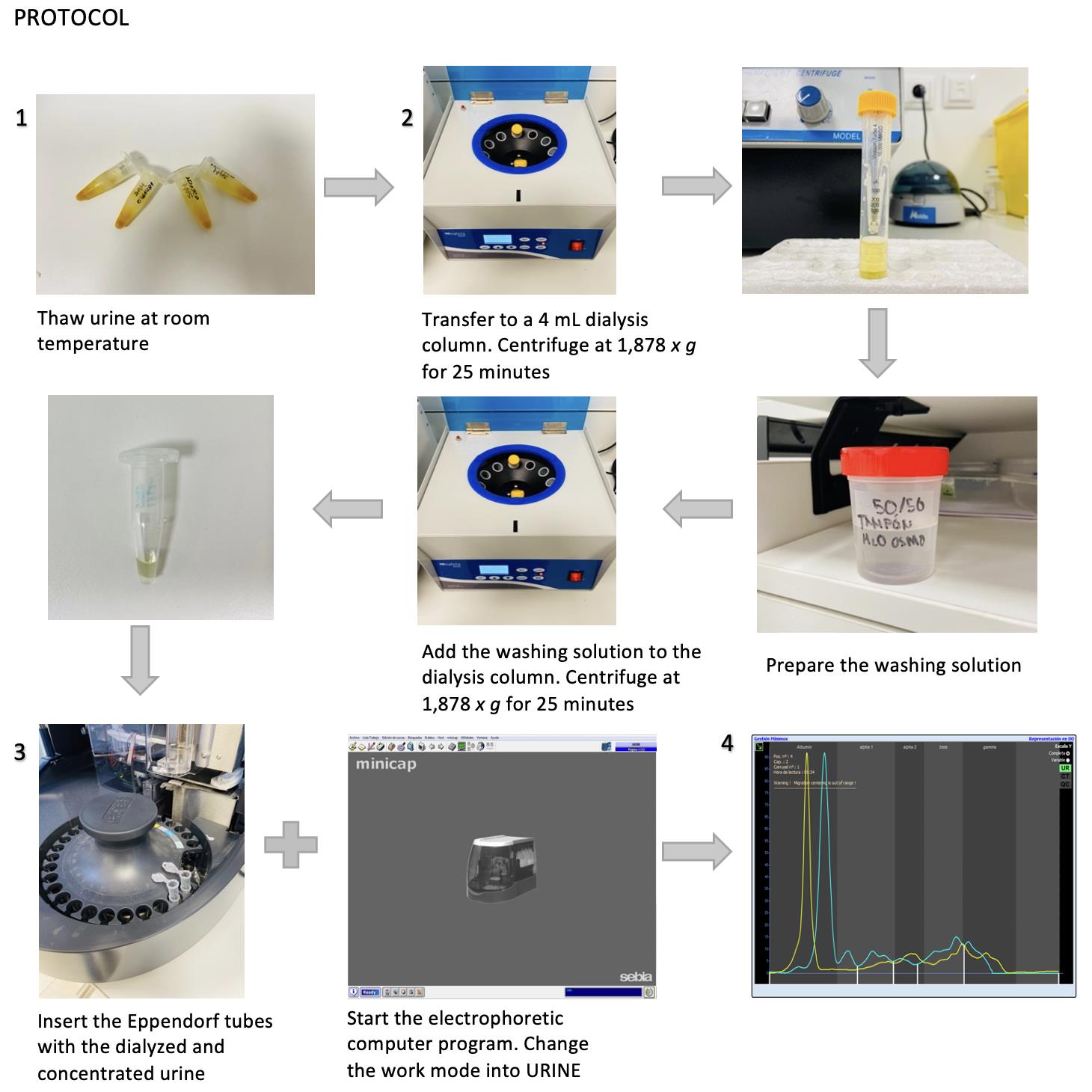
Procedure
文章信息
版权信息
© 2022 The Authors; exclusive licensee Bio-protocol LLC.
如何引用
Navarro, P. F., Gil, L. and Férnandez-Barredo, S. (2022). Evaluation of Urine Proteins by Capillary Electrophoresis. Bio-protocol 12(15): e4466. DOI: 10.21769/BioProtoc.4466.
分类
生物化学 > 蛋白质 > 电泳
您对这篇实验方法有问题吗?
在此处发布您的问题,我们将邀请本文作者来回答。同时,我们会将您的问题发布到Bio-protocol Exchange,以便寻求社区成员的帮助。