

Quantitative Live Confocal Imaging in Aquilegia Floral Meristems
白葵花分生组织的定量实时共聚焦成像
(*contributed equally to this work) 发布: 2022年06月20日第12卷第12期 DOI: 10.21769/BioProtoc.4449 浏览次数: 3634
评审: Vivien Jane Coulson-ThomasBeatriz GoncalvesAnonymous reviewer(s)
参见作者原研究论文
The authors used this protocol in:
Feb 2022
Advertisement


Abstract
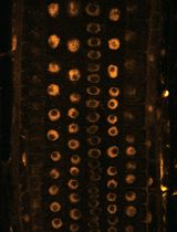
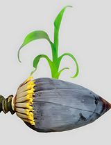
In this study, we present a detailed protocol for live imaging and quantitative analysis of floral meristem development in Aquilegia coerulea, a member of the buttercup family (Ranunculaceae). Using confocal microscopy and the image analysis software MorphoGraphX, we were able to examine the cellular growth dynamics during floral organ primordia initiation, and the transition from floral meristem proliferation to termination. This protocol provides a powerful tool to study the development of the meristem and floral organ primordia, and should be easily adaptable to many plant lineages, including other emerging model systems. It will allow researchers to explore questions outside the scope of common model systems.
Keywords: Live imaging (实时成像 )Background
Meristems are groups of pluripotent stem cells, typically located at the tips of shoots (Steeves and Sussex, 1989). Many fundamental features of meristems are shared across all vascular plants, e.g., the maintenance of a pool of undifferentiated cells, regulated cell proliferation and expansion, and control of post-embryonic organogenesis. However, there remains a great deal of unexplored variation in meristem structure and behavior across land plants. Exploring this diversity is hampered by the reliance on common developmental techniques, such as fixed tissue sectioning and imaging, that do not allow processes such as spatial and temporal patterns of cell division and expansion to be directly observed. In model systems such as Arabidopsis thaliana, genetic and molecular tools have been coupled with advancements in live imaging techniques to allow analyses of both cell behaviors and gene expression in real time. These tools have provided considerable progress in our understanding of meristem development (e.g., Prunet et al., 2016; Geng and Zhou, 2019; Shi et al., 2020; Caggiano et al., 2021; Silveira et al., 2022). However, these advancements are currently limited to a small number of model species, and there is a pressing need to develop quantitative live imaging techniques in non-model systems, and specifically, approaches that may be broadly practical across a range of plant taxa. Here, we present a detailed protocol for live imaging and analysis of floral meristems in Aquilegia coerulea, a member of the buttercup family (Ranunculaceae). This protocol provides a powerful tool to study the development of the meristem and initiation of floral organs, and should be easily adaptable to many plant lineages, including other emerging model systems. This protocol will allow researchers to explore questions outside the scope of common model systems.
Materials and Reagents
35 × 10 mm Petri dish (Corning, product number: 430588)
100 × 15 mm Petri dish (Fisher Scientific, catalog number: FB0875712)
1.5 mL microcentrifuge Eppendorf tubes
Parafilm
Kimwipe
Microscope slides (no special coating required)
Aluminum foil
128 cell plug trays and 4-inch plant plots
Seeds of Aquilegia × coerulea ‘Kiragami’ can be purchased from Swallowtail Garden Seeds (Santa Rosa, CA, USA)
Agar (Invitrogen, catalog number: 16500-100)
Linsmaier & Skoog medium (Caisson Labs, catalog number: L2P03)
Sucrose (Macron Fine Chemicals, catalog number: 57-50-1)
NaOH (Sigma-Aldrich, catalog number: 221465)
Kinetin (Sigma, catalog number: K0753-1G)
Gibberellic Acid (Sigma, catalog number: G7645-1G)
100% EtOH
Sterile ddH2O
Bleach
Propidium Iodide (Sigma-Aldrich, catalog number: P4864)
Note: Potential carcinogen; avoid contact with skin and eyes; wear suitable protective clothing, gloves, and eye/face protection.
Glue (any type of glue that can glue Petri dish to the glass slides)
Culture medium (see Recipes)
Equipment
Scalpel with No. 10 blade (BioQuip Products, catalog number: 2723A)
Straight dissecting needle (Carolina, catalog number: 627201)
Precision Watchmaker's Forceps, Extra-Fine Point (Carolina, catalog number: 624791)
0.5 mm Glass beads (Sigma-Aldrich, catalog number: 18406)
Razor blades
Microscope (Zeiss Stemi DV4 Stereo)
Microscope (LSM 980 NLO Multi-photon with a water immersion lens W Plan-Apochromat 20×/1.0 DIC UV-IR M27 75mm)
Software
FIJI (https://imagej.net/software/fiji/) or ImageJ (https://imagej.net/software/imagej/)
MorphographX (MGX) https://morphographx.org/software/.
Procedure
文章信息
版权信息
© 2022 The Authors; exclusive licensee Bio-protocol LLC.
如何引用
Min, Y., Conway, S. J. and Kramer, E. M. (2022). Quantitative Live Confocal Imaging in Aquilegia Floral Meristems. Bio-protocol 12(12): e4449. DOI: 10.21769/BioProtoc.4449.
分类
细胞生物学 > 细胞成像 > 共聚焦显微镜
发育生物学 > 形态建成 > 器官形成
植物科学 > 植物发育生物学 > 形态建成
您对这篇实验方法有问题吗?
在此处发布您的问题,我们将邀请本文作者来回答。同时,我们会将您的问题发布到Bio-protocol Exchange,以便寻求社区成员的帮助。