

CRISPR/Cas9 Gene Editing of HeLa Cells to Tag Proteins with mNeonGreen
mNeonGreen对 HeLa 细胞进行 CRISPR/Cas9 基因编辑以标记蛋白质
发布: 2022年05月20日第12卷第10期 DOI: 10.21769/BioProtoc.4415 浏览次数: 4302
评审: Antoine de MorreeOlli MatilainenAnonymous reviewer(s)
参见作者原研究论文
The authors used this protocol in:
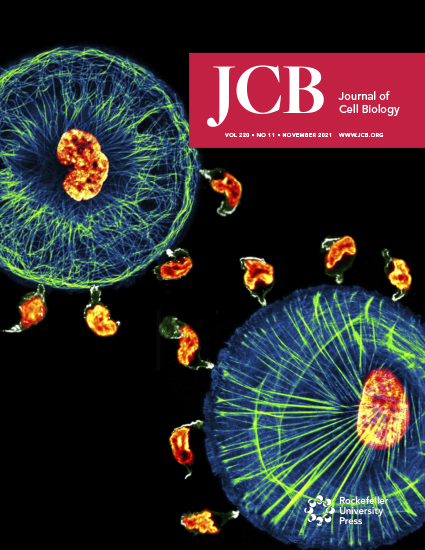
Nov 2021
Advertisement


Abstract
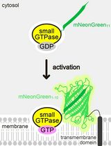
Subcellular localization dynamics of proteins involved in signal transduction processes is crucial in determining the signaling outcome. However, there is very limited information about the localization of endogenous signaling proteins in living cells. For example, biochemical mechanisms underlying the signaling pathway from epidermal growth factor (EGF) receptor (EGFR) to RAS-RAF and ERK1/2/MAPK are well understood, whereas the operational domains of this pathway in the cell remain poorly characterized. Tagging of endogenous components of signaling pathways with fluorescent proteins allows more accurate characterization of their intracellular dynamics at their native expression levels controlled by endogenous regulatory mechanisms, thus avoiding possible tainting effects of overexpression and mistargeting. In this study, we describe methodological approaches to label components of the EGFR-RAS-MAPK pathway, such as Grb2, KRAS, and NRAS, with the fluorescent protein mNeonGreen (mNG) using CRISPR/Cas9 gene-editing, as well as generation of homozygous single-cell clones of the edited target protein.
Keywords: mNeonGreen (mNeonGreen)Background
The epidermal growth factor (EGF) receptor (EGFR) is activated by EGF or other ligands at the cell surface, which triggers the RAS-RAF-MAPK/ERK1/2 signaling pathway involved in cell proliferation, differentiation, survival, and motility (Karnoub and Weinberg, 2008). Activated EGFR is endocytosed and accumulated in early endosomes, where it is either recycled back to the plasma membrane or targeted for lysosomal degradation. Whether active EGFR continues to signal through the RAS-ERK1/2 axis from endosomes to sustain ERK1/2 activation remains controversial and is under debate in the literature (reviewed in von Zastrow and Sorkin, 2021). Evidence for the endosomal signaling to ERK1/2 is based on the detection of components of the ERK1/2 pathway in endosomes in cells stimulated with EGF (Pol et al., 1998; Howe et al., 2001; Jiang and Sorkin, 2002; Lu et al., 2009; Schmick et al., 2014). However, these observations were made either with overexpressed recombinant proteins or with chemically fixed cells or by subcellular fractionation, approaches that may not correctly report subcellular localization of endogenous proteins. We initially studied the spatio-temporal dynamics of the endogenous tagged EGFR and components of the ERK1/2 pathway by fluorescently tagging these proteins using gene editing techniques utilizing zinc finger nucleases (ZFN) and transcription activator-like effector nucleases (TALEN) (Pinilla-Macua et al., 2017, 2016). Subsequently, we switched to using the CRISPR-Cas9 system (Surve et al., 2019; Surve et al., 2021) for gene-editing. We selected for these studies a subvariant of HeLa cells that we consistently used in the laboratory because they express EGFR at levels similar to those in many mammalian cells in vivo, and because we have rigorously characterized the EGFR endocytic trafficking system in these cells.
CRISPR-Cas9 has proven to be versatile and efficient method of gene editing compared to ZFNs and TALEN, and is widely used for gene knockouts, gene activation, gene inhibition, and knock-ins. The fascinating discovery and evolution of CRISPR technology is beyond scope of this article, and excellent reviews on this topic can be found elsewhere. We used this technology to tag KRAS, NRAS, and Grb2. Generally, there are two main components of CRISPR-Cas9 technology: i) Cas9—a RNA guided endonuclease, ii) a short non-coding guide RNA consisting of a target complementary CRISPR RNA (crRNA) and a trans-activating crRNA (tracrRNA) that shuttles Cas9 to a specific site. These components are delivered inside the cells via plasmids or via direct delivery of an in vitro generated complex of purified Cas9 and purified sgRNA (Ran et al., 2013). Following the delivery, gRNA through Watson-Crick base pairing binds to its target DNA sequence, and Cas9 makes a double stranded break (DSB) near the PAM site of gRNA. The DSB is either repaired by the error prone non-homologous end joining (NHEJ) mechanism or by precise homology dependent repair (HDR) mechanism. In case of gene tagging, an additional component is provided in the form of a repair template, i.e., a donor DNA containing sequences of a fluorescent protein or small tags such as His, HA, or MYC, which through HDR allows the insertion of the tag of interest. HDR mechanism of repair is inherently inefficient, resulting in lower yields of clones of cells with the in-frame incorporated tag compared to generation of knockout clones, which generally involves NHEJ. The protocol here describes the variations (Table 1) in the approach by which gene tagging (knock-in) can be achieved in transformed cell lines like HeLa.
Materials and Reagents
CellTrics 50 μm sterile Disposable Filters (Sysmex, catalog number: 04-004-2327)
96-well plate (Ibidi, 15 μ-Plate 96 Well Black)
12-well cell culture plate (Corning, catalog number: 3513)
25 or 75 cm2 cell culture flasks (Corning, catalog number: 430639 or 430641U)
15 mL tube conical base (Sarstedt, catalog number: 62.554.205)
DNA oligonucleotide primers from IDT
GeneArt Precision gRNA Synthesis kit (Invitrogen, catalog number: A29377)
Tracr Fragment + T7 Primer Mix (contains the universal PCR amplification primers and the 80-nt constant region of the crRNA/tracrRNA) (Invitrogen, catalog number: A29377)
PhusionTM High-Fidelity PCR Master Mix (2×) (Invitrogen, catalog number: A29377)
Nuclease free water
dNTP mix (10 mM each of dATP, dGTP, dCTP, dTTP) (NEB, catalog number: N0447)
5× TranscriptAidTM Reaction Buffer (Invitrogen, catalog number: A29377)
TranscriptAidTM Enzyme Mix (Invitrogen, catalog number: A29377)
DNase I (Invitrogen, catalog number: A29377)
Single stranded DNA (ssDNA) oligos from IDT
NEBuilder® HiFi DNA Assembly Master Mix (NEB, catalog number: E2621)
pSpCas9(BB)-2A-Puro (PX459) plasmid (Addgene plasmid # 62988)
BbsI (NEB, catalog number: R0539)
NEB Buffer 2 (NEB, catalog number: B7002S)
NEB® 5-α Competent E. coli (High Efficiency) (NEB, catalog number: C2987)
Ampicillin Agar plates and Kanamycin Agar plates
NEB buffer 2.1 (NEB, catalog number: B7202)
QIAquick PCR Purification Kit (Qiagen, catalog number: 28104)
Synthetic double stranded donor DNAs from IDT
Phusion High-Fidelity DNA Polymerase (NEB, catalog number: M0530)
Phusion GC buffer 5× (NEB, catalog number: B0519)
Zero BluntTM TOPOTM PCR Cloning Kit (Invitrogen, catalog number: 45-1245)
EcoRI-HF (NEB, catalog number: R3101)
CutSmart Buffer 10× (NEB, catalog number: B6004)
QIAquick Gel Extraction Kit (Qiagen, catalog number: 28704)
XbaI (NEB, catalog number: R0145)
Easy-Fusion Halo plasmid (Addgene plasmid # 112850)
EcoRI-HF (NEB, catalog number: R3101)
AvaI (NEB, catalog number: R0152)
HincII (NEB, catalog number: R0103)
DMEM (Gibco, catalog number: 11965-092) with 10% FBS (Invitrogen, catalog number: 16140071)
DPBS (without Ca2+ or Mg2+) (Gibco, catalog number: 14190-144)
Trypsin (Gibco, catalog number: 25200-056)
Neon Transfection System (Invitrogen, catalog number: MPK5000)
Neon Transfection System 10 μL Kit (Invitrogen, catalog number: MPK1025)
Buffer R
Puromycin (Sigma-Aldrich, catalog number: P8833)
Platinum Cas 9 (Invitrogen, catalog number: A36498)
BD FACSAria III sorter fitted with 488 100 mW Trigon laser and 100 μm nozzle (BD Biosciences)
Nocodazole (Sigma-Aldrich, catalog number: M1404)
Lipofectamine 3000 (Invitrogen, catalog number: L3000001)
OptiMEM (Invitrogen, catalog number: 31985062)
Recombinant KRAS rabbit monoclonal antibody (Thermo Fisher Scientific, catalog number: 703345)
Cell Sorting buffer (see Recipes)
TGH buffer (see Recipes)
Software
https://www.benchling.com/. Benchling is a cloud-based informatics platform that is free to academic researchers. It provides design tools for DNA, oligonucleotides, and amino acids.
https://portals.broadinstitute.org/gppx/crispick/. The website is hosted by the Genetic Perturbation Platform of the Broad institute, MA, USA.
http://chopchop.cbu.uib.no/. CHOPCHOP is a web-based tool, designed by the Valen group at the University of Bergen, Norway. The website is for non-profit and academic use only.
https://nebuilder.neb.com. The web tool can be used to design primers for HiFi DNA assembly or Gibson assembly.
Procedure
文章信息
版权信息
© 2022 The Authors; exclusive licensee Bio-protocol LLC.
如何引用
Readers should cite both the Bio-protocol article and the original research article where this protocol was used:
- Surve, S. and Sorkin, A. (2022). CRISPR/Cas9 Gene Editing of HeLa Cells to Tag Proteins with mNeonGreen. Bio-protocol 12(10): e4415. DOI: 10.21769/BioProtoc.4415.
- Surve, S., Watkins, S. C. and Sorkin, A. (2021). EGFR-RAS-MAPK signaling is confined to the plasma membrane and associated endorecycling protrusions. J Cell Biol 220(11).
分类
细胞生物学 > 细胞工程 > CRISPR-cas9
细胞生物学 > 细胞信号传导 > 胞内信号传导
您对这篇实验方法有问题吗?
在此处发布您的问题,我们将邀请本文作者来回答。同时,我们会将您的问题发布到Bio-protocol Exchange,以便寻求社区成员的帮助。