

Analysis of Heterocyst and Akinete Specific Glycolipids in Cyanobacteria Using Thin-layer Chromatography
使用薄层色谱法分析蓝藻中的异囊体和动体特异性糖脂
发布: 2022年03月20日第12卷第6期 DOI: 10.21769/BioProtoc.4355 浏览次数: 2493
评审: Dennis J NürnbergAmberley D. StephensKumiko Okazaki
参见作者原研究论文
The authors used this protocol in:

Apr 2021
Advertisement


Abstract
Several filamentous cyanobacteria like Nostoc differentiate specialized cells in response to changes in environmental factors, such as low light or nutrient starvation. These specialized cells are termed heterocysts and akinetes. Under conditions of nitrogen limitation, nitrogen-fixing heterocysts form in a semi-regular pattern and provide the filament with organic nitrogen compounds. Akinetes are spore-like dormant cells, which allow survival during adverse unfavorable conditions. Both cell types possess multilayered thick envelopes mainly composed of an outermost polysaccharide layer and inner layers of glycolipids, that are important for stress adaptation. To study these envelope glycolipids, a method for the isolation, separation and analysis of lipids from heterocysts and akinetes is essential. The present protocol describes a method involving the extraction of lipids from cyanobacteria using solvents and their separation and visualization on silica plates, to render analysis simple and easy. This protocol is relevant for studying mutants that are defective in glycolipid layer formation and for the comparison of glycolipid composition of heterocysts and akinetes under different environmental stresses.
Keywords: Cyanobacteria (蓝藻)Background
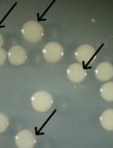
Filamentous cyanobacteria, such as Anabaena variabilis ATCC 29413 and Nostoc punctiforme ATCC 29133, are widely used as model organisms for the study of multicellularity and their capability to differentiate heterocysts and akinetes. Under favorable conditions, these photosynthetic Gram-negative bacteria grow in the form of filaments, which are composed of hundreds of vegetative cells. In response to insufficient supply of organically combined nitrogen, about 10% of semi-randomly spaced cells can differentiate into nitrogen-fixing heterocysts, which provide the filaments with nitrogen (Figure 1A) (Fay, 1992; Muro-Pastor and Maldener, 2019). Akinetes are spore-like resting cells that differentiate from the vegetative cells in response to diverse environmental conditions, including changes in light intensity and quality, temperature, and nutrient deficiency (Figure 1B).

Figure 1. Light micrographs showing cell differentiation in A. variabilis. (A) Vegetative filament containing heterocyst, indicated by black arrowhead. (B) Akinetes induced under low light condition. Scale bars, 10 µm.
Both cell types—heterocysts and akinetes—are characterized by the presence of a thick multilayered envelope, mainly composed of exopolysaccharides and glycolipids (HGLs) (Maldener et al., 2014; Perez et al., 2016; Garg and Maldener, 2021b). The glycolipids, which form laminated layers in the heterocyst and akinete envelopes, are of great interest due to their different functions in these two cell types. In heterocysts, they act as an oxygen diffusion barrier to protect the oxygen labile enzyme nitrogenase from oxygen (Fay, 1992; Wolk et al., 1994), while in akinetes, glycolipids protect the cells from various stress factors, such as freezing, desiccation, and lysozyme attack (Garg and Maldener, 2021a).
Mutants with aberrantly formed heterocyst envelopes are unable to grow without alternative sources of combined nitrogen. Many of those lose the ability to synthesize HGLs or to transport them beyond the cell wall to the heterocyst envelope (Ernst et al., 1992; Fiedler et al., 1998; Fan et al., 2005). The akinete envelope shares some structural similarities with the heterocyst envelope, suggesting that akinetes are the evolutionary ancestors of heterocysts (Wolk et al., 1994; Perez et al., 2018). To better understand this evolutionary relationship, a prerequisite is the elucidation of the composition of these special cells’ envelopes. Additionally, mutants showing phenotypes related to survival under environmental stress or starvation could be checked for the correct laminated layer composition using the protocol presented here.
Here, we use the thin-layer chromatography (TLC) protocol that was previously established by Nichols and Wood in 1968; with this method, they identified glycolipids, which are specific for the heterocyst forming cyanobacteria (Nichols and Wood, 1968). Later, the Wolk group showed that these glycolipids constitute the laminated layer in the envelope of heterocysts (Winkenbach et al., 1972). The two major heterocyst specific glycolipids in Anabaena species are characterized as 1-α-glucosyl-3,25-hexacosanediol (HG26-diol) and its 3-ketotautomer (HG26-keto-ol). Their separation via TLC is dependent on their polarity. We utilized this method with a few modifications for the analysis of the akinete specific glycolipids (Perez et al., 2018; Garg and Maldener, 2021a). We present a reproducible and reliable TLC protocol to analyze the glycolipids present in the heterocyst and akinete envelopes. This protocol includes the easy extraction of lipids using regular solvents, such as methanol and chloroform, and their clear separation and visualization on the TLC plate, which can be scanned directly. Furthermore, this method allows the extraction of specific glycolipids from the TLC plate in a manner that subsequent analysis of their chemical composition by HPLC-MS analysis is possible, as previously described (Perez et al., 2018; Shvarev et al., 2018).
Materials and Reagents
50 mL of cyanobacterial liquid cultures containing heterocysts or akinetes
Pipette tips (10 µL, 200 µL, 1,000 µL) (LTS, RAININ, Mettler Toledo. Gießen)
1.5 mL microcentrifuge tubes
50 mL conical tubes
Soft graphite pencil
30 cm ruler
Paper tape (masking tape)
Methanol ≥99.9% (HPLC grade) (Sigma, Merck, Darmstadt)
Chloroform (HPLC grade) (Roth, Karlsruhe)
Aluminium plate coated with silica gel (Macherey-Nagel, catalog number: 818033)
Acetic acid (Sigma, Merck, Darmstadt)
MilliQ water
25% Sulfuric acid (H2SO4) (Carl Roth, catalog number: 0967.1)
Equipment
Pipettes (P1000, P200, P20 and P10) (RAININ, Mettler Toledo)
100 mL Erlenmeyer flasks
50 mL glass beaker
Centrifuges:
Centrifuge (Eppendorf, 5417C) for microcentrifuge tubes
Centrifuge (Eppendorf, Centrifuge 5804 R) for conical tubes
Vortex Genie® 2 (Scientific Industries, Inc., catalog number: 6235684)
Fume hood
Hair dryer
Incubator or oven for TLC plate development (This can be any incubator that reaches the desired temperature of 180°C)
Flat-bed Scanner (e.g., EPSON Perfection V550 Photo)
Developing Chamber for TLC (Macherey-Nagel GmbH & Co. KG, catalog number: 9003500)
TLC sprayer with Erlenmeyer flask (e.g., DWK Life Sciences KimbleTM KontesTM Reagent Sprayers Head Only)
Software
Scanner software (e.g., EPSON Scan)
Procedure
文章信息
版权信息
© 2022 The Authors; exclusive licensee Bio-protocol LLC.
如何引用
Garg, R., Perez, R. and Maldener, I. (2022). Analysis of Heterocyst and Akinete Specific Glycolipids in Cyanobacteria Using Thin-layer Chromatography. Bio-protocol 12(6): e4355. DOI: 10.21769/BioProtoc.4355.
分类
生物化学 > 脂质 > 脂质分离
微生物学 > 微生物生理学 > 适应
细胞生物学 > 细胞结构 > 细胞表面
您对这篇实验方法有问题吗?
在此处发布您的问题,我们将邀请本文作者来回答。同时,我们会将您的问题发布到Bio-protocol Exchange,以便寻求社区成员的帮助。