

A Rapid Protocol for Direct Isolation of Osteoclast Lineage Cells from Mouse Bone Marrow
一种直接从小鼠骨髓中快速分离破骨细胞的方法
(*contributed equally to this work) 发布: 2022年03月05日第12卷第5期 DOI: 10.21769/BioProtoc.4338 浏览次数: 4406
评审: Salma MerchantKuo-Ching "KC" MeiYogita Jethmalani
参见作者原研究论文
The authors used this protocol in:

Apr 2021


Abstract
Osteoclast lineage cells (OLCs), including osteoclast precursors (OCPs) and mature osteoclasts (MOCs), participate in bone remodeling and mediate pathologic bone loss. Thus, it is essential to obtain OLCs for exploring their molecular features in both physiological and pathological conditions in vivo. However, the conventional protocols for obtaining OLCs ex vivo are not only time-consuming, but also unable to capture the cellular status of OLCs in vivo. In addition, the current antibody-based isolation approaches, such as fluorescence-/ magnetic-activated cell sorting, are not able to obtain pure osteoclasts because no unique surface antigen for osteoclasts has been identified. Here, we develop a rapid protocol for directly isolating OLCs from mouse bone marrow through magnetic-activated cell sorting (MACS). This protocol can rapidly enrich OCPs and MOCs, respectively, depending on the expression of the distinctive surface markers at their differentiation stages. It is optimized to isolate OLCs from four mice concurrently, of which sorting procedure could be completed within ~5 h.
Keywords: Magnetic-activated cell sorting (磁激活细胞分选)Background
Osteoclast lineage cells (OLCs) derived from bone marrow monocytes/macrophages (BMMs) govern bone resorption to regulate bone remodeling (Quinn et al., 1998). BMMs first differentiate into the committed osteoclast precursors (OCPs), which further fuse together to form mature osteoclasts (MOCs) (Asagiri and Takayanagi, 2007). Mounting evidence demonstrates that the dysregulation of osteoclasts could cause skeletal disorders, such as osteoporosis and Paget’s disease (Feng and McDonald, 2011; Novack and Mbalaviele, 2016). The excessive osteoclast activity and aberrant osteoclastic resorption in subchondral bone could also contribute to the progression of osteoarthritis (Zhen et al., 2013; Zhu et al., 2019; Liu et al., 2021). Thus, it is essential to directly isolate live OLCs in vivo, for studying their biological features and pathologic changes.
The laboratory mouse is a commonly used animal model for investigating osteoclast function (Elefteriou and Yang, 2011). The conventional protocol for obtaining osteoclasts relies on stimulating the primary mouse BMMs from bone marrow cells (BMCs) with macrophage colony stimulating factor (M-CSF) and receptor activator of NFκB-ligand (RANKL) in vitro (Glantschnig et al., 2003; Marino et al., 2014). This protocol requires more than one week to form the MOCs in vitro, which could not capture their real-time status in vivo. Alternatively, fluorescence-activated cell sorting (FACS) has been previously employed to directly isolate the live OLCs from BMCs in vivo (Atkins et al., 2006; Zhang et al., 2012; Madel et al., 2018). Nevertheless, the disadvantages of FACS, including time-consuming procedures, low yield, and high cost, significantly limit its widespread use (Lee and Lufkin, 2012; Almeida et al., 2014). Therefore, it is necessary to introduce more efficient isolation techniques for directly obtaining live OLCs in vivo.
Magnetic-activated cell sorting (MACS) is another frequently used technique for sorting live cells in vivo, which enables rapid cell isolation with high yield and low cost (Lee and Lufkin, 2012; Almeida et al., 2014). This technique has been previously applied to recognize single surface markers of osteoclasts, for isolating OLCs from chicken or mouse BMCs in vivo (Collin-Osdoby and Osdoby, 2012; Liu et al., 2015). Since no specific surface marker for defining OCPs or MOCs has been found until now, it is not feasible to directly distinguish OCPs and MOCs from OLCs through one single surface marker. Calcitonin receptor (CTR), which is highly expressed on osteoclasts, is commonly used for characterizing MOCs, while osteoclast-associated receptor (OSCAR) was considered a typical cell surface marker for OLCs. We have previously employed MACS to harvest the OSCAR+ OLCs from mouse bone marrow in vivo for downstream gene and miRNA expression assay (Liu et al., 2015; Li et al., 2016). Considering the distinctive expression of OSCAR and CTR throughout the differentiation stages of OLCs, OSCAR may serve as an ideal surface marker for identifying OLCs among BMCs, while CTR could be used to distinguish the MOCs (OSCAR+CTR+) from the OCPs (OSCAR+CTR–) within the OSCAR+ OLCs. Therefore, we further developed a two-step MACS method for distinguishing OCPs and MOCs from OLCs in BMCs, which demonstrated high efficiency in the direct capture of OCPs and MOCs from mice that had undergone surgery-induced osteoarthritis in our most recent study (Liu et al., 2021).
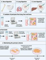
Here, we described this novel two-step MACS protocol for rapidly isolating OCPs and MOCs directly from mouse bone marrow with high yield and purity. This protocol consists of three main steps: BMC preparation, antibody-microbead labelling and magnetic sorting, and osteoclast characterization (Figure 1). In brief, the CTR+ and CTR– cell fractions were first separated from mouse BMCs by magnetic positive and negative selection. Thereafter, the OSCAR+CTR+ MOCs, among the CTR+ cell fractions, and the OSCAR+CTR– OCPs, among the CTR– cell fractions, were enriched by another magnetic positive selection. We employed this protocol to rapidly isolate the MOCs and OCPs from both ovariectomized (OVX) and sham-operated (Sham) mice. The sorting procedures can be carried out in four mice concurrently within ~5 h.

Figure 1. Schematic diagram of isolation for osteoclast lineage cells (OLCs) from mouse bone marrow
Materials and Reagents
1.5 mL microtubes (Biopioneer, catalog number: CNT-1.5F)
15 mL Falcon tubes (Corning, catalog number: 430791)
50 mL Falcon tubes (Corning, catalog number: 352070)
MS columns (Miltenyi Biotech, catalog number: 130-042-201, column capacity: up to 107 magnetically labeled cells from up to 2 × 10⁸ total cells)
LS columns (Miltenyi Biotech, catalog number: 130-042-041, column capacity: up to 108 magnetically labeled cells from up to 2 × 10⁹ total cells)
0.2-μm pore sized filter, bottle top (Fisher Scientific, catalog number: 595-4520)
1 mL syringe with 25G × 25 mm needle (Terumo)
100 mm Culture dish (Corning, catalog number: 354600)
30 μm pre-separation filters (Miltenyi Biotech, catalog number: 130-041-407,)
Cell strainer (BD Falcon, 40 μm, catalog number: 352340)
75% (vol/vol) Ethanol
CAUTION! Flammable. Keep away from flame and sources of ignition. Store in dry and well-ventilated areas.
QuadroMACS Starting Kit (Miltenyi Biotech, catalog number: 130-091-051)
Dulbecco's modified Eagle's medium (DMEM; Thermo Fisher, catalog number: 11965)
Penicillin/streptomycin (Thermo Fisher, catalog number: 15140122)
Fetal bovine serum (FBS) (Thermo Fisher, catalog number: 16170-078)
Bovine serum albumin solution (BSA) (MP Biomedicals, catalog number: 810531)
Sodium chloride (NaCl) (Fisher Scientific, catalog number: 7647-14-5)
Potassium chloride (KCl) (Fisher Scientific, catalog number: 7447-40-7)
Sodium phosphate (Na2HPO4) (Fisher Scientific, catalog number: 7778-77-0)
Potassium bicarbonate (KHCO3) (Fisher Scientific, catalog number: 298-14-6)
10× red blood cell (RBC) lysis solution (Miltenyi Biotech, catalog number: 130-094-183)
Dead Cell Removal Kit (Miltenyi Biotech, catalog number: 130-090-101), including the dead cell removal MicroBeads and 20× dead cell binding (DCB) buffer stock solution
Ethylenediaminetetraacetic acid (EDTA) (Sigma, catalog number: E6758-100G)
Paraformaldehyde (Sigma-Aldrich, catalog number: 158127)
Methanol (Sigma-Aldrich, catalog number: 34860)
Triton-X100 (Sigma-Aldrich, catalog number: X100-500ML)
Toluidine Blue (Sigma-Aldrich, catalog number: 198161)
OSCAR antibody (R&D Systems, catalog number: MAB1633, Isotype: monoclonal rat IgG2A)
Biotin-conjugated CTR antibody (LifeSpan BioSciences, catalog number: LS-C523811, Isotype: Biotin-conjugated polyclonal rabbit IgG)
Anti-Rat IgG MicroBeads (Miltenyi Biotech, catalog number: 130-048-502)
Anti-Biotin Multisort Kit (Miltenyi Biotech, catalog number: 130-091-256), including the Anti-Biotin Multisort MicroBeads, MultiSort Release Reagent, and MultiSort Stop Reagent
Monoclonal Rat IgG2A Isotype Control (R&D Systems, catalog number: MAB006)
Biotin-conjugated polyclonal rabbit IgG Isotype Control (LifeSpan BioSciences, catalog number: LSC351735)
Alexa Fluor 488 goat anti-rat IgG (H+L) Cross-Adsorbed secondary antibody (Thermo Scientific, catalog number: A11006)
PE goat anti-rabbit IgG (H+L) Cross-Adsorbed secondary antibody (Thermo Scientific, catalog number: P-2771MP)
DPBS (see Recipes)
MACS buffer (see Recipes)
Complete DMEM Medium (see Recipes)
1× RBC Lysis Solution (see Recipes)
1× DCB buffer (see Recipes)
1% Toluidine Blue solution (see Recipes)
Equipment
Dissection tools (surgical scissors, microforceps)
4°C Swinging-bucket centrifuge (e.g., Allegra X-15 R, Beckman Coulter)
Hemocytometer/counting chamber (e.g., Hausser Scientific Partnership, catalog number: 3200)
FACS (e.g., BD FACS, Aria III Cell-Sorting System, BD Biosciences)
Procedure
文章信息
版权信息
© 2022 The Authors; exclusive licensee Bio-protocol LLC.
如何引用
Dang, L., Li, N., Wu, X., Li, D., Zhang, Z., Zhang, B. T., Lyu, A., Chen, L., Zhang, G. and Liu, J. (2022). A Rapid Protocol for Direct Isolation of Osteoclast Lineage Cells from Mouse Bone Marrow. Bio-protocol 12(5): e4338. DOI: 10.21769/BioProtoc.4338.
分类
细胞生物学 > 细胞分离和培养 > 细胞分离 > 免疫磁珠
生物工程 > 生物医学工程
您对这篇实验方法有问题吗?
在此处发布您的问题,我们将邀请本文作者来回答。同时,我们会将您的问题发布到Bio-protocol Exchange,以便寻求社区成员的帮助。