

An Alternative Technique for Monitoring the Live Interaction of Monocytes and Tumor Cells with Nanoparticles in the Mouse Lung
一种监测小鼠肺中单核细胞和肿瘤细胞与纳米颗粒实时相互作用的替代技术
发布: 2022年01月20日第12卷第2期 DOI: 10.21769/BioProtoc.4293 浏览次数: 3379
评审: Alessandro DidonnaEVANGELOS THEODOROUAnonymous reviewer(s)
参见作者原研究论文
The authors used this protocol in:
Oct 2020
Advertisement


Abstract
Nanomaterials are increasingly used for the diagnosis and treatment of cancer, including lung cancer. For the clinical translation of nano-based theranostics, it is vital to detect and monitor their accumulation in the tumor, as well as their interaction with tumor, immune cells, and the tumor microenvironment (TME). While high resolution microscopy of fixed tumor specimens can provide some of this information from individual thin slices, it cannot capture cellular events over time and lacks 3D information of the tumor tissue. On the other hand, in vivo optical procedures either fall short of providing the necessary cellular resolution, as in the case of epifluorescence optical imaging, or are very demanding, as for instance intravital lung microscopy. We describe an alternative approach to investigate nanoparticle-cell interactions in entire mouse lung lobes, by longitudinal live cell confocal microscopy at nanometer resolution. By filling the lung ex vivo with 1% agarose, we were able to stabilize the lung lobes and visualize the interaction of fluorescent cells and nanoparticles for at least 4 hours post mortem. This high resolution ex vivo live cell imaging approach is an easy 4D tool for assessing several dynamic processes in tumor tissue, such as the traffic of cells, shedding of extracellular vesicles (EVs), and the accumulation of nanoparticles in tumor tissue.
Graphic abstract:
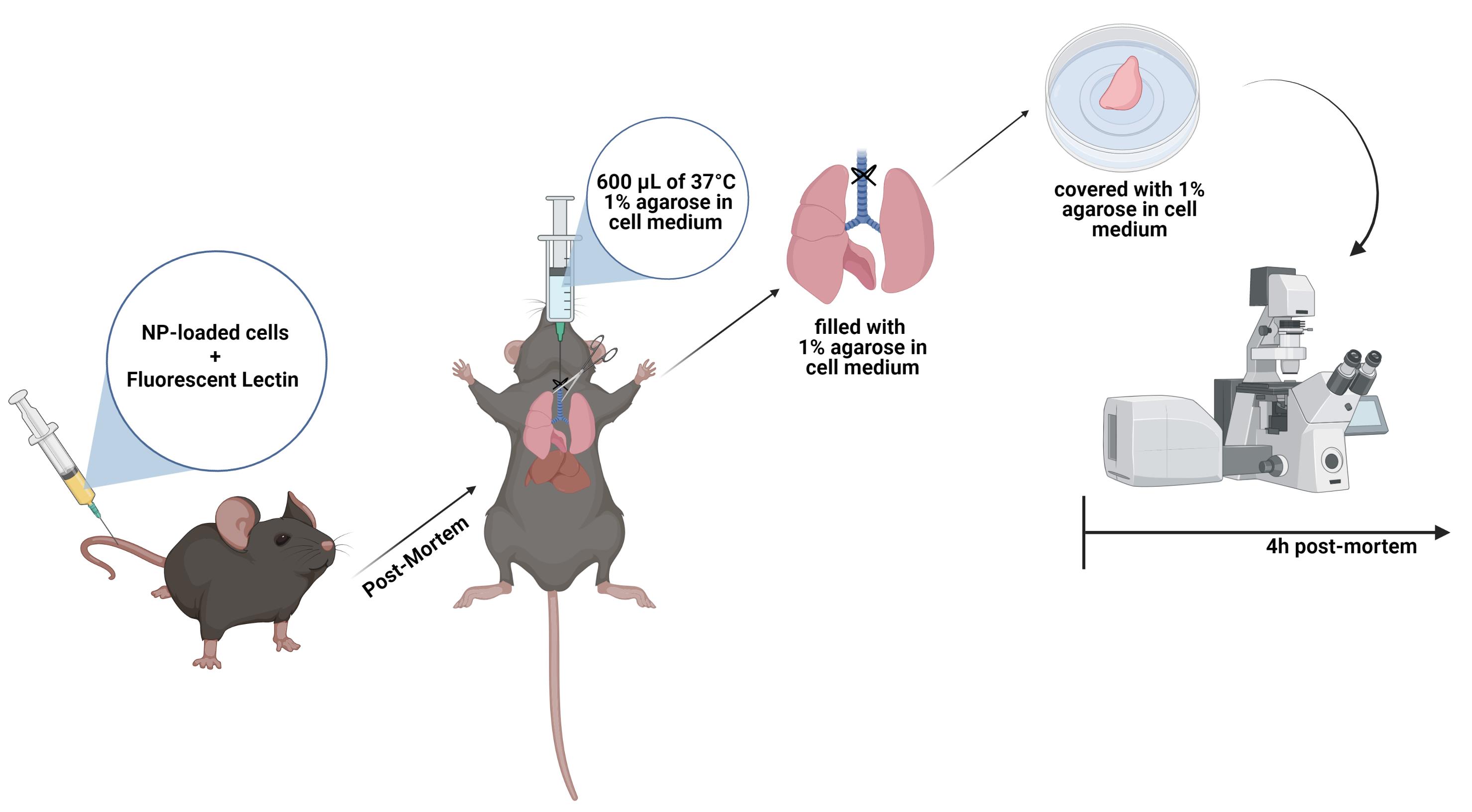
Schematic of the workflow for live cell imaging in the mouse lung.
Background
The advancement of the nanotechnology field has enabled the exploitation of nanoparticle (NP)-based cancer therapies (Jain and Stylianopoulos, 2010; Shi et al., 2017). Compared to the standard chemotherapeutic approach, NPs confer several advantages as they improve drug solubility and stability, prolong drug half-lives in plasma, minimize off-target effects, and concentrate drugs at the target site (Mudshinge et al., 2011). Combining all these characteristics, some NPs have been successfully approved by the FDA or are currently in clinical trials (Dinndorf et al., 2007; Ediriwickrema and Saltzman, 2015). Despite these achievements, several details have to be considered in novel nanoparticle formulations, as most NP-based drugs have failed to improve patient outcomes. One of the most important is the relation of NPs with the tumor microenvironment (TME), which includes tumor cells, immune cells, and blood vessels (Blank et al., 2017).
Traditionally, the characterization of the cellular uptake and interaction of NPs in the TME has been evaluated by 2D tissue sections. Despite the nanoscale resolution, immunohistochemistry and histology fail to demonstrate the complex organization of biological specimens and only provide static snapshots of events. With the development of new volumetric microscopy techniques, the understanding of cell-cell interactions in intact samples has become clearer. Furthermore, in comparison with other live-cell or in vivo technologies, confocal microscopy enables real-time imaging from general tissue architecture at nanoscale resolution (Ramos-Gomes et al., 2020).
Recently, we published a protocol for live high-resolution imaging of the interaction between monocytes and tumor cells with nanoparticles in the mouse lung (Ramos-Gomes et al., 2020). Here, we were able to demonstrate the dynamics between tumor and immune cells, the reaction of the macrophages/monocytes towards NPs, and characterize the NP deposition in the tumor. This approach opens new avenues to dynamically understand how nanotherapies are going to affect the TME and reach target sites.
Materials and Reagents
6-well plates
20 G blunt cannula with the tips cut-off (BD Microlance 3, catalog number: 301300)
1 mL syringe ( BD Plastipak, catalog number: 303172)
U100 Insulin syringe (Braun, catalog number: 9151125)
Uncoated Ibidi 35 mm cell culture dishes (ibidi GmbH, catalog number: 80131)
Cotton sowing thread (not too thin; Wenco, catalog number: 140720)
Surgical disposable scalpel (Braun, catalog number: 5518075)
Kimtech paper (Kimtech Science, catalog number: 05511)
25 mm round cover slips, 170 µm thick (Menzel-Gläser; VWR, catalog number: 631-1346)
Fluorescent-Labeled Lectin (Isolectin B4 from Bandeiraea simplicifolia, Sigma GmbH)
1× Phosphate buffered saline (PBS, Gibco, catalog number: 14190-144)
Isoflurane (Forene, abbvie)
70% ethanol (Honeywell, catalog number: 32205-2.5L-GL)
1% agarose (BioFroxx, catalog number: 16500-500) in DMEM without phenol red (Gibco, catalog number: 31053-028) (see Recipes)
Cells
For visualization, tumor cells need to be fluorescent or preloaded with fluorescent NPs. Here, we use:
LLC-red fluorescence protein (RFP) lung tumor cells
human A549-mCherry lung tumor cells, which were prelabeled with fluorescent Atto488-Barium-NPs
Fluorescent reporter mouse for visualizing immune cells or tumor cells. Here we use C57BL/6-Tg(CD68-EGFP)1Drg/J transgenic mouse (The Jackson Laboratory, Stock No: 026827)
NMRI-Foxn1nu mice (Charles River Laboratories Inc, Strain code: 639)
Equipment
Surgical Wagner scissors (F.S.T., catalog number: 14068-12)
Tissue forceps (F.S.T., catalog number: 11021-12)
Standard pattern forceps (F.S.T., catalog number: 11000-14)
Water bath at 40°C (Memmert GmbH)
Isoflurane Vaporizer (VetEquip, catalog number: 911104)
Confocal/superresolution laser-scanning microscope system equipped with a tunable laser (470-670 nm) and GaAsP-PMT/Spectral detectors (Carl Zeiss, model: LSM880 equipped with Airyscan detection mode)
Microwave (Sharp)
Software
Zen Black (Carl Zeiss)
Imaris (https://imaris.oxinst.com) (Bitplane, version 9.1.2)
Fiji (freeware; https://fiji.sc/)
Procedure
文章信息
版权信息
© 2022 The Authors; exclusive licensee Bio-protocol LLC.
如何引用
Ramos-Gomes, F., Ferreira, N., Alves, F. and Markus, M. A. (2022). An Alternative Technique for Monitoring the Live Interaction of Monocytes and Tumor Cells with Nanoparticles in the Mouse Lung . Bio-protocol 12(2): e4293. DOI: 10.21769/BioProtoc.4293.
分类
癌症生物学 > 肿瘤免疫学 > 药物发现和分析
生物工程 > 生物医学工程
您对这篇实验方法有问题吗?
在此处发布您的问题,我们将邀请本文作者来回答。同时,我们会将您的问题发布到Bio-protocol Exchange,以便寻求社区成员的帮助。