

Extraction and Electrophoretic Analysis of Bacterial Lipopolysaccharides and Outer Membrane Proteins
细菌脂多糖和外膜蛋白的提取和电泳分析
发布: 2021年12月20日第11卷第24期 DOI: 10.21769/BioProtoc.4263 浏览次数: 5812
评审: Alexandros AlexandratosLionel SchiavolinAnonymous reviewer(s)
参见作者原研究论文
The authors used this protocol in:

Jan 2021
Advertisement


Abstract
Lipopolysaccharides (LPS) (or lipooligosaccharides [LOS], which lack the O-antigen side chains characteristic of LPS), and outer membrane proteins (OMP) are major cell-surface molecules in the outer membrane (OM) of gram-negative bacteria. The LPS is responsible for causing endotoxic shock in infected hosts and, in conjunction with some OMPs, provides protection to the bacterium against host innate immune defenses and attachment to host cells. Electrophoretic analysis can provide valuable information regarding the size, number, and variability of LPS/LOS and OMP components between bacterial strains and mutants, which aids in understanding the basic biology and virulence factors of a particular species. Furthermore, highly purified extracts are normally not required if only electrophoretic analysis is to be done, and various methods have been established for such procedures. Here, we review ameliorated procedures for fast and convenient extraction of LPS/LOS and protein-enriched outer membranes (PEOM) for optimal electrophoretic resolution. Specifically, we will describe the phenol-based micro-method for LPS/LOS extraction, a differential extraction procedure with sodium lauryl sarcosinate for PEOM, and gel preparation for electrophoretic analysis of LPS/LOS samples in detail.
Graphic abstract:

Workflow for the preparation and analysis of LPS/LOS and PEOM.
Background
The outer membrane (OM) of Gram-negative bacteria is a unique lipid bilayer with two asymmetric membrane leaflets. While the inner leaflet of the OM consists of conventional phospholipids, the outer leaflet predominately contains the lipid A component of lipopolysaccharides (LPS) or lipooligosaccharides (LOS) (Silhavy et al., 2010). LPS molecules typically contain three domains: a hydrophobic membrane anchor known as lipid A (the endotoxic component of LPS), a non-repeating “core” oligosaccharide, and a variable repeating chain of glycoses that make up the O-antigen. LOSs lack the repeating O-antigen of LPS, the core oligosaccharide often branches off different heptose residues, and may phase vary in composition. Furthermore, the terminal oligosaccharide of some species’ LOS can mimic host cell surface oligosaccharides and/or is decorated with sialic acid (Tsai, 2001). LOSs are often present in bacteria that reside predominately on host mucosal surfaces (Preston et al., 1996). The proteins of the OM (OMP) represent about 50% of the OM mass, and can be divided into integral membrane proteins and lipoproteins. Unlike the integral IM proteins spanning the membrane in the form of hydrophobic α-helices, integral OM proteins have β sheets that fold into cylinders (Bos et al., 2007). Collectively, LPS/LOS and OMP contribute to the OM’s ability to serve as a selective permeability barrier that prevents the entry of harmful substances and allows the influx of nutrient molecules (Nikaido, 2003). Due to their important roles as virulence factors, antigenic factors, and targets for molecular typing, these components are of great interest and have been examined in many biomedical studies (Cloeckaert et al., 1996; Inzana et al., 1997; Giordano et al., 2020) for their roles as adhesins, (endo)toxins, protective barriers to host immunity, and enzymatic proteins.
For detailed studies of the composition of these macromolecules embedded in the OM, it is imperative to prepare highly purified materials and to differentiate subtle changes with sensitive screening methods. LPS/LOS has been successfully extracted with a variety of reagents, including pyridine (Goebel et al., 1945), trichloroacetic acid (Ribi et al., 1961), EDTA (Leive, 1965), phenol (Westphal and Jann, 1965), water (Roberts et al., 1967), ether (Galanos et al., 1969), sodium dodecyl sulfate (SDS) (Darveau and Hancock, 1983), butanol (Morrison and Leive, 1975), chloroform-methanol (Raetz et al., 1985), and methanol (Nurminen and Vaara, 1996). A commonly used method is a variation of the phenol-water extraction procedure (Johnson et al., 1976), which applies EDTA and enzymatic treatment prior to harsh chemical extraction. However, micro versions of the phenol-water protocol (Inzana, 1983) or treatment with proteinase K (Hitchcock and Brown, 1983) are adequate when highly purified materials are not required, such as for electrophoretic gel analysis (Figure 1).
One critical step in the isolation of protein-enriched outer membranes (PEOM) is to separate the OM from the inner (cytoplasmic) membrane (IM) and from proteins in the periplasmic space. Due to differences in the densities of the OM and IM, techniques such as sucrose density gradient centrifugation have been developed for the separation of OM and IM of different bacterial species (Miura and Mizushima, 1968; Osborn et al., 1972; Hancock and Carey, 1979; Loeb et al., 1981). Assays for enzymes specific to the IM (succinic dehydrogenase) can be used to confirm the OM is not contaminated with non-OM proteins (Loeb et al., 1981). However, the IM and OM also differ in their susceptibility to solubilization by detergents. Therefore, a simpler and more rapid approach to isolating PEOM is to use differential extraction with detergents, such as sodium lauryl sarcosinate (Filip et al., 1973; Barenkamp et al., 1981) or Triton X-100 (Schnaitman, 1971; Loeb et al., 1981). Differential gradient centrifugation is ideal for the initial characterization of OM proteins, but extraction with detergent is more time-efficient, sufficient for PEOM preparations, and appropriate for general qualitative and quantitative electrophoretic analysis (Filip et al., 1973; Murakami et al., 2002; Hobb et al., 2009). Different detergents and specific procedures are likely to be more effective for different bacterial species. For example, compared to Triton X-100, we have found sodium lauryl sarcosinate to be more effective for the extraction of Histophilus somni PEOM (Figure 2), which can be determined by comparison to PEOM isolated by sucrose density gradients.
The most common and cost-effective analytical method for examination of polysaccharides and proteins is SDS-polyacrylamide gel electrophoresis (SDS-PAGE). Previously, we have reported that optimal gel resolution of LPS/LOS can be achieved by using a bilayer stacking gel and high-quality reagents (Inzana and Apicella, 1999). Other methods have also been described that improve the electrophoretic resolution of LPS/LOS, such as replacing glycine with tricine in the cathode running buffer (Lesse et al., 1990). However, we have not noticed any improvement in resolution using tricine in the cathode buffer compared to the use of two stacking gels. When combined with staining techniques such as silver staining for LPS/LOS (Tsai and Frasch, 1982), and Coomassie blue (Diezel et al., 1972) or silver staining (Merril et al., 1981) for proteins, SDS-PAGE analyses can provide sensitive and relatively rapid information about changes in the structure of LPS/LOS or the expression of OMP. Here, we present protocols for rapid extraction of LPS/LOS and PEOM preparations for electrophoretic analysis. One modification of the protocol previously described for electrophoretic analysis of LPS/LOS (Inzana and Apicella, 1999), is the use of the solubilization buffer described by Tsai and Frasch (1982), which incorporates sucrose in place of glycerol and a lower concentration of 2-mercaptoethanol. These protocols should be applicable to most Gram-negative bacteria.
Materials and Reagents
Notes:
Acrylamide and bisacrylamide are highly neurotoxic. Phenol, sodium lauryl sarcosinate, SDS, ammonium persulfate, TEMED, bromophenol blue, β-mercaptoethanol, and glacial acetic acid are irritants to the eyes, skin, or respiratory tract. Avoid eye/skin contact and inhalation. Handle these compounds while wearing personal protective equipment.
Ethanol, glacial acetic acid, and TEMED are highly flammable. Phenol is combustible. Use these chemicals in a chemical fume hood.
Microcentrifuge tubes (1.5-2-ml) (Thermo Fisher Scientific, catalog number: 50-751-5024 or 07-200-210)
Conical centrifuge tubes (15- or 50-ml) (Thermo Fisher Scientific, catalog number: 14-959-49B, 06-443-21)
Ultra-centrifuge tubes (open-top, thinwall, ultra-clear) and adapters (16 mm diameter) (Thermo Fisher Scientific, catalog numbers: NC9570324, NC1532017)
Glass vials (1-fluid dram or smaller size) (Thermo Fisher Scientific, DWK Life Sciences WheatonTM, catalog number: 06-408B)
One-milliliter syringe (Thermo Fisher Scientific, Air-TiteTM, catalog number: 14-817-173)
Needles (18, 20 and 22 gauge) (Thermo Fisher Scientific, catalog numbers: 14-826-5G, 14-826-5C, 14-826-5A)
Plastic tubing (1/32" I.D. × 3/32" O.D. × 1/32" Wall; Thermo Fisher Scientific, catalog number: 14-387-347)
Acid-washed glass plates (18 cm × 16 cm; Thermo Fisher Scientific, HoeferTM, catalog number: 03-500-204)
Columbia broth (Thermo Fisher Scientific, BD DifcoTM, catalog number: DF0944-17-0), or any medium suitable for growth of the bacteria to be used
Columbia blood agar, 5% sheep, 15 × 100 mm plate (Hardy Diagnostics, catalog number: A16), or any agar plate suitable for growth of the bacteria to be used
High-grade distilled water, such as from Milli-Q Integral water purification system
HPLC-grade water (VWR, catalog number: 87003-652)
Sodium chloride (NaCl) (Thermo Fisher Scientific, J.T. BakerTM, catalog number: 02-004-045)
Sodium phosphate monobasic (NaH2PO4) (Sigma-Aldrich, catalog number: S3139-250G)
Sodium phosphate dibasic heptahydrate (Na2HPO4·7H2O) (Thermo Fisher Scientific, catalog number: S373-500)
Magnesium chloride (MgCl2) (Sigma-Aldrich, catalog number: M8266-100G)
Trizma® base (Sigma-Aldrich, catalog number: T1503-1KG)
HEPES (Sigma-Aldrich, catalog number: H3375-250G)
Liquid phenol (90%) (Thermo Fisher Scientific, catalog number: A931I-4)
Ethanol (Thermo Fisher Scientific, catalog number: BP2818500)
Protease inhibitor (Thermo Fisher Scientific, PierceTM, catalog number: PIA32955)
DNase I (10,000-25,000 units/mg) (Thermo Fisher Scientific, InvitrogenTM, catalog number: 18-047-019)
RNase A (≥5000 U/mg protein) (Thermo Fisher Scientific, catalog number: FEREN0531)
Sodium lauryl sarcosinate (Sigma-Aldrich, catalog number: L5777-100G)
BCA (bicinchoninic acid) Protein Assay Kit (Thermo Fisher Scientific, PierceTM, catalog number: PI23227)
Acrylamide (VWR, catalog number: VWRV0341-500G)
N,N′-Methylene-bisacrylamide (VWR, catalog number: VWRV0172-100G)
Urea (VWR chemicals BDH, catalog number: BDH4602-500G)
Tetramethylethylenediamine (TEMED) (Sigma-Aldrich, catalog number: T9281-25ML)
Sodium dodecyl sulfate (SDS) (Sigma-Aldrich, catalog number: L3771-1KG)
Glycine (Sigma-Aldrich, catalog number: G7403-1KG)
Ammonium persulfate (Sigma-Aldrich, catalog number: A3678-25G)
Sucrose (Sigma-Aldrich, catalog number: S0389)
Glycerol (Sigma-Aldrich, catalog number: G5516-500ML)
2-Mercaptoethanol (Sigma-Aldrich, catalog number: M3148-100ML)
Bromophenol blue (Sigma-Aldrich, catalog number: B8026-5G)
4-12% Bis-Tris gels (Thermo Fisher Scientific, catalog number: NP0336BOX)
Protein staining solution (Thermo Fisher Scientific, PageBlueTM, catalog number: PI24620)
Glacial acetic acid (Thermo Fisher Scientific, catalog number: A38-212)
Periodic acid (Sigma-Aldrich, catalog number: P0430-25G)
Silver nitrate (Sigma-Aldrich, catalog number: 209139)
Calcium chloride dihydrate (CaCl2·2H2O) (Thermo Fisher Scientific, catalog number: C79-500)
Ammonium hydroxide solution (NH4OH) (Sigma-Aldrich, catalog number: 338818-100ML)
Sodium Hydroxide (NaOH) (Thermo Fisher Scientific, catalog number: S318-1)
Citric Acid monohydrate (Sigma-Aldrich, catalog number: C1909-500G)
Formalin (37% formaldehyde) (Sigma-Aldrich, catalog number: 252549)
Phosphate buffered saline (PBS, see Recipes)
5 M NaCl (see Recipes)
2 M MgCl2 (see Recipes)
Tris buffers (see Recipes)
HEPES buffer (see Recipes)
Acrylamide (30%) stock (see Recipes)
9 M Urea solution (see Recipes)
SDS solution (10%, see Recipes)
10× Tris/glycine running buffer (see Recipes)
1× SDS Tris/glycine running buffer (see Recipes)
5% Ammonium persulfate solution (see Recipes)
2× Solubilization buffer (see Recipes)
Fixative (see Recipes)
Oxidizing solution (see Recipes)
Silver nitrate solution (20%, see Recipes)
Silver stain reagent (see Recipes)
Developer solution (see Recipes)
Equipment
Milli-Q Integral water purification system (MilliporeSigma, catalog number: ZIQ7010T0)
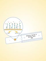
Colorimeter CO7000 (VWR, catalog number: WAPR80-3000-42)
Incubator shaker (Thermo Fisher Scientific, HoeferTM, catalog number: SHKE5000 S)
Orbital/rocking shaker (Thermo Fisher Scientific, catalog number: 11-676-681)
Hot plate with magnetic stiring capability (Thermo Fisher Scientific, catalog number: SP88850200)
Sonicator (Qsonica, catalog number: Q500-110)
Fume hood (Labconco, catalog number: 100600040)
Centrifuges and rotors for handling 0.5-50-ml phenol-resistant centrifuge tubes or bottles [e.g., Beckman Avanti J-E centrifuge (Beckman Coulter, catalog number: A20699) with JA-20 (Beckman Coulter, catalog number: 334831) and JLA-10.500 rotors (Beckman Coulter, catalog number: 369681); Sorvall Legend XTR centrifuge (Thermo Scientific, catalog number: 75210061) with TX-750 rotor (Thermo Scientific, catalog number: 75003180), F13-14x50cy rotor (Thermo Scientific, catalog number: 75003661), round buckets (Thermo Scientific, catalog number: 75003608), and conical adapters (Thermo Scientific, catalog number: 75003639)]
Microcentrifuge with a temperature-control system (Eppendorf, catalog number: 5401000137)
Ultracentrifuge and rotors (e.g., Optima XE-90 with Type 70.1 Ti rotor, Beckman Coulter, catalog numbers: A94471, 342184)
Bottles (250-ml) and adapters for Beckman Avanti J-E centrifuge (Thermo Fisher Scientific, catalog number: NC9304619 and NC1476800)
Nephelo culture flasks (Thermo Fisher Scientific, catalog numbers: 50-194-5583 for 300-ml and 50-194-5576 for 500-ml)
Filtering flasks with side arm (vacuum flask) (Thermo Fisher Scientific, KIMAXTM, catalog number: 10-181B)
Erlenmeyer Flasks (Thermo Fisher Scientific, PYREXTM, catalog number: 10-090C)
Micro stir bars (1/16 × 5/16 inch) (Thermo Fisher Scientific, catalog number: 14-512-150)
Spacers (0.75 mm thick, 16 cm long, 2 cm wide; Thermo Fisher Scientific, HoeferTM, catalog number: 03-500-209)
Gel casting stand (Thermo Fisher Scientific, HoeferTM, catalog number: 03-500-247)
Gel casting frames (Thermo Fisher Scientific, HoeferTM, catalog number: 03-500-241)
Gel combs (15 Wells, 0.75 mm thick; Thermo Fisher Scientific, HoeferTM, catalog number: 03-500-188)
Polyacrylamide vertical gel electrophoresis system (for LOS/LPS: Hoefer, HoeferTM SE 600, catalog number: SE600-15-1.5) (for PEOM: Thermo Fisher Scientific, InvitrogenTM, catalog number: EI0002)
Power supply that is capable of loads from 5 mA and adjustable in 1 mA steps (Bio-Rad, PowerPacTM, catalog number: 1645056)
Acid-washed glass dishes or trays (20.1 × 20.1 × 5.5 cm; Thermo Fisher Scientific, catalog number: 15-242B)
Gel imaging system (Bio-Rad, ChemiDocTM, catalog number: 17001402)
Procedure
文章信息
版权信息
© 2021 The Authors; exclusive licensee Bio-protocol LLC.
如何引用
Lee, Y. J. and Inzana, T. J. (2021). Extraction and Electrophoretic Analysis of Bacterial Lipopolysaccharides and Outer Membrane Proteins. Bio-protocol 11(24): e4263. DOI: 10.21769/BioProtoc.4263.
分类
微生物学 > 微生物生物化学 > 糖类
微生物学 > 微生物蛋白质组学 > 膜蛋白
生物化学 > 糖类 > 多糖
您对这篇实验方法有问题吗?
在此处发布您的问题,我们将邀请本文作者来回答。同时,我们会将您的问题发布到Bio-protocol Exchange,以便寻求社区成员的帮助。