

Pericyte Mapping in Cerebral Slices with the Far-red Fluorophore TO-PRO-3
用远红荧光体TO-PRO-3绘制大脑切片中的周细胞地图
发布: 2021年11月20日第11卷第22期 DOI: 10.21769/BioProtoc.4222 浏览次数: 3610
评审: Miao HeSilvia Olivera-BravoMartine Cohen-Salmon
参见作者原研究论文
The authors used this protocol in:
May 2021
Advertisement


Abstract
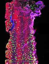
This protocol describes a method for high-resolution confocal imaging of pericytes with the far-red fluorophore TO-PROTM-3 Iodide 642/661 in cerebral slices of murine. Identification of pericytes with TO-PRO-3 is a short time-consuming, high cost-effective and robust technique to label pericytes with no need for immunostaining or generation of reporter mice. Since the TO-PRO-3 stain resists immunofluorescence, and lacks spectral overlap, the probe is well suited for multiple labelling. Our procedures also combine TO-PRO-3-staining of pericytes with fluorescent markers for astrocytes and vessels in brain slices. These approaches should enable the assessment of pericyte biology in gliovascular unit.
Keywords: Pericyte imaging (包皮细胞成像)Background
Fluorescence imaging at the cellular level offers an exceptional tool to track pericytes under confocal or bi-photonic microscopy. Tagging specific pericyte surface antigens, such as chondroitin sulphate proteoglycan neuron-glial 2 (NG2) and platelet-derived growth factor receptor beta (PDGFRβ), proved to be an excellent approach to identify pericytes in the cerebral microvasculature. Antigen labelling is achieved via immunofluorescence with specific antibodies or through fluorescent protein expression under the control of specific promoters for NG2 and PDGFRβ (Ozerdem et al., 2001; Mishra et al., 2014; Hartmann et al., 2015a and 2015b; Jung et al., 2018; Smyth et al., 2018). Notwithstanding, immune techniques involve several steps that take place over hours or days, whereas generation of reporter mice is costly and laborious, mainly in studies employing transgenic mouse models. Herein, we describe a simple, robust and rapid (e.g., min) fluorescent labelling assay to image pericytes in murine brain slices with the far-red fluorophore TO-PROTM-3 Iodide 642/661. This carbocyanine monomer probe has recently been recognized as a pericyte biomarker in both ex vivo (Mai-Morente et al., 2021) and in vivo (Tong et al., 2021) conditions. TO-PRO-3 stains nucleus in fixed tissue (Van Hooijdonk et al., 1994; de Mazière et al., 1996; Suzuki et al., 1997), but is selectively incorporated by living pericytes ex vivo when applied into the physiological saline or in vivo after topical administration (Lacar et al., 2012; Mai-Morente et al., 2021; Tong et al., 2021). Identification of murine brain pericytes by TO-PRO-3 is unambiguous in the tested age range (P06-P90) and, as reported (Mai-Morente et al., 2021), TO-PRO-3-stained pericytes express the classical pericyte immunomarkers NG2 and PDGFRβ and incorporate the pericyte dye NeuroTrace 500/525 (Damisah et al., 2017). Only a subset of TO-PRO-3 pericytes expresses the contractile protein alpha-smooth muscle actin (α-SMA) (Mai-Morente et al., 2021). The far-red emitting TO-PRO-3 dye exhibits negligible autofluorescence and phototoxicity (Suseela et al., 2018), which favours its use in live imaging; additionally, TO-PRO-3-loaded slices can be fixed and processed for immunolabelling (Lacar et al., 2012; Mai-Morente et al., 2021). Since TO-PRO-3 resists immunostaining and fluoresces far from the green and red fluorophores in the light spectrum, it is appropriate for multiple labelling with fluorescent-conjugated probes and antibodies or green fluorescent protein (GFP) reporters. The protocols described here include procedures to identify vessels and astroglia intimately associated with TO-PRO-3-labelled pericytes. Given the ease and reliability of the technique, mapping pericytes with TO-PRO-3 should facilitate future research on pericyte structure and function in cerebral slices.
Materials and Reagents
96-well plate
6-well plate
12/24-well plate
Ice plastic tray with silicone bottom for domestic use
Nylon mesh of a tea plastic strainer
Plastic transfer pipettes (Biologix, catalog number: 30-0138)
Sartorius mLINE® mechanical Biohit pipettors 2, 20, 200 and 1,000 µl
Six-well, twenty-four-well and ninety-six-well multidishes (DeltaLab, catalog numbers: 657160, 662160 and 655180, respectively)
Custom-made strainer
Perfusion chamber
Transparent (glass or polypropylene) cylindrical test tubes with rounded bottom
Conventional 21 gauge (21 G) syringe needles
BD IntramedicTM Polyethylene Tubing, 100 ft × 0.034" × 0.050" (Becton Dickinson, catalog number: 427421)
Microscope glass slides (Deltalab, catalog number: D 100001)
Microscope glass coverslips (Deltalab, catalog number: D 102440) and N1.5 (Knittel Glass, catalog number: VM52440Y1A0.1)
Aluminium foil
Absorbent tissue
Adhesive tape
Permanent marker pen
Fine-tipped paintbrushes
Nail varnish
Hippocampal and cortical slices (300-400 µM thick) from P06-P90 male and female mice [Mus musculus on a C57BL/6 background (Jackson Laboratory, RRID: IMSR_JAX: 000664)] and Rattus norvegicus [Sprague-Dawley (Charles River Laboratories, Strain code 400)]
MilliQ-water or double-distilled water (ddH2O)
Quinolinium, 4-[3-(3-methyl-2(3H)-benzothiazolylidene)-1-propenyl]-1-[3-(trimethylammonio) propyl]-, diiodide/157199-63-8 or TO-PROTM-3 Iodide 642/661 (Life Thermo Fisher Scientific, catalog number: T3605)
NeurotraceTM 500/525 Green Fluorescent Nissl (Life Thermo Fisher Scientific, catalog number: N21480)
Poly-L-Lysine (Sigma-Aldrich, catalog number: P4832)
Lycopersicon Esculentum (Tomato) Lectin DyLight 488 (LEL-DyLight 488) (Life Thermo Fisher Scientific, catalog number: L32470)
Isolectin B4 conjugated to fluorescein isothiocyanate (FITC-ISOB4) (Sigma-Aldrich, catalog number: L2895)
Rabbit anti-GFAP-Cy3TM (Sigma-Aldrich, catalog number: C9205)
2-[4-(Aminoiminomethyl) phenyl]-1H-Indole-6-carboximidamide hydrochloride (DAPI) (Sigma-Aldrich, catalog number: D09542)
Hoechst 33342 (Sigma Aldrich, catalog number: 23491-45-4)
Bovine serum albumin (BSA) (Sigma-Aldrich, catalog number: 048-46-8)
Glycine (Sigma-Aldrich, catalog number: 56-40-6)
Glycerol or Fluoromont-GTM Mounting Medium (Life Thermo Fisher Scientific, catalog number: 00-4958-02)
NaCl (Sigma-Aldrich, catalog number: 7647-14-5)
KCl (Sigma-Aldrich, catalog number: 7447-40-7)
NaHCO3 (Sigma-Aldrich, catalog number: S5761)
NaH2PO4·H2O (Sigma-Aldrich, catalog number: 10049-21-5)
Na2HPO4·H2O (Sigma-Aldrich, catalog number: S9763)
KH2PO4 (Sigma-Aldrich, catalog number: P0662)
Glucose (Sigma-Aldrich, catalog number: G5767)
MgSO4 (Sigma-Aldrich, catalog number: M7506)
CaCl2·2H2O (Sigma-Aldrich, catalog number: C3881)
Paraformaldehyde powder (PFA) (Sigma-Aldrich, catalog number: 158127)
NaOH and HCl
Artificial cerebrospinal fluid solution (ACSF) (see Recipes)
Blocking/permeabilizing solution (see Recipes)
Diluting solution for antibodies (see Recipes)
Fixing solution (see Recipes)
Phosphate buffered saline (PBS) (see Recipes)
PBST (see Recipes)
Equipment
Gas tank 5% CO2, 95% O2
Digital pHmeter (ORION, model: 410A)
Digital Analytical Balance (RadWag, AS82/220.R2)
Thermolyne Type 16700 Mixer Maxi-Mix1 vortex mixer
TS-2000A VDRL Shaker
Fume hood 1300 Series A2 Class II, Type A2 Bio Safety Cabinets
Thermo ScientificTM CimarecTM Basic Stirring Hotplates SP13132033 (ThermoFisher Scientific)
Confocal Laser Scanning Microscope (Leica TCS SP5 TANDEM SCANNER) equipped with:
A 40× oil immersion objective Leica N.A 1,3 with UV correction.
405 nm diode laser, argon gas laser emission at 488 nm and HeNe lasers for 543 nm and 633 emission.
Coverslip Clamp Chamber (ALA Scientific Instruments Inc.)
HCT-10 Temperature Controller (ALA Scientific Instruments Inc.)
Peristaltic Pump (Scientific Industries Inc., Model 203)
Refrigerator and freezer
Thermostatic water bath
Software
Image acquisition and storage system (LAS AF Lite Software)
Image analysis software (Fiji, ImageJ version 1.53c)
Photo editing software (Adobe Photoshop CS6 13.0 × 64 and Adobe Illustrator CS6 16.0.0)
Procedure
文章信息
版权信息
© 2021 The Authors; exclusive licensee Bio-protocol LLC.
如何引用
Mai-Morente, S. P., Irigoyen, J. P., Carriquiry, V. M., Marset, V. M., Di Doménico, M., Isasi, E. and Abudara, V. (2021). Pericyte Mapping in Cerebral Slices with the Far-red Fluorophore TO-PRO-3. Bio-protocol 11(22): e4222. DOI: 10.21769/BioProtoc.4222.
分类
细胞生物学 > 细胞成像 > 共聚焦显微镜
神经科学 > 基础技术
生物科学 > 生物技术
您对这篇实验方法有问题吗?
在此处发布您的问题,我们将邀请本文作者来回答。同时,我们会将您的问题发布到Bio-protocol Exchange,以便寻求社区成员的帮助。