- EN - English
- CN - 中文
Proteoliposomes for Studying Lipid-protein Interactions in Membranes in vitro
体外研究膜脂-蛋白相互作用的蛋白质脂质体
发布: 2021年10月20日第11卷第20期 DOI: 10.21769/BioProtoc.4197 浏览次数: 3346
评审: Khyati Hitesh ShahSheng-Yang Wu

相关实验方案

利用SP3珠和稳定同位素质谱技术优化蛋白质合成速率:植物核糖体的案例研究
Dione Gentry-Torfer [...] Federico Martinez-Seidel
2024年05月05日 2853 阅读

基于活性蛋白质组学和二维聚丙烯酰胺凝胶电泳(2D-PAGE)鉴定拟南芥细胞间隙液中的靶蛋白酶
Sayaka Matsui and Yoshikatsu Matsubayashi
2025年03月05日 1966 阅读
Abstract
Lipids in biomembranes can control the structure and, therefore, the functionality of membrane-embedded protein complexes. Unraveling how the lipid composition determines the mode of operation of membrane proteins provides mechanistic insights into their functionality. We applied a proteoliposome technique for studying how proteins function in biomembranes. The incorporation of isolated membrane proteins in preformed liposomes made from a well-defined lipid composition (proteoliposomes) is a powerful tool for studying lipid-protein interactions. Over several decades, the proteoliposome technique was employed for many different membrane proteins. Recently, it was recognized that different lipid compositions control the light-harvesting functionality of the major photosynthetic light-harvesting complex II (LHCII) isolated from plant thylakoid membranes in vitro. This technique allows systematic examination of the role of so-called non-bilayer lipids on light-harvesting characteristics of LHCII. This protocol describes the isolation of LHCII from leaves and details a four-step procedure to incorporate the detergent-solubilized membrane protein in large unilamellar vesicles (LUV). The protocol was optimized to ensure a very high lipid/protein ratio, designed to specifically examine lipid-protein interactions by minimizing LHCII aggregation. The procedure provides structurally and functionally highly intact LHCII in a detergent-free lipid bilayer with a defined composition.
Keywords: Photosynthesis (光合作用)Background
Biological membranes fulfill a battery of essential functions in living cells. These functions are defined and regulated by specialized membrane proteins that are embedded in the hydrophobic matrix of a lipid-bilayer. This lipid matrix is not just an inert solvation space for membrane proteins but can adopt an active role in controlling their structure and function by specific lipid-protein interactions (Mourtisen and Bloom, 1993; Cantor, 1997; Mondal et al., 2014). In particular, a special lipid class called non-bilayer lipids can have a strong impact on membrane protein structure and function (van den Brinck-van der Laan et al., 2004). Non-bilayer lipids are abundant in biomembranes in general and in photosynthetic thylakoid membranes in particular (Boudiere et al., 2014). A concept that explains lipid-protein interactions in biomembranes and the role of non-bilayer lipids is the lateral membrane pressure hypothesis (Cantor, 1997; van den Brinck-van der Laan et al., 2004; Anishkin et al., 2014). This hypothesis predicts that the characteristic hydrostatic pressure profile along the z-axis of the lipid bilayer is dependent on the physical and chemical nature of the lipid mixture and that this pressure controls and modifies the structure of membrane-integral protein complexes (Cantor, 1997; van den Brinck-van der Laan et al., 2004; Anishkin et al., 2014). Studying lipid-protein interactions is challenging partially because native biomembranes are complex, composed of multi-component systems with different proteins and many different lipid types (diversity of headgroups and fatty acids). However, proteoliposomes are a system that allows examination of lipid-protein interactions under compositionally well-defined conditions. Proteoliposomes are composed of large unilamellar vesicles (LUVs) made of a lipid bilayer in which isolated membrane proteins have been incorporated (Rigaud et al., 1995; Rigaud and Levy, 2003). They have been used for studying energy-transducing membrane proteins (Rigaud et al., 1995), including photosynthetic membrane proteins (McDonnel and Staehelin, 1980; Moya et al., 2001; Yang et al., 2006; Natali et al., 2016; Crisafi and Pandit, 2017; Tietz et al., 2020). Different methods exist to integrate isolated detergent-solubilized membrane protein complexes into the lipid bilayer matrix of LUVs (Rigaud et al., 1995; Rigaud and Levy, 2003). Here, a detergent-based reconstitution protocol (Rigaud et al., 1995; Rigaud and Levy, 2003) was optimized to study lipid-protein interactions of photosynthetic light-harvesting complex II (LHCII) isolated from spinach leaves (Tietz et al., 2020). LHCII is a ~25 kDa-sized trimeric protein complex embedded in the thylakoid membranes of plants and green algae that binds 42 chlorophylls and four carotenoids (Barros and Kühlbrandt, 2009). It is the most abundant membrane protein complex on earth and optimized for highly efficient harvesting of sunlight. The LHCII-proteoliposomes preparation consists of four steps (Rigaud et al., 1995; Rigaud and Levy, 2003; Tietz et al., 2020): (i) preparation of LUVs with defined lipid composition and diameter, (ii) destabilization of the LUVs by doping them with a maximal amount of detergent without formation of mixed lipid-detergent micelles, (iii) incorporation of detergent-solubilized LHCII in the destabilized LUVs, and (iv) detergent removal by polystyrene beads (Biobeads) and purification by gel filtration. Compared to other protocols, the procedure introduced here generates LHCII-proteoliposomes with a very low protein density (lipid/protein ratio ~60,000), specifically allowing the study of lipid-protein interactions. At the same time, the structural and functional integrity of the LHCII is highly preserved during the proteoliposome preparation, as indicated by CD- and low-temperature fluorescence spectroscopy (Tietz et al., 2020). The protocol is versatile since it is expected to work for other membrane protein complexes and lipid mixtures as well.
Materials and Reagents
For LHCII protein isolation
2 × 250 ml glass beakers (cooled on ice)
8 × glass centrifuge tubes with thick walls to hold the low temperature (cooled on ice)
3 × 150 ml glass beakers (with magnetic stir bar)
4 × 50 ml measuring cylinders
2 × 100 ml measuring cylinders
6 × SS34 rotor tubes (cooled on ice)
6 × SW28 rotor tubes (cooled on ice)
2 × gauzes (mesh-size 20 μm) (10 × 10 cm) with funnel for 250 ml beaker
Soft paint brush
2 × ice buckets with lids (to keep samples dark)
Magnetic stirrer with stir bars
Standard glass test tubes
20 ml glass pipettes
1.5 ml Eppendorf® cups
80% acetone
Solid KCl salt
Stock solution (see Recipes)
Solution A (see Recipes)
Solution B (see Recipes)
Solution NET (see Recipes)
Solution SE (see Recipes)
Solution S (see Recipes)
0.1 M and 1 M HCl (see Recipes)
Solution KCl (see Recipes)
For proteoliposomes preparation
Isolated thylakoid membrane lipids dissolved in chloroform. Store in small glass vial with Teflon® sealed lid at -20°C to minimize evaporation. Lipids can be purchased from Avanti® Polar Lipids (Alabaster, Alabama, USA) or can be isolated from leaves. For isolation of thylakoid lipids, see Hara and Radin (1978), with further purification described in Kotapati and Bates (2020). Lipid concentrations: monogalactosyldiacylglycerol (MGDG), 70 mM; digalactosyldiacylglycerol (DGDG), 30 mM; sulfoquinovosylgalactosyldiacylglycerol (SQDG), 20 mM; and phosphatidyldiacylglycerol (PG) 20 mM
Ice bucket with lid
5 ml glass grinding tube with cap
200 nm and 400 nm 13 mm (diameter) nucleopore membrane filters (Whatman®)
PETE mesh spacer 13 mm (Sterlitech®)
3-ml glass standard spectrometer glass cuvette (side length 1 cm) with magnetic micro stir bar
Magnetic stirrer with micro stir bar (for spectrometer glass cuvette)
Sephadex G-25 M PD10 gel filtration column (GE Healthcare®) with fraction collector filled with standard glass test tubes
Chloroform
Nitrogen gas tank with regulator, hose, and attached Pasteur pipette
SM2 BioBeads (Bio-Rad®)
Dewar with liquid nitrogen
Glass tubes for liquid nitrogen measurements
Catalase bovine liver (10,000-40,000 units per mg, Sigma-Aldrich)
Glucose oxidase from Aspergillus (550 units per ml, Sigma-Aldrich)
Glucose (200 mM, Sigma-Aldrich)
10 mM HEPES (see Recipes)
73.6 mM alpha-DM solubilized (see Recipes)
Equipment
LHCII Isolation
1 L Waring commercial blender (Thermo Fisher Scientific)
pH meter (Mettler Toledo, SevenEasy)
Gradient mixer with stirrer (home-made)
Eppendorf Table centrifuge 5810
Sorvall RC 5CPlus centrifuge with SS34 rotor (pre-chilled to 4°C)
Beckman L8-70M ultra-centrifuge with swing bucket SW28 rotor (pre-chilled to 4°C)
Proteoliposomes
250 ml glass beaker with thermometer
High-pressure extruder (Lipex® Extruder), requires N2 gas
Nitrogen tank with regulator attached to extruder
UV-Vis spectrometer (U-3900 spectrophotometer (Hitachi®))
Fluorescence spectrometer (Fluromax-4 spectrofluorometer (Horiba®)) with liquid nitrogen Dewar assembly.
Rotor evaporator (Büchli® Rotavapor R) with heated water bath with glass grinding to connect 5 ml glass tube and ventilation for N2 gas
Fraction collector (Gilson® FC 205)
Oven (set to 60°C) (Fisher Scientific®, catalog number: 3510S)
Procedure
文章信息
版权信息
© 2021 The Authors; exclusive licensee Bio-protocol LLC.
如何引用
Readers should cite both the Bio-protocol article and the original research article where this protocol was used:
- Kirchhoff, H. (2021). Proteoliposomes for Studying Lipid-protein Interactions in Membranes in vitro. Bio-protocol 11(20): e4197. DOI: 10.21769/BioProtoc.4197.
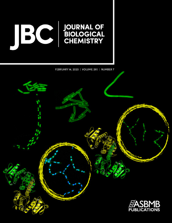
- Tietz, S., Leuenberger, M., Höhner, R., Olson, A.H., Fleming, G.R., Kirchhoff, H. (2020). A proteoliposome-based system reveals how lipids control photosynthetic light harvesting. J Biol Chem 295: 1857-1866.
分类
植物科学 > 植物生物化学 > 蛋白质 > 分离和纯化
植物科学 > 植物生理学 > 新陈代谢
生物化学 > 其它化合物
您对这篇实验方法有问题吗?
在此处发布您的问题,我们将邀请本文作者来回答。同时,我们会将您的问题发布到Bio-protocol Exchange,以便寻求社区成员的帮助。
Share
Bluesky
X
Copy link


