

Isolation and Electrophysiology of Murine Sympathetic Postganglionic Neurons in the Thoracic Paravertebral Ganglia
小鼠胸椎旁神经节交感神经节后神经元的分离与电生理研究
发布: 2021年10月20日第11卷第20期 DOI: 10.21769/BioProtoc.4189 浏览次数: 4944
评审: Mary L. PhillipsAnonymous reviewer(s)
参见作者原研究论文
The authors used this protocol in:

May 2019
Advertisement


Abstract
The thoracic paravertebral sympathetic chain postganglionic neurons (tSPNs) represent the predominant sympathetic control of vascular function in the trunk and upper extremities. tSPNs cluster to form ganglia linked by an interganglionic nerve and receive multisegmental convergent and divergent synaptic input from cholinergic sympathetic preganglionic neurons of the spinal cord (Blackman and Purves, 1969; Lichtman et al., 1980). Studies in the past have focused on cervical and lumbar chain ganglia in multiple species, but few have examined the thoracic chain ganglia, whose location and diminutive size make them less conducive to experimentation. Seminal studies on the integrative properties of preganglionic axonal projections onto tSPNs were performed in guinea pig (Blackman and Purves, 1969; Lichtman et al., 1980), but as mice have become the accepted mammalian genetic model organism, there is need to reproduce and expand on these studies in this smaller model. We describe an ex vivo approach that enables electrophysiological, calcium imaging, and optogenetic assessment of convergence, divergence, and studies on pre- to postganglionic synaptic transmission, as well as whole-cell recordings from individual tSPNs. Preganglionic axonal connections from intact ventral roots and interganglionic nerves across multiple segments can be stimulated to evoke compound action potential responses in individual thoracic ganglia as recorded with suction electrodes. Chemical block of synaptic transmission differentiates spiking of preganglionic axons from synaptically-recruited tSPNs. Further dissection, including removal of the sympathetic chain, enables whole-cell patch clamp recordings from individual tSPNs for characterization of cellular and synaptic properties.
Keywords: Mouse (小鼠)Background
Thoracic sympathetic postganglionic neurons (tSPNs) are housed in bilateral paravertebral chain ganglia. Comprising a significant component of the final motor output of the sympathetic nervous system, paravertebral tSPNs contribute to autonomic homeostatic mechanisms by directly innervating effector tissues, including vasculature, adipose tissue, sweat glands, and piloerector muscles (Janig, 2006; Bartness et al., 2010). Thoracic sympathetic ganglia contain a distinct composition of genetically separable postganglionic neuron groups as compared to more rostral cervical and lower thoracic segments (Furlan et al., 2016). Their morphological and electrophysiological properties may also differ from sympathetic postganglionic neurons (SPNs) elsewhere (Jobling and Gibbins, 1999). Our understanding of the electrophysiological properties of paravertebral sympathetic chain postganglionic neurons has been largely derived from studies in cervical and lumbar ganglia (Eccles, 1935; Erulkar and Woodward, 1968; Lichtman et al., 1979; Purves and Wigston, 1983; Cassell et al., 1986; Li and Horn, 2006; Bratton et al., 2010) with intracellular recordings undertaken using sharp microelectrode approaches (Blackman and Purves, 1969; Lichtman et al., 1980; Jobling and Gibbins, 1999) (however, see Springer et al., 2015) with an impalement injury conductance that alters basic membrane properties (Staley et al., 1992; Rall, 2011; Springer et al., 2015).
Despite their physiological importance, our understanding of the function of paravertebral ganglia is based largely on work undertaken in larger mammalian models (Blackman and Purves, 1969; Lichtman et al., 1980; Rubin and Purves, 1980; Purves and Lichtman, 1985). Greater understanding of their operational properties has not benefited from powerful molecular toolkits provided by molecular genetic approaches in mouse models (Furlan et al., 2016). Unlike cervical and lumbar chain ganglia, tSPNs are situated in a protected paraspinal and subpleural location on the ventral wall of the thoracic cavity. This makes in vivo electrophysiological studies difficult and explains the need for ex vivo approaches (Blackman and Purves, 1969; Lichtman et al., 1980; Rubin and Purves, 1980). Moreover, as the largely transparent individual ganglia are very small (<200 μm in length) with the entire thoracic chain being <2 cm, dissection for study is challenging.
We also developed the ex vivo preparation to enable whole-cell recordings of tSPNs for assessment of membrane properties independent of impalement leak produced with sharp microelectrodes. Results revealed an order of magnitude higher membrane resistivity and associated amplified excitability, with greater intrinsic capacity for synaptic integration and the ability for maintained firing (McKinnon et al., 2019).
To undertake whole-cell patch recordings, the chain is removed entirely from the vertebral column using fine iridectomy microdissection scissors and glass probes (McKinnon et al., 2019). With this method, one can investigate multisegmental preganglionic actions in tSPNs in mice (cp Lichtman et al., 1980), including via optogenetic recruitment of preganglionic cholinergic axons using ChAT::CHR2 mice (Llewellyn et al., 2010). As there are several genetically distinct populations of sympathetic preganglionic neurons (Deuchars and Lall, 2015; Blum et al., 2020), various cre-based driver approaches can be leveraged to study multisegmental actions arising from different preganglionic populations. Capturing individual or population activity of genetically distinct tSPNs is also possible with the use of cre-based reporters or genetically-encoded Ca2+ indicators (Furlan et al., 2016). In summary, the described approach enables the use of mice for highly accessible ex vivo studies for electrophysiology, calcium imaging, optogenetics, and pharmacology for cellular and circuit studies on input-output relations of the thoracic paravertebral chain.
Materials and Reagents
2 mm glass probes made from stringer (Bullseye Glass, catalog number: 000147-0272-F-Tube)
½ ml Monoject Insulin Syringe, 29 G × ½” (Covidien, catalog number: 8881600350)
Needle 25 G (BD PrecisionGlide, catalog no: 14-826G)
1.5 ml MCT Graduated Mixed Tubes (Fisherbrand, catalog number: 05-408-137)
0.20 mm Stainless Steel Insect pins (Fine Science Tools, catalog number: 26002-20 )
0.22 μm Syringe Filter (Fisherbrand, catalog number: 09-719C)
100 mm × 15 mm Petri Dish (VWR, catalog number: 25384-070)
Square plastic weighing dish (Dyn-A-Med, catalog number: 80055)
Electrode Storage (World Precision Instruments, catalog number: E215)
VWR micro cover glass 24 × 50 mm (VWR, catalog number: 48393081)
Adult C57Blk/6 mice (6+ weeks old, alternative animal strains can be used)
Collagenase Type III (Worthington Biochemical Corporation, catalog number: LS004180)
Isoflurane, USP (Piramal Critical Care, catalog number: 400648037)
Urethane (Sigma-Aldrich, catalog number: U2500)
Ketamine (Henry Schein, catalog number: 056344)
Xylazine (Sigma-Aldrich, catalog number: X1251)
Custom-built Sylgard-coated dissecting dish (see Procedure D)
SYLGARDTM 170 Silicone Elastomer Kit (DOW Inc., catalog number: 4026157)
High vacuum grease (DOW Corning, catalog number: H051J89018)
Sodium chloride (Fisher Scientific, catalog number: S642-500)
Potassium chloride (Sigma-Aldrich, catalog number: P9541-1KG)
Magnesium sulfate Heptahydrate (Fisher Scientific, catalog number: BP213-1)
Calcium chloride dihydrate (Fisher Scientific, catalog number: C69-500)
Potassium phosphate monobasic (Sigma-Aldrich, catalog number: P5655-500G)
D-(+)-Glucose (Sigma-Aldrich, catalog number: G7528-1KG)
Sodium bicarbonate (Sigma-Aldrich, catalog number: S6297-1KG)
95% O2, 5% CO2 gas (Nexair, catalog number: UN3156)
Potassium D-gluconate, 99% (Alfa Aesar, catalog number: B25135)
EGTA (Sigma-Aldrich, catalog number: E-3889)
HEPES (Sigma-Aldrich, catalog number: H3375-250G)
ATP (Sigma-Aldrich, catalog number: A9187)
GTP (Sigma-Aldrich, catalog number: G9002)
Artificial Cerebrospinal Solution (aCSF) for Electrophysiological Recordings (see Recipes)
High Magnesium Low Calcium Microdissection Solution (see Recipes)
Patch Electrode Solution (see Recipes)
Equipment
Surgical tools
2.5 mm Vannas spring scissors (Fine Science Tools, catalog number: 15002-08)
Wagner scissors (Fine Science Tools, catalog number: 14068-12)
9 mm Castroviejo microdissecting spring scissors (Roboz, catalog number: Rs-5658)
Dumont #5 Forceps (Fine Science Tools, catalog number: 11251-20)
pH meter (Denver Instruments, model: UB-10)
Osmometer (Vapro, model: Model 5600)
Magnetic stir plate (IKA Works USA, model: CERAMAG Midi)
Magnetic stir bar (VWR, catalog number: 76006-400)
600 ml beaker (Pyrex, catalog number: CLS1000600)
Recording equipment for whole-cell recordings
Generally, there are multiple commercially available components that can be used to undertake visually guided whole-cell recordings from microdissected nervous tissue. We used an upright microscope containing 40× liquid immersion objectives with differential interference contrast and infrared imaging for image enhancement. Conventional electrophysiology recording methods were employed, including an experimental chamber with oxygenated aCSF, air table, micromanipulator with attached electrode holder, high impedance low noise patch clamp amplifier, A/D converter, and associated specialty software for data capture. Details are provided in various reviews. Those used by our lab are described in a recent publication (McKinnon et al., 2019) as listed below (Figure 1).
Upright DIC fluorescence microscope (Olympus, model: BX51WI) affixed with a low-light camera (Olympus, model: OLY-150)
Vertical Electrode Puller (Narishige, model: PP-83)
Micro-forge (Narshige, model: MF-9)
MultiClamp 700A and Digidata 1322A (Molecular Devices)
Custom Sylgard-coated recording chamber and perfusion system
Controllable blue light laser (custom built but are also commercially available; e.g., Thorlabs S1FC473MM Multimode Fiber-Coupled Laser)
Electrical stimulation can be provided by commercially available stimulators (e.g., A-M Systems, model: 2200 Analog Stimulus Isolator)
Gravity fed superfusion with Masterflex pump for recirculation (Cole Palmer, model: 77200-50)
Tubing for perfusion (Masterflex, catalog number: 96412-16)
1.5-mm outer diameter filamented, borosilicate glass capillaries (World Precision Instruments, catalog number: TW150F-4)
Coated silver wire (A-M Systems, catalog number: 787000)
Connector Socket 20-24AWG gold crimp (Digi-Key, catalog number: 205090-1)
Connector D-Sub Pin 20-24 AWG AU (Digi-key, catalog number: 205089-1)
Silicone tubing for electrode (Masterflex, catalog number: 96410-10)
Cotton Swab with plastic shaft (Just The Basics, catalog number: PINSB0300DLJB01)
Quick-Setting Epoxy Syringe (J-B Weld, catalog number: 50112)
4-Indent D-Sub Crimper 26-20 AWG (Paladin Tools, model: PA1440)
Multipurpose wire stripper and cutter (Klein Tools Inc., model: 1010)
Three-Way, Stopcock with Male Luer Lock, Non-Sterile (Cole-Palmer, catalog number: UX-30600-02)
5 ml disposable syringe with Luer Lock (BD, catalog number: 309646)
Electrical tape (VWR, catalog number: 470020-186)
Insulated shielded Copper Wire 20AWG 300V BLK 25' (CNC Tech, catalog number: 1430-20-1-0500-001-1-TS)
Ground electrode probe (WPI, model: Ep2)
Communication Cable (Belden, catalog number: 8441)
Double banana plug Connectors for amplifier (Pomona Electronics, catalog number: 1330)
Single banana Plug (Pomona Electronics, catalog number: 1325)
Precision screwdriver (Westward, model: 401L69)
Clorox bleach (VWR, catalog number: 89501-620)
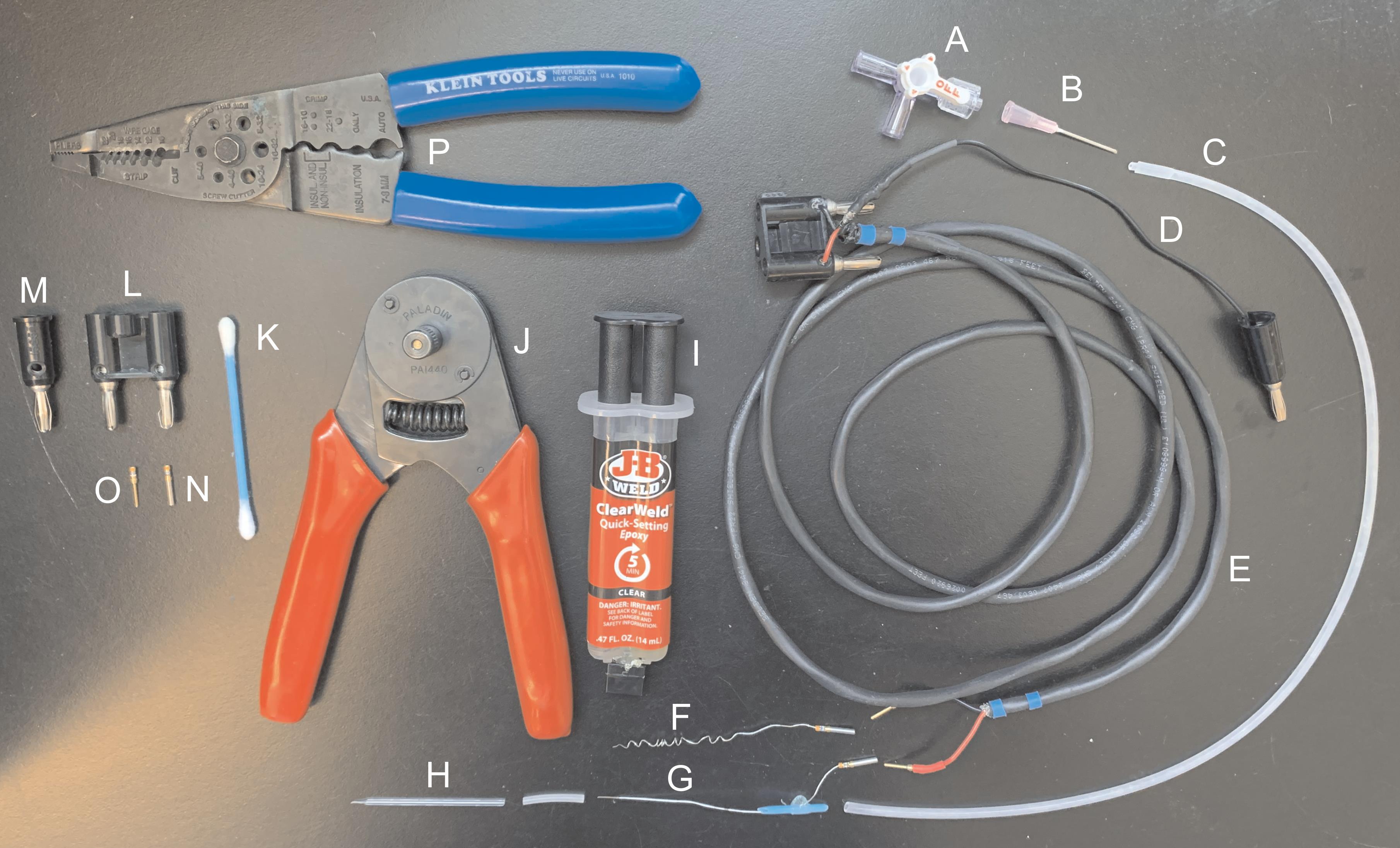
Figure 1. Materials for manufacturing suction electrodes. (A) Three-way stopcock with Luer lock. (B) Blunt 25 G needle. (C) Silicone tubing. (D) Insulated copper wire. (E) Communication cable. (F-G) Coated silver wire. (H) Glass electrode. (I) Quick-setting epoxy syringe. (J) D-sub crimper. (K) Cotton swab with plastic shaft. (L) Double banana plug. (M) Single banana plug. (N) Connector socket. (O) Connector D-Sub pin. (P) Wire stripper and cutter.Recording equipment for studies involving multisegmental preganglionic and postganglionic compound action potentials
Stereo dissecting microscope with 0.8-5.6× zoom range and DFPL 0.5× objective (Olympus, model: SZX7)
Multichannel differential amplifier (custom-built but are also commercially available (e.g., A-M Systems, model: 1700 Differential AC Amplifier)
Constant current stimulator (custom built but are also commercially available; e.g., A-M Systems, model: 2200 Analog Stimulus Isolator)
Controllable blue light laser (custom built but are also commercially available; e.g., Thorlabs, S1FC473MM Multimode Fiber-Coupled Laser)
Magnetic stand with 3-axis manual micromanipulator control (Kanetec, model: MB-PSL)
Gravity fed superfusion with Masterflex pump for recirculation (Cole Palmer, catalog number: 77200-50)
Tubing for perfusion (Masterflex, catalog number: 96412-16)
Digidata 1322A (Molecular Devices)
1.5-mm outer diameter filamented, borosilicate glass capillaries (World Precision Instruments, catalog number: TW150F-4)
Coated silver wire (A-M Systems, catalog number: 787000).
Connector Socket 20-24AWG gold crimp (Digi-Key, catalog number: 205090-1).
Connector D-Sub Pin 20-24 AWG AU (Digi-key, catalog number: 205089-1).
Ground electrode probe (WPI, model: Ep2)
Communication Cable (Belden, catalog number: 8441)
Double banana plug Connectors for amplifier (Pomona Electronics, catalog number: 1330)
Single banana Plug (Pomona Electronics, catalog number: 1325)
Image Capture Equipment for Calcium Imaging
AC/DC differential amplifier (A-M Systems, model: 51249)
Master 8 system and an ISO-Flex stimulus isolator (A.M.P.I, model: 1955)
Inverted microscope (Olympus, model: IX70)
Xenon lamp housing (Olympus, model: U-ULS75XE)
Power supply (Olympus, model: AH2-RX-T)
Neutral density filter (Olympus, catalog number: Chroma ND-50)
Uniblitz shutter (Vincent Associates, model: VCM-D1)
400 nm dichroic mirror (Olympus, model: Chroma NC474265)
490-520nm Emission filter (Olympus, model: Chroma U-MF2)
CCD camera (Stanford Photonics, model: XR/ABF with XR/M camera)
Trinitron color video monitor (Sony, model: PVM-135MD)
Software
Clampex software (Molecular Devices, RRID: SCR_011323)
PCI software (Hamamatsu, Sunayama-cho, Naka-ku, Hamamatsu City, Shizuoka, Japan)
Fiji (ImageJ from NIH)
Procedure
文章信息
版权信息
© 2021 The Authors; exclusive licensee Bio-protocol LLC.
如何引用
Readers should cite both the Bio-protocol article and the original research article where this protocol was used:
- Halder, M., McKinnon, M. L., Li, Y., Wenner, P. and Hochman, S. (2021). Isolation and Electrophysiology of Murine Sympathetic Postganglionic Neurons in the Thoracic Paravertebral Ganglia. Bio-protocol 11(20): e4189. DOI: 10.21769/BioProtoc.4189.
- McKinnon, M. L., Tian, K., Li, Y., Sokoloff, A. J., Galvin, M. L., Choi, M. H., Prinz, A. and Hochman, S. (2019). Dramatically Amplified Thoracic Sympathetic Postganglionic Excitability and Integrative Capacity Revealed with Whole-Cell Patch-Clamp Recordings.eNeuro 6(2): ENEURO.0433-0418.2019.
分类
神经科学 > 周围神经系统 > 坐骨神经
细胞生物学 > 基于细胞的分析方法 > 电生理技术
您对这篇实验方法有问题吗?
在此处发布您的问题,我们将邀请本文作者来回答。同时,我们会将您的问题发布到Bio-protocol Exchange,以便寻求社区成员的帮助。