

Isolation, Culture, and Identification of Primary Müller Cells from Human Retina
人视网膜原代 Müller 细胞的分离、培养和鉴定
发布: 2021年10月05日第11卷第19期 DOI: 10.21769/BioProtoc.4179 浏览次数: 3959
评审: Gal HaimovichPhilipp WörsdörferAnonymous reviewer(s)

参见作者原研究论文
The authors used this protocol in:

Apr 2019
Advertisement


Abstract
Müller cells, the major glial cells of the retina, play vital roles in maintaining redox homeostasis and retinal metabolism. An immortalized human Müller cell line (MIO-M1) is widely used as an in vitro model to study Müller cells’ function, but they may not be exactly the same as primarily cultured human Müller cells. The use of human primary Müller cells (huPMCs) in culture has been limited by the requirement for complicated culture systems or particular age ranges of donors. We have successfully grown huPMCs using our established protocol. The cell type was pure, and cultured cells expressed Müller cell-specific markers strongly. The cultured huPMCs were used for morphologic, metabolic, transcriptomic, and functional studies.
Graphic abstract:
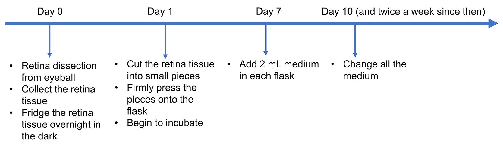
Timeline for human primary Müller cell (huPMC) culture
Background
Müller cells, the principal macroglial cells of the vertebrate retina, are responsible for retinal redox homeostasis and provide metabolic support for retinal neurons (Reichenbach and Bringmann, 2013). Müller cells are also a promising target for therapeutic regeneration (Ahmad et al., 2011). An immortalized human Müller cell line (MIO-M1) was characterized in 2002 and has been widely used as an in vitro model to study human Müller cells (Limb et al., 2002). Primary cultures of human cells may offer a more appropriate in vitro model than immortalized cell lines for single-cell studies (Schnichels, 2020). The use of human primary Müller cells (huPMCs) in culture has been limited by the requirement for complicated culture systems (Giannelli et al., 2011) or particular age ranges of donors (Lupien and Salesse, 2007). Human retina tissue from donors cultured using our modified protocol was refrigerated overnight in the dark before digestion. The tissue was then cut into small pieces and firmly pressed to the base of the cell culture flask. These pieces were cultured with a minimum amount of medium for a week then provided with a regular amount of medium. Cell colonies emerged after culturing for two to three weeks in donors from almost all age groups. Our culture system does not require growth factors or Matrigel. Firmly pressing retina tissue to the base of the flask greatly increased the success rate, even in donor samples where the post-mortem time exceeded 24 h. We have successfully detected the following Müller cell markers: CRALBP, VIMENTIN, SOX2, GS, and AQP4.
Materials and Reagents
Pipette tips
Corning® CellBIND® 25 cm2 (T25) rectangular canted neck cell culture flask with vent cap (Corning, catalog number: 3289)
5 ml flat bottom tube (screw cap) (Techno Plas, catalog number: P5016SU)
Sterile gauze swab (LIVINGSTONE, catalog number: GSS075X5PL)
1 ml disposable syringe (LIVINGSTONE, catalog number: DSL001MLS)
18 G sterile hypodermic needle (LIVINGSTONE, catalog number: DN18GX1.5LV)
Falcon® 60 mm TC-treated Easy-Grip style cell culture dish (Corning, catalog number: 353004)
FisherbrandTM Pasteur Pipets (Fisher Scientific, catalog number: 22-209361)
Foil
Serological pipet (Corning, Costar, catalog number: 4487)
15 ml centrifuge tube (Corning, catalog number: 430791)
50 ml centrifuge tube (Corning, catalog number: 430291)
DMEM (Dulbecco's modified Eagle medium), high glucose, GlutaMAXTM supplement, pyruvate (ThermoFisher, Gibco, catalog number: 10569-010)
Fetal bovine serum (FBS) (Sigma-Aldrich, catalog number: F9423)
Penicillin-streptomycin (Sigma-Aldrich, catalog number: P4333)
TrypLETM select enzyme (1×) (ThermoFisher, Gibco, catalog number: 12563-029)
PBS tablets (Medicago, catalog number: 09-2051-100)
CO2 independent medium (ThermoFisher, Gibco, catalog number: 18045-088)
2% PFA in PBS
EZ Slides (Millicell, catalog number: PEZGS0816)
Microscope cover glass, rectangular (Knittel, catalog number: G417)
Triton X-100 (Merck, catalog number: 30632.4N)
Donkey serum (Millipore, Sigma-Aldrich, catalog number: S30-100ML)
Primary antibodies:
CRALBP (Abcam, catalog number: ab15051)
VIMENTIN (abcam, catalog number: ab92547)
SOX2 (Millipore, catalog number: AB5603)
Secondary antibodies:
Donkey anti-Rabbit IgG (H+L), Alexa Fluor 488 (Invitrogen, ThermoFisher, catalog number: A-21206)
Donkey anti-Mouse IgG (H+L), Alexa Fluor 594 (ThermoFisher, Invitrogen, catalog number: A-21203)
Hoechst 33342 nucleic acid stain (ThermoFisher, catalog number: H3570)
Vectashield Antifade Mounting Medium (Vector Laboratories, catalog number: H-1000)
DMSO (Dimethyl sulfoxide) (Sigma-Aldrich, catalog number: D2650)
Cryopreservation tubes (Nunc CryoTubes) (Nunc, catalog number: V7634)
Isopropyl alcohol (Sigma-Aldrich, catalog number: W292907)
Sterile specimen jar (Livingstone, catalog number: TP5744SLN)
Complete medium (see Recipes)
Sterile PBS (see Recipes)
Ethanol (80%, v/v) (see Recipes)
Freezing medium (see Recipes)
Blocking solution (see Recipes)
Dilution solution (see Recipes)
Equipment
Forceps (blink medical, katena, model: HR142, Forcep Jewellers No.5)
Scissors (Eyeline, ACROfine, model number: AB-9921D)
Kelly forceps (MediTools, catalog number: SHF-2718)
Autoclave
37°C water bath
CO2 incubator (37°C, 5% CO2)
Dissection hood
Dissection microscope
Fume cupboard
Inverted microscope
Confocal microscope
Shaking bed
Pipette
4°C fridge
Centrifuge compatible with 15 ml conical tubes
Freezing container (Thermo Scientific, catalog number: 5100-0001)
-80°C freezer
Liquid nitrogen storage
Procedure
Collect and pre-treat retina tissue (DAY 0)
Eyes are transported from the eye bank after the cornea is removed for transplantation. Use CO2 Independent Medium to transporting the tissue under atmospheric conditions. Autoclave the dissection tools are beforehand. Use 80% ethanol (v/v) for general sterilizing in the tissue culture room. Dissect donors’ eyes and detach the retina from the eyecup. Starve the retina in DMEM without FBS or growth factors in the fridge and keep in the dark overnight. Rod photoreceptors consume more energy to maintain resting membrane potential in the dark (Okawa et al., 2008); thus, most of them are assumed to have died after being kept in the dark overnight.
Transport human donor eyes (cornea already removed) with a sterile specimen jar in a CO2-independent medium.
Using scissors and forceps, dissect and remove iris, lens, and vitreous from the eyecup and separate neural retina gently from the underlying retinal pigment epithelium (Video 1).
 Video 1. Dissect the neural retina
Collect 1 cm2 of human neural retina in a sterile flat bottom tube containing 5 ml DMEM (for 1-2 T25 flasks).
Wrap the tube with foil to avoid light and keep it at 4°C overnight.
Digest and seed cells (DAY 1)
Digestion destroys the tissue structure. The small pieces of retinal tissue, needle fixation to the flask, and CellBIND® surface facilitate cell attachment.
Pre-warm the TrypLE digestion solution and complete medium (see Recipes) in a 37°C water bath before the experiment.
Transfer the tissue to a new sterile flat bottom tube containing 5 ml pre-warmed TrypLE to digest the retina. Incubate in a CO2 incubator at 37°C for 60 min. Invert the tube every 20 min during digestion.
Use sterile forceps to transfer the digested retina into a 60 mm cell culture dish containing 10 ml complete medium.
Cut the tissue into small pieces (around 1 × 1 mm) with scissors and forceps in the medium under a dissection microscope in the dissection hood.
Transfer the retinal pieces into T25-cell culture flasks using sterile Pasteur glass pipettes with a minimum volume of complete medium (Figure 1A and 1B; Video 2).
Video 2. Transfer, distribute and fix the retinal piecesBend the sterile 18 G needle to a 90° angle using hemostatic forceps. Separate the dissected retinal pieces and distribute them around the bottom of the flask using the angled needle to prevent clumping (Video 2).
Use an 18 G angled needle to firmly press the retinal pieces onto the bottom of the T25 flasks to enhance the success rate of primary human Müller cell culture; lay down the flask under the microscope, find the retina pieces and firmly press down the syringe so that the sharp needle can fix the tiny retina pieces onto the base. (Figure 1C and 1D; Video 2). Place the flask vertically and add 2 ml complete medium into each flask. Avoid washing off the attached retinal pieces.
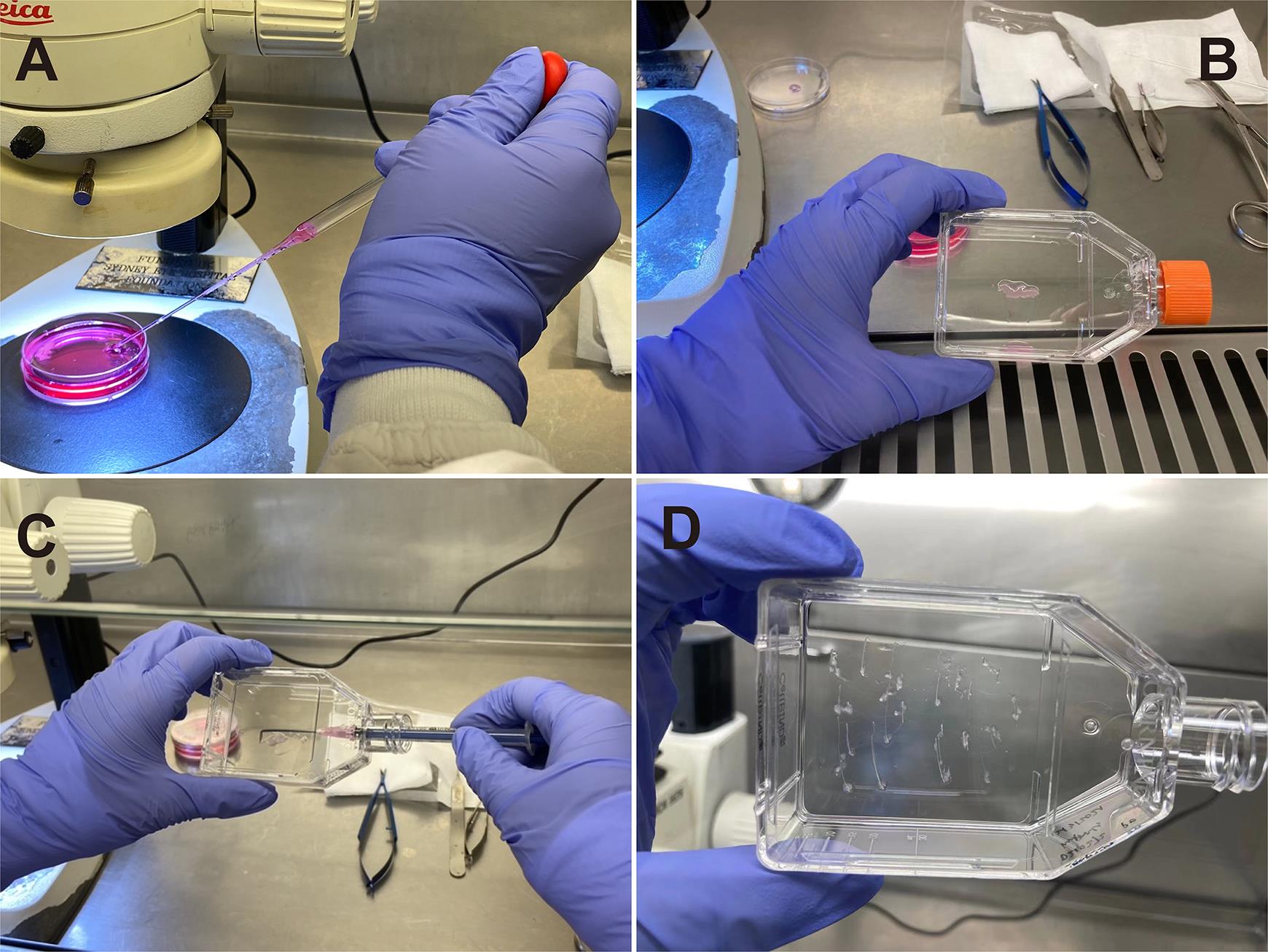
Figure 1. Transfer the retinal pieces to the T25 flask (A and B) and firmly press the retinal pieces to the bottom (C and D)Place the flask vertically in the cell culture incubator (CO2, 37°C) for 15 min to allow better attachment of retinal pieces to the bottom of T-25 flasks.
Place the T25 flasks down horizontally. The medium (2 ml) is enough to cover the bottom of the flask and will minimize the chance of tissue detachment during culture.
Minimize the disturbance of the flasks.
Increase medium volume (DAY 7)
Add another 2 ml complete medium in each flask.
Minimize the disturbance of the flasks.
Routine culture (from DAY 10)
It takes approximately 2-3 weeks before huPMCs colonies emerge from the tissue and another 2-3 weeks before reaching 90-100% confluency (Figure 2).
Change the medium with 4 ml fresh complete medium twice a week.
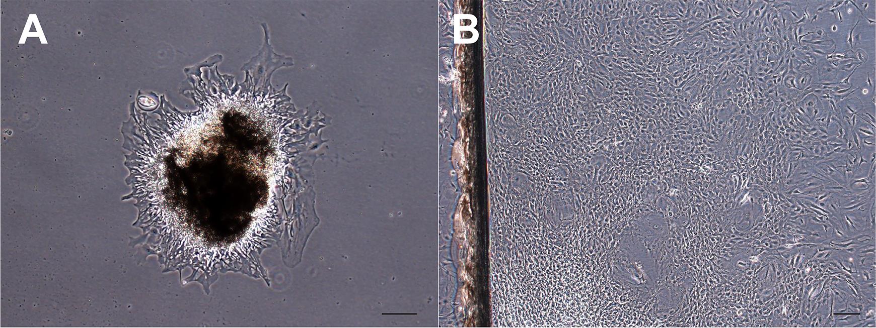
Figure 2. Morphology of cultured colonies. (A) Colony after 2 weeks of culture. (B) Colony after 6 weeks of culture, showing 90-100% confluency. Scale bars: 200 μm.
Passage
Digestion of huPMCs usually takes longer than for most cell lines (including MIO, ARPE19, or HEK293T). Passage 1:1 to begin. Apply 1:2 to 1:3 passaging after P1. Our lab uses P3 huPMCs for experiments, although passaging to P10 is generally possible (depending on donors).
Remove the medium and rinse with 3 ml sterile PBS.
Remove the PBS and add 2 ml TrypLE in each T25 flask to digest.
Incubate the flask with TrypLE in a cell culture incubator 6-8 min (when more than half of the cells detach from the bottom under the microscope).
Terminate the digestion by adding 2 ml complete medium in the flask.
Detach all the cells by pipetting and transfer all the liquid to a 15 ml centrifuge tube.
Pellet the huPMCs by centrifuging (200 × g, 5 min, and 20°C).
Resuspend the pellet in 1 ml complete medium and transfer to a new T25 flask with 3 ml complete medium.
Place the flasks back in the cell culture incubator.
Cryopreserve and revive
The huPMCs cultured in our system can be cryopreserved and revived.
Digest the huPMCs and pellet the cells by centrifuging as described above.
Resuspend the cells in freezing medium. For each confluent T25 flask, the pellets can be resuspended in a 1 ml freezing medium in cryopreservation tubes.
The cryopreservation tubes are transferred to a freezing container filled with isopropyl alcohol at room temperature. The container is then transferred to a -80°C freezer. The cryopreservation tubes can be stored in liquid nitrogen from the next day.
For the revival, thaw the huPMCs in a 37°C water bath with minimum time (normally 1 min). Gently transfer the cells with a freezing medium to a 15 ml centrifuge tube. Add 4 ml pre-warmed complete culture medium into the tube drop by drop.
Centrifuge at 200 × g for 5 min at 20°C and decant the supernatant. Resuspend the pellet in 4 ml complete culture medium and seed the cells into a T25 flask.
Keep the flask in the incubator. The huPMCs usually reach 100% confluency within 3 days.
Identification
A group of Müller cell markers (CRALBP, VIMENTIN, SOX2, GS, and AQP4) can be detected from P3. Immunocytochemistry is suggested for identification.
Digest P2 huPMCs as described above and seed the P3 huPMCs on EZ slides, 5,000 cells per well, with 500 ul complete medium. Culture the huPMCs in the 8-well EZ slide in the incubator at 37°C for 2 days.
Replace the medium with 500 μl pre-warmed (37°C) PBS and leave the slide on a shaking bed (25 rpm) for 5 min.
Replace the PBS with an additional 500 μl PBS and leave the slide on a shaking bed (25 rpm) for 5 minutes.
(In a fume cupboard) Remove the PBS and fix the cells in 2% PFA for 30 minutes at room temperature.
(In a fume cupboard) Remove the PFA. Wash the cells with PBS three times (as in Step G3), and the cells are ready for blocking.
Replace PBS with 120 μl blocking solution per well. Block for 1 h at room temperature.
Dilute primary antibodies with dilution solution (CRALBP 1:100, VIMENTIN 1:400, SOX2 1:200, GS 1:50, and AQP4 1:100). Centrifuge (13,000 × g, 5 min, 20°C) before application. Replace blocking solution with 120 μl of the desired antibody solution in each well. Incubate the slide with primary antibodies overnight on a shaking bed (25 rpm) at 4°C.
Remove the primary antibodies. Rinse the cells with PBS three times (Step G3).
Dilute secondary antibodies (donkey anti-mouse or rabbit, both 1:1,000) with dilution solution. Centrifuge (13,000 × g, 5 min, 20°C) before application. Replace PBS with 120 μl of the desired antibody solution in each well. Incubate the slide with secondary antibodies in the dark for 4 h at room temperature.
Remove the secondary antibodies. Rinse the cells with PBS three times (Step G3).
Stain with Hoechst 33342 (5.6 mg/L) for 5 min at room temperature. Avoid light.
Remove the Hoechst. Rinse the cells with PBS three times (Step G3).
Break the walls of the EZ slide as described in the manual. Mount the slide with Vectashield Antifade Mounting Medium. Immunofluorescence images can be taken using confocal microscopy immediately.
Notes
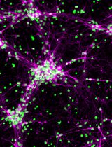
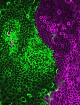
We have successfully cultured human primary Müller cells from retina donors aged 41 to 79 years old, following this protocol. The cultured cells were VIMENTIN/CRALBP/SOX2 positive (Figure 3). Other Müller cell markers positively tested include GS and AQP4 (data not shown). In most cases, huPMCs can be passaged successfully before P10 without a change of morphology and remain CRALBP positive.
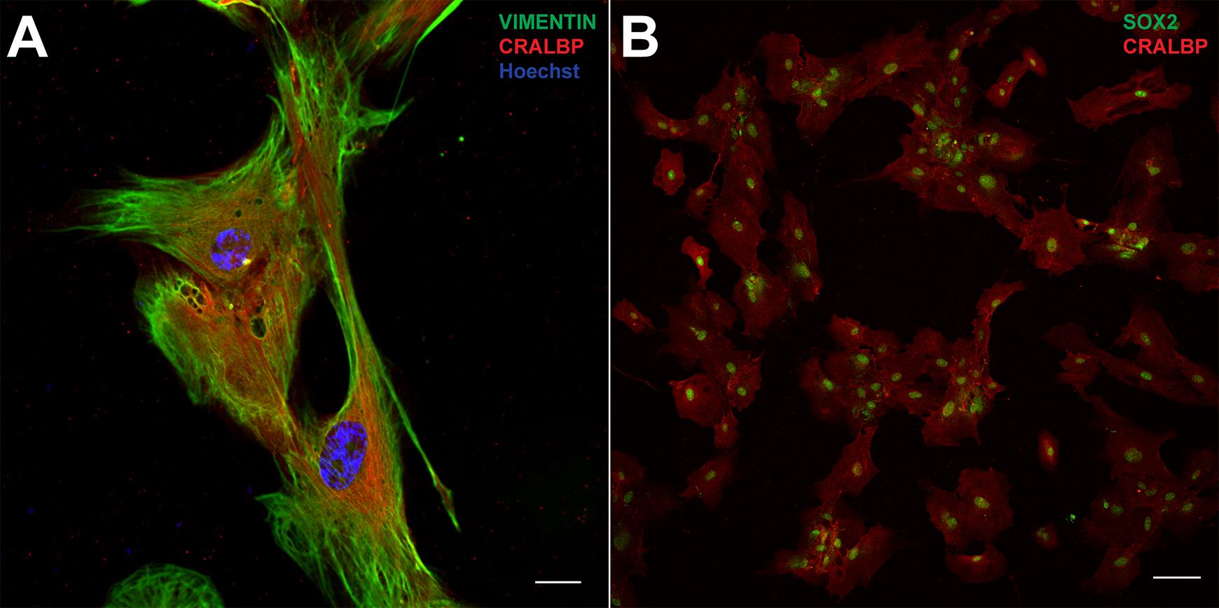
Figure 3. Immunofluorescent staining of huPMCs (P3) with VIMENTIN (green), CRALBP (red) and Hoechst (blue) (A); CRALBP (red) and SOX2 (green) (B). Scale bar in A: 20 μm; B: 100 μm.
Recipes
Complete medium
DMEM supplemented with 10% heat-inactivated fetal bovine serum (FBS) and 1% Penicillin-Streptomycin.
Aliquot to 50 ml centrifuge tubes and store at 4°C. Avoid light and repeated opening. Use within a month.
Sterile PBS
Dissolve 5 PBS tablets in MQ water and fill up to 500 ml before autoclaving (0.14 M NaCl, 0.0027 M KCl, 0.010 M phosphate buffer pH 7.4).
Aliquot in 50 ml centrifuge tubes and store at 4°C. Use within 3 months.
Ethanol (80%, v/v)
Add 1.6 L ethanol in a glass jar, then fill up to 2 L with MQ water.
Freezing medium
20% FBS and 10% DMSO in DMEM
Blocking solution
5%(v/v) Normal Donkey Serum, 0.5% Triton X-100 in PBS
Dilution solution
1% Normal Donkey Serum, 0.5% Triton X-100 in PBS
Acknowledgments
We acknowledge the Lions NSW Eye Bank and Australian Ocular Biobank tissue coordinators and scientists and eye donors and their families for the human donor eye tissue used in this project. This study was supported by the Ophthalmic Research Institute of Australia and a grant from the Lowy Medical Research Institute. This protocol was derived from Zhang et al. (2019) (DOI: 10.7554/eLife.43598).
Competing interests
The authors declare no competing interests.
Ethics
Human subjects: Human retinas were obtained from post-mortem donor eyes with ethical approval from the Human Research Ethics Committee of the University of Sydney (HREC#:16/282).
References
- Ahmad, I., Del Debbio, C. B., Das, A. V. and Parameswaran, S. (2011). Müller glia: a promising target for therapeutic regeneration. Invest Ophthalmol Vis Sci 52(8): 5758-5764.
- Giannelli, S. G., Demontis, G. C., Pertile, G., Rama, P. and Broccoli, V. (2011). Adult human Müller glia cells are a highly efficient source of rod photoreceptors. Stem Cells 29(2): 344-356.
- Limb, G. A., Salt, T. E., Munro, P. M., Moss, S. E. and Khaw, P. T. (2002). In vitro characterization of a spontaneously immortalized human Müller cell line (MIO-M1). Invest Ophthalmol Vis Sci 43(3): 864-869.
- Lupien, C. B. and Salesse, C. (2007). Characterization of two spontaneously generated human Müller cell lines from donors with type 1 and type 2 diabetes. Invest Ophthalmol Vis Sci 48(2): 874-880.
- Okawa, H., Sampath, A. P., Laughlin, S. B. and Fain, G. L. (2008). ATP consumption by mammalian rod photoreceptors in darkness and in light. Current Biol 18(24): 1917-1921.
- Reichenbach, A. and Bringmann, A. (2013). New functions of Müller cells. Glia 61(5): 651-678.
- Schnichels, S., Paquet-Durand, F., Löscher, M., Tsai, T., Hurst, J., Joachim, S. C. and Klettner, A. (2020). Retina in a dish: Cell cultures, retinal explants and animal models for common diseases of the retina. Prog Retin Eye Res. doi: 10.1016/j.preteyeres.2020.100880.
- Zhang, T., Zhu, L., Madigan, M. C., Liu, W., Shen, W., Cherepanoff, S., Zhou, F., Zeng, S., Du, J. and Gillies, M. C. (2019). Human macular Müller cells rely more on serine biosynthesis to combat oxidative stress than those from the periphery. eLife 8: e43598.
文章信息
版权信息
![]() Chen et al. This article is distributed under the terms of the Creative Commons Attribution License (CC BY 4.0).
Chen et al. This article is distributed under the terms of the Creative Commons Attribution License (CC BY 4.0).
如何引用
Readers should cite both the Bio-protocol article and the original research article where this protocol was used:
- Chen, Y., Zhang, T., Zeng, S., Yam, M., Gillies, M. C. and Zhu, L. (2021). Isolation, Culture, and Identification of Primary Müller Cells from Human Retina. Bio-protocol 11(19): e4179. DOI: 10.21769/BioProtoc.4179.
- Zhang, T., Zhu, L., Madigan, M. C., Liu, W., Shen, W., Cherepanoff, S., Zhou, F., Zeng, S., Du, J. and Gillies, M. C. (2019). Human macular Müller cells rely more on serine biosynthesis to combat oxidative stress than those from the periphery. eLife 8: e43598.
分类
神经科学 > 感觉和运动系统 > 视网膜
神经科学 > 感觉和运动系统 > 细胞分离和培养
细胞生物学 > 细胞分离和培养 > 单层培养
您对这篇实验方法有问题吗?
在此处发布您的问题,我们将邀请本文作者来回答。同时,我们会将您的问题发布到Bio-protocol Exchange,以便寻求社区成员的帮助。