

Measuring DNA Damage Using the Alkaline Comet Assay in Cultured Cells
培养细胞DNA损伤的碱性彗星试验
发布: 2021年08月20日第11卷第16期 DOI: 10.21769/BioProtoc.4119 浏览次数: 6653
评审: Marion HoggAnonymous reviewer(s)
参见作者原研究论文
The authors used this protocol in:

Mar 2020


Abstract
Maintenance of DNA integrity is of pivotal importance for cells to circumvent detrimental processes that can ultimately lead to the development of various diseases. In the face of a plethora of endogenous and exogenous DNA-damaging agents, cells have evolved a variety of DNA repair mechanisms that are responsible for safeguarding genetic integrity. Given the relevance of DNA damage and its repair in disease, measuring the amount of both aspects is of considerable interest. The comet assay is a widely used method that allows the measurement of both DNA damage and its repair in cells. For this, cells are treated with DNA-damaging agents and embedded into a thin layer of agarose on top of a microscope slide. Subsequent lysis removes all protein and lipid components to leave so-called ‘nucleoids’ consisting of naked DNA remaining in the agarose. These nucleoids are then subjected to electrophoresis, whereby the negatively charged DNA migrates toward the anode depending on its degree of fragmentation and creates shapes resembling comets, which can be subsequently visualized and analyzed by fluorescence microscopy. The comet assay can be adapted to assess a wide variety of genotoxins and repair kinetics, in addition to both DNA single-strand and double-strand breaks. In this protocol, we describe in detail how to perform the alkaline comet assay to assess single-strand breaks and their repair using cultured human cell lines. We describe the workflow for assessing the amount of DNA damage generated by agents such as hydrogen peroxide (H2O2) and methyl-methanesulfonate (MMS) or present endogenously in cells, and how to assess the repair kinetics after such an insult. The procedure described herein is easy to follow and allows the cost-effective assessment of single-strand breaks and their repair kinetics in cultured cells.
Keywords: DNA damage (DNA损伤)Background
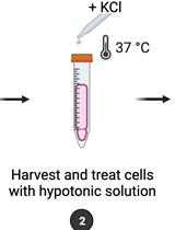
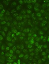
Maintaining the integrity of DNA is a pivotal prerequisite for cells to ensure that all physiological processes function impeccably. Cells are constantly exposed to a plethora of exogenous and endogenous agents that can damage their DNA. Examples of exogenous noxious agents include ultraviolet light, ionizing radiation, and reactive chemical compounds, while endogenous damage can arise due to, e.g., reactive oxygen- and nitrogen species derived from cellular metabolism (Van Loon et al., 2010). If left unrepaired, damage to DNA can lead to mutations, which in turn can alter the functionality of the affected DNA. This can potentially result in altered protein abundance or activity and give rise to diseases such as cancer, neurodegeneration, and aging (Hoeijmakers, 2009; Markkanen, 2017). To counteract the deleterious effects of DNA damage, cells have evolved a series of intricate DNA repair mechanisms that detect and repair such insults (Jackson and Bartek, 2009; Ciccia and Elledge, 2010). Given the relevance of DNA damage in disease, it is of considerable interest to be able to measure levels of DNA damage that are induced by exposure of cells to particular agents or to assess whether the repair kinetics of such damage is altered through specific treatments or genetic backgrounds. Methods to measure DNA damage and repair range from PCR-based methods and enzyme-linked immunosorbent assays to more elaborate next-generation sequencing-based methods (Li and Sancar, 2020). Among this multitude of assays, the comet assay is a widely used method to measure both the amount of DNA damage and its repair in cells (Olive, 2009; Collins, 2014). For this, cells are treated with DNA-damaging agents and embedded into a thin layer of agarose on top of a microscope slide (Figure 1). Subsequent lysis removes all protein and lipid components to leave so-called ‘nucleoids’ consisting of naked DNA remaining in the agarose. These nucleoids are then subjected to electrophoresis, whereby the negatively charged DNA migrates toward the anode depending on its degree of fragmentation and creates shapes resembling comets, which can be subsequently visualized and analyzed by fluorescence microscopy. The comet assay can be adapted to assess a wide variety of genotoxins and repair kinetics, in addition to both DNA single-strand and double-strand breaks. In this protocol, we describe in detail how to perform the alkaline comet assay to assess single-strand breaks and their repair using cultured human cell lines. In addition to cultured human cell lines, the assay can be used for cells directly derived from tissues, provided that they can be dissociated into single-cell suspensions. This exact protocol was used in our recent publication (Clementi et al., 2020). For a detailed description of the neutral comet assay that can be used to detect DNA double-strand breaks, please refer to the separate protocol published in Bio-protocol.

Figure 1. Flowchart of the comet assay. Overview of the sequence of events as discussed in the text.
Materials and Reagents
Superfrost microscopy slides (ThermoScientic, Menzel-Gläser, Superfrost Plus, catalog number: J1800AMNZ), store at room temperature. The use of charged slides is necessary to ensure proper adhesion of agarose to the slides and to minimize sample loss during handling
Rectangular coverslips (Coverslips 22 × 50 mm; ThermoScientic, Menzel-Gläser, catalog number: MA062210), store at room temperature
24-well cell culture plates (TPP, catalog number: 92024), store at room temperature
15 ml Falcon tubes (Greiner, catalog number: 188271 Zentrifugenröhrchen 15 ml, Producer: Huberlab AG, catalog number: 7.188 271), store at room temperature
Trypsin 10× stock solution (Gibco, catalog number: 15090-046), store stock solution at -20°C. Dilute 1:10 in 1× PBS for a working solution; this is stable at 4°C for several weeks
Potassium chloride (KCl) (Merck, catalog number: 1.04936.1000), store at room temperature
Normal melting point agarose (Standard Agarose-Type LE; BioConcept, catalog number: 7-01P02-R), store at room temperature
Low melting point agarose (Low Melt Agarose 100 g; Bio & Sell, catalog number: BS20.47.100), store at room temperature
Na2HPO4·7H2O (Sigma-Aldrich, catalog number: S9390-1Kg), store at room temperature
KH2PO4 (Sigma, catalog number: 602187), store at room temperature
NaCl (Sigma, catalog number: 71380-1KG), store at room temperature
EDTA disodium salt dihydrate (C10H14N2Na2O8·2H2O) (Sigma-Aldrich, catalog number: 03685-1KG), store at room temperature
Tris base (Sigma, Life science, catalog number: T1503-500G), store at room temperature
Sodium hydroxide pellets (NaOH) (Merck, catalog number: 1.06498.100), store at room temperature
DMSO (Sigma-Aldrich, catalog number: D5879-1L), store at room temperature
Triton X-100 (MP-biomedicals/LLC, catalog number: 807426), store at room temperature
SYBR Gold nucleic acid gel stain (Life Technologies, catalog number: S11494), aliquot and store at -20°C protected from light
30% hydrogen peroxide (H2O2) (Roth, catalog number: 8070.2), store at 4°C
Methyl-methanesulfonate (MMS) (Sigma, catalog number: 129925), make 10 M solution in sterile dH2O, aliquot, and store at -20°C
Lysis buffer for the alkaline comet assay (see Recipes)
Electrophoresis buffer for the alkaline comet assay (see Recipes)
Comet staining solution (see Recipes)
PBS (phosphate-buffered saline) (see Recipes)
Equipment
Trevigen CometAssay ESII apparatus (Trevigen, catalog number: 4250-050-ES)
Fluorescence microscope capable of excitation at 470-530 nm to image SYBR-Gold (excitation maximum around 495 nm, emission approx. 537 nm)
Water bath capable of maintaining a constant temperature of 37°C and large enough to hold a 250-ml glass bottle
Big styrofoam box or similar, large enough to hold a glass plate for 10-12 comet slides on ice
Small mechanic’s level
Coplin jars capable of holding 5 or 10 slides
‘Humid chamber’ consisting of a plastic box with a lid, large enough to hold 10-12 slides on a wet paper towel and be placed in an incubator at 37°C
Software
Fiji image processing package (https://imagej.net/Fiji)
OpenComet plugin for Fiji (http://www.cometbio.org/) (Gyori et al., 2014)
Procedure
文章信息
版权信息
© 2021 The Authors; exclusive licensee Bio-protocol LLC.
如何引用
Clementi, E., Garajova, Z. and Markkanen, E. (2021). Measuring DNA Damage Using the Alkaline Comet Assay in Cultured Cells. Bio-protocol 11(16): e4119. DOI: 10.21769/BioProtoc.4119.
分类
癌症生物学 > 基因组不稳定性及突变 > 细胞生物学试验 > DNA结构和改变
发育生物学 > 细胞生长和命运决定
细胞生物学 > 细胞分离和培养 > 单层培养
您对这篇实验方法有问题吗?
在此处发布您的问题,我们将邀请本文作者来回答。同时,我们会将您的问题发布到Bio-protocol Exchange,以便寻求社区成员的帮助。