

A Pulse–chase EdU Method for Detection of Cell Division Orientation in Arabidopsis and Juncus prismatocarpus Leaf Primordia
脉冲追踪EdU法检测拟南芥和笄石菖叶原基细胞分裂方向


发布: 2021年01月05日第11卷第1期 DOI: 10.21769/BioProtoc.3882 浏览次数: 4864
评审: Takumi HigakiAleksandr GavrinAnonymous reviewer(s)
参见作者原研究论文
The authors used this protocol in:
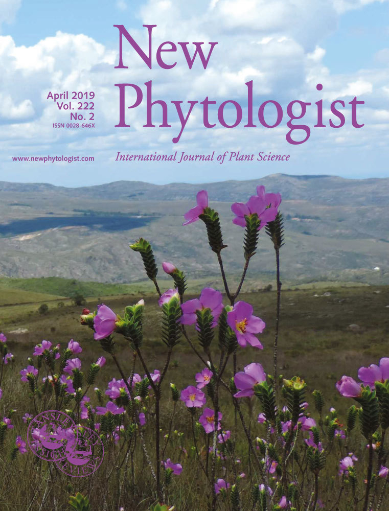
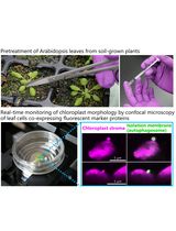
Apr 2019
Advertisement


Abstract
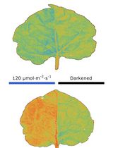
In plants, the morphological diversity of leaves is largely determined by cell division, especially cell division orientation. Whereas cell division itself is easily monitored, the detection and quantification of cell division orientation are difficult. The few existing methods for detection and quantification of cell division orientation are either inefficient or laborious. Here, we describe a pulse-chase strategy using a 5-ethynyl-2’-deoxyuridine (EdU) labeling assay. Plant tissues are first incubated with EdU for a short period (pulse), followed by a long incubation without EdU (chase). Using this method, the positions of daughter cells are easily detected and can be used to quantify cell division orientation. Our protocol is rapid and very efficient for quantitative analysis of cell division orientation, and can be applied to both model and non-model plant species.
Graphic abstract
Plant cell division pairs clearly visualized by a pulse-chase EdU method
Background
Plant cells are attached to each other by cell walls and cannot migrate. Therefore, organized, oriented cell division during the early stages of leaf development largely determines the shapes of mature leaves. Thus far, no method for efficient and rapid detection and quantification of cell division orientation has been reported. Existing methods include visualization of daughter nuclei (in telophase) using a pCYCB1;1::GUS (β-glucuronidase) reporter line (Horiguchi et al., 2011) or visualization of spindle equators (in metaphase) using 4’,6-diamidino-2-phenylindole (DAPI) staining (Fukushima et al., 2015). However, these methods require considerable processing time and effort to identify cells from a particular phase. Recently, 5-ethynyl-2’-deoxyuridine (EdU), a thymidine analog, has become the agent of choice for monitoring cell division activity (Kotogany et al., 2010; Ichihashi et al., 2011; Nakayama et al., 2012, 2014 and 2015; Furuya et al., 2018; Koga et al., 2020). By combining a pulse-chase strategy and EdU staining, we developed a method that can rapidly detect large numbers of cell pairs (two daughter cells after a cell division event) over a short period of time (Yin and Tsukaya, 2016). In brief, plant tissues are first incubated with EdU for a short period (pulse), during which EdU is incorporated in cells in S phase. The pulse period is followed by a long incubation without EdU (chase), allowing EdU-carrying cells to proceed through the subsequent G2 and M phases. By optimization of the lengths of pulse and chase periods, pairs of daughter cells can be detected, because they contain identical amounts of EdU. Cell division orientation can be deduced according to the positions of the two daughter cells. Using this method, we showed that cell division orientation is locally non-stochastic in leaf primordia of Arabidopsis thaliana (Arabidopsis), a model plant species for which leaf morphogenesis has been extensively studied (Yin and Tsukaya, 2016). Furthermore, we combined this method with conventional paraffin sectioning and analyzed cell division orientation in leaves with more complex structures such as those of Juncus prismatocarpus, a non-model plant species (Yin and Tsukaya, 2019).
Materials and Reagents
1.5- or 5-ml tube
APS-coated glass slide (Matsunami, catalog number: SAPS )
Coverslip (Matsunami, catalog number: C025601 )
Razor blade
Aluminum foil
Gloves
Arabidopsis seed
J. prismatocarpus seed
10x phosphate buffered saline (PBS) (pH 7.4) (Nacalai Tesque, catalog number: 27575-31 ) (store at room temperature)
0.5% (v/v) Triton X-100 in 1x PBS (pH 7.4) (store at room temperature)
Click-iTTM EdU Cell Proliferation Kit for Imaging (Thermo Fisher Scientific, catalog number: C10337 ) (store at 4 °C and protect unopened components from light; store stock and working solutions following the manufacturer’s specifications)
DAPI (Thermo Fisher Scientific, catalog number: D1306 ) (store at 4 °C and protect from light)
Höechst 33342 (Component G of 11)
Ethanol (Wako, catalog number: 057-00451 )
Ethanol series (30%, 50%, 70%, 90%, and 100%) (store at room temperature)
Xylene (Wako, catalog number: 244-00081) (store at room temperature, preferably in a fume hood)
(Optional) Histo-Clear (National Diagnostics, catalog number: HS-200 )
Paraffin (McCormick Scientific, catalog number: 39502004 ) (store at room temperature and avoid moisture)
Distilled water (dH2O)
Sucrose (Nacalai Tesque, catalog number: 30403-55 )
Vitamin B5 (Wako ASB-00022727-001 )
Agar (Wako, catalog number: 016-11875 )
1 M KOH
Formaldehyde (Wako, catalog number: 064-00406 )
Glacial acetic acid (Wako, catalog number: 017-00251 )
Murashige and Skoog (MS) plant salt mixture (Wako, catalog number: 392-00591 )
MS plates and liquid MS medium (see Recipes) (store at 4°C)
EdU detection cocktail (see Recipes)
FAA (see Recipes) (store at room temperature)
Equipment
Forceps
Shaker (AS ONE, model: SRR-2 )
Confocal laser scanning microscope (Zeiss, model: LSM 780 )
Incubator (Taitec, model: HB-80 )
Paraffin mold (multiple)
Rotary microtome (Leica, model: Microm HM 355S )
Slide warmer (Tissue Tek, model: PS-52S )
Slide holder
Glass tank (multiple)
Software
Zen 2012 blue edition
Procedure
文章信息
版权信息
© 2021 The Authors; exclusive licensee Bio-protocol LLC.
如何引用
Yin, X. and Tsukaya, H. (2021). A Pulse–chase EdU Method for Detection of Cell Division Orientation in Arabidopsis and Juncus prismatocarpus Leaf Primordia. Bio-protocol 11(1): e3882. DOI: 10.21769/BioProtoc.3882.
分类
植物科学 > 植物发育生物学 > 形态建成
植物科学 > 植物细胞生物学 > 细胞成像
您对这篇实验方法有问题吗?
在此处发布您的问题,我们将邀请本文作者来回答。同时,我们会将您的问题发布到Bio-protocol Exchange,以便寻求社区成员的帮助。